Impetigo
LAST UPDATED: May 28, 2022
Introduction
Impetigo is a contagious bacterial infection of the superficial skin, predominantly occuring in children. There are two forms, the more common non-bullous impetigo, often referred to simply as impetigo, and bullous impetigo.
This chapter is set out as follows:
Aetiology
-
Pathogens
-
Impetigo - in the UK Staphylococcus aureus is the most common organism, Streptococcus pyogenes is the other pathogen involved, and on occasions both organisms can be found together. In warmer climates Streptococcus pyogenes is more prevalent
-
Bullous impetigo - is nearly always caused by Staphylococcus aureus
-
Methicillin-resistant Staphylococcus aureus (MRSA) is an increasingly common cause of impetigo, and is associated more often with the non-bullous form
-
Transmission
-
Impetigo - may be passed on from an infected individual, or arise with no clear source of infection. It often enters the skin at the site of a minor skin injury, or secondarily to another skin condition such as chickenpox
-
Bullous impetigo - can affect intact skin
History
-
Impetigo can affect people of all races
-
Although impetigo can affect any age, the non-bullous form is most common in children 2-5 years of age, and bullous impetigo under the age of 2 years
-
Males and females are affected equally, except in adults where male involvement predominates
-
Impetigo is usually asymptomatic or mildly itchy
Clinical findings
Impetigo
-
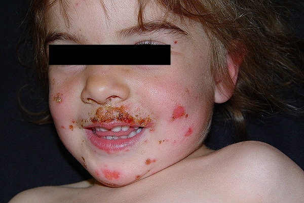
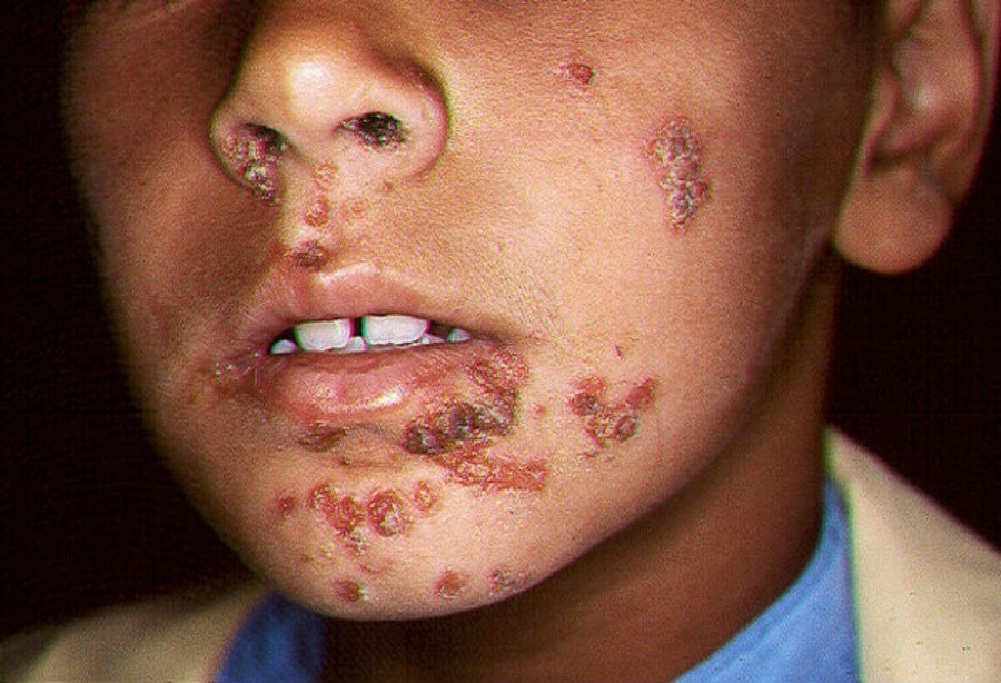
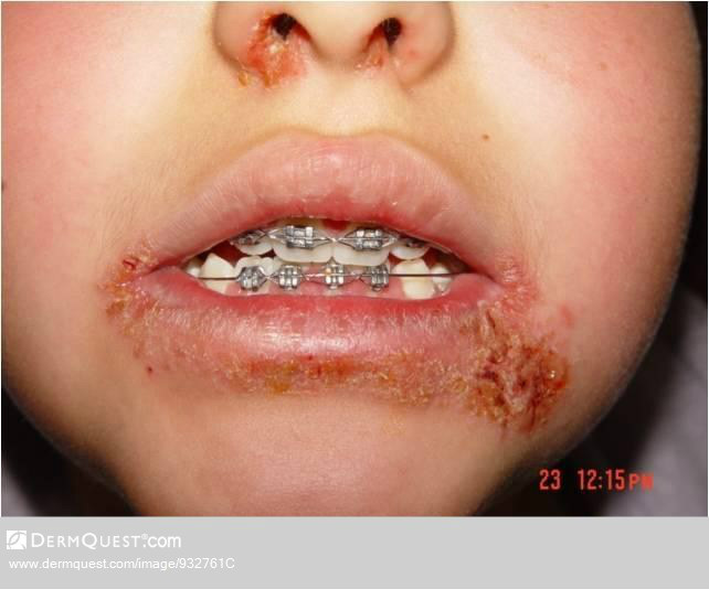
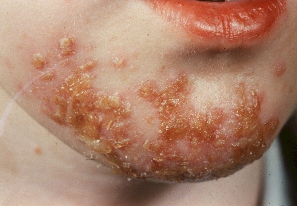
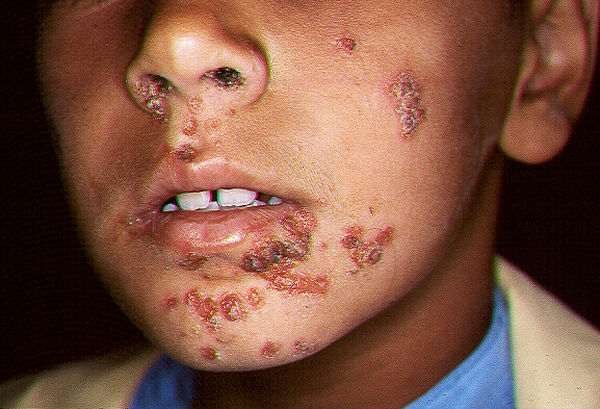
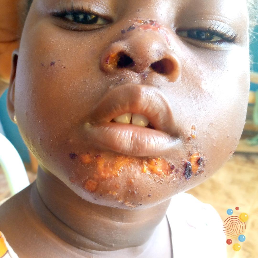
Multiple lesions arise, most commonly on exposed sites such as the face (particularly around the nose and mouth) and limbs, or in the flexures, especially the axillae
-
The initial lesion is a very thin-walled vesicle on an erythematous base, which ruptures easily and is seldom observed
-
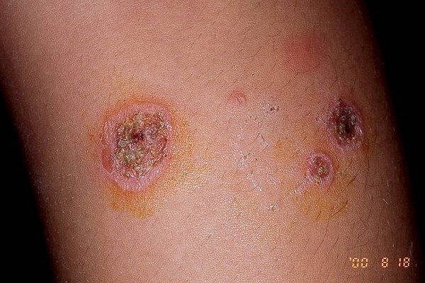
The exudate dries to form golden yellow or yellow-brown crusts, which tend to be thicker in streptococcal infections
-
Lesions extend gradually without central healing
-
Lesions resolve without scaring within 2-3 weeks
-
As with bullous impetigo, if there is an underlying skin condition such as eczema, lesions can become more widespread. Indeed patients with eczema herpeticum may become impetiginised (ie develop secondary impetigo), which can result in a confusing clinical picture, although a patient with primary eczema herpeticum that becomes impetiginised is likely to be more unwell then a child with primary impetigo
-
Mucosal involvement is uncommon
Bullous impetigo
-
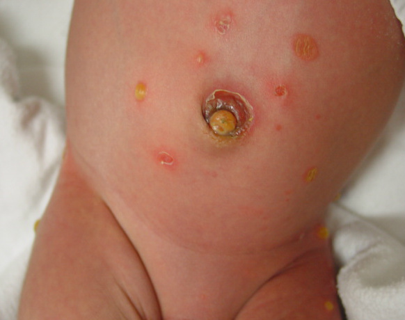
Small or large bullae arise over a short period of time, usually spreading locally on the face, trunk, extremities, buttocks, or perineal regions and may reach distal areas
-
Bullae, which are less easily ruptured than in the non-bullous form, initially contain clear fluid, which then becomes cloudy. Once ruptured, brown-yellow crusts develop
-
Unlike with the non-bullous form, extension occurs with central clearing
-
The buccal mucous membrane can be involved
Clinical Images
Please refer to notes on image rights at bottom of the page with regards to individual image
ownership.
Investigations
-
Impetigo is usually diagnosed on the basis of the clinical appearance
-
Poorly responsive or recurrent cases of impetigo should be swabbed for C&S to identify possible methicillin-resistant Staphylococcus aureus (MRSA). Swabs are best taken from a moist lesion, or, in cases of bullous impetigo from a de-roofed blister
Management
Step 1: general measures
-
Provide a patient information leaflet on impetigo
-
Cover affected areas where possible
-
Wash hands regularly
-
Use separate towels and flannels
-
Avoid school until the lesions are healed or crusted over, or 48 hours after antibiotics are started
Step 2: mild / local infections
-
Consider hydrogen peroxide 1% cream for people with localised non-bullous impetigo who are not systemically unwell or at high risk of complications
-
Other treatments:
-
Topical fusidic acid for 7-10 days
-
In cases of fusidic acid resistance use topical mupirocin
Step 3: more widespread infection
-
Use a systemic antibiotic for 7 days, either flucloxacillin or erythromycin / clarithromycin. Reasons for choosing the latter include cases of penicillin allergy, or if there are concerns with regards to compliance with flucloxacillin given its unpleasant taste and QDS dosing regime
Step 4: MRSA infection
-
Refer to local guidelines
Step 5: persistent or recurrent impetigo
-
The nose is one of the most common sites of carriage for Staphylococcus aureus, so treatment of recurrent cases should include the application of nasal mupirocin up both nostrils BD for 5 days
-
Wash the whole body daily with antibacterial emollient, eg the Dermol range, or anti-septic, eg chlorhexidine
-
Consider a prolonged course of oral antibiotics for up to six weeks
-
Identify and treat other carriers and possible sources of re-infection - it may be useful to take nasal swabs from other household contacts even if they do not have any cutaneous symptoms
Complications
-
Are uncommon and most cases of impetigo settle fully within 2-3 weeks
-
Streptococcal infection occasionally causes acute glomerulonephritis
Disclaimer - the author PCDS cannot accept responsibility for any misleading or incorrect statements, and the management of individual patients remains the direct responsibility of the individual doctor. We do however hope that visitors to this site can contact us regarding comments that are considered misleading or incorrect so that we can continue to improve the site.
Image Rights - The PCDS would like to thank Dermatoweb, DermQuest (Galderma), and others who have contributed images. All named individuals and organisations maintain copyright for the relevant images.