Pyogenic granuloma
LAST UPDATED: Oct 29, 2023
Introduction
A pyogenic granuloma is a common, benign and rapidly growing vascular lesion, possibly resulting from trauma.
This chapter is set out as follows:
Aetiology
-
Trauma is often an important initiating factor, although a clear history cannot always be ascertained
History
-
Most commonly occur in children, young adults and pregnancy, but can arise at any age
-
The lesion is sudden in onset, grows rapidly and bleeds after minimal trauma
Clinical findings
Distribution
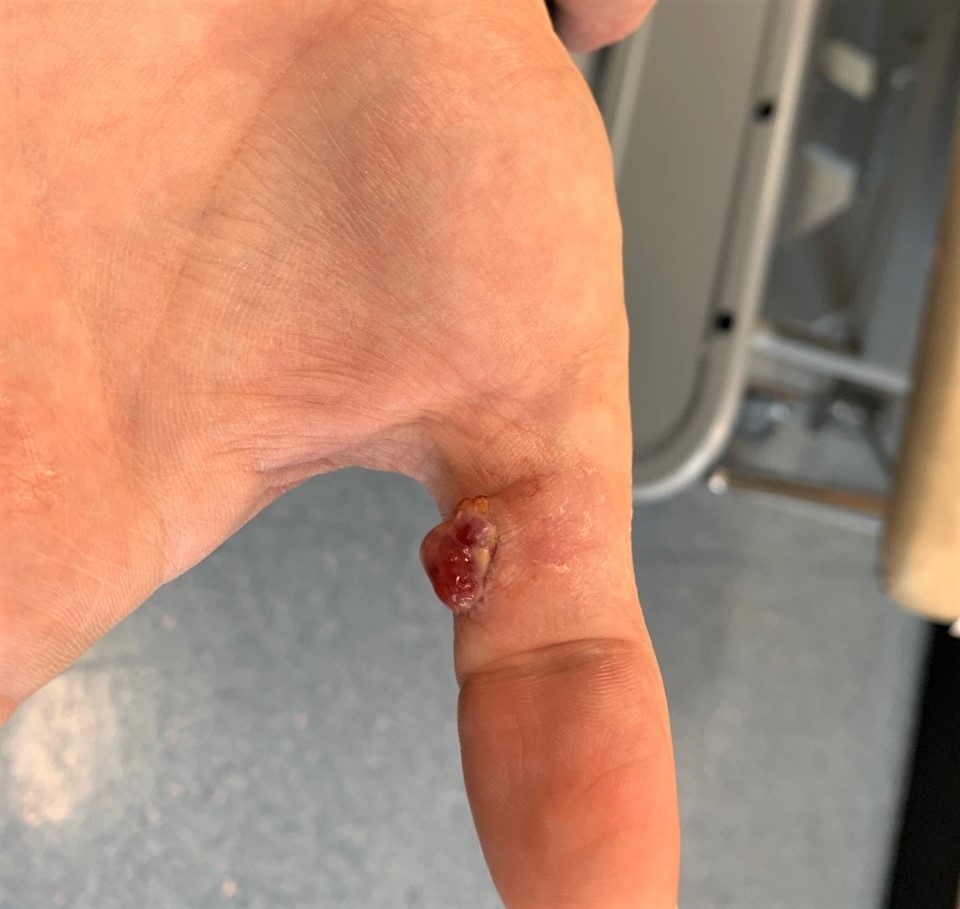
- Can arise on any part of the body but the most common sites are the fingers / hands, head and upper trunk
Morphology
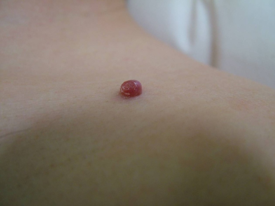
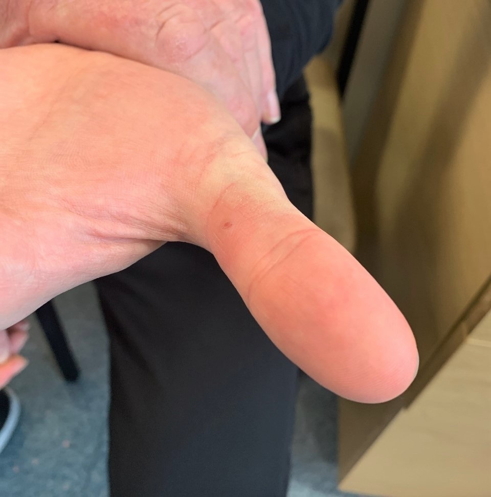
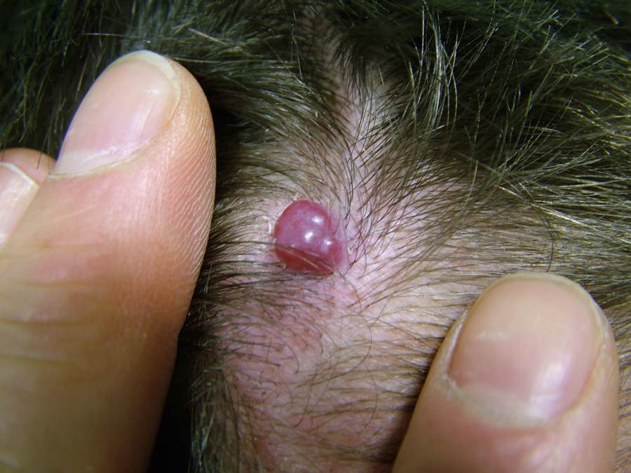
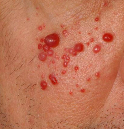
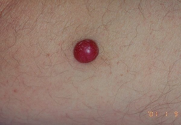
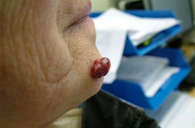
- Starts as a small red spot but quickly enlarges into a nodule
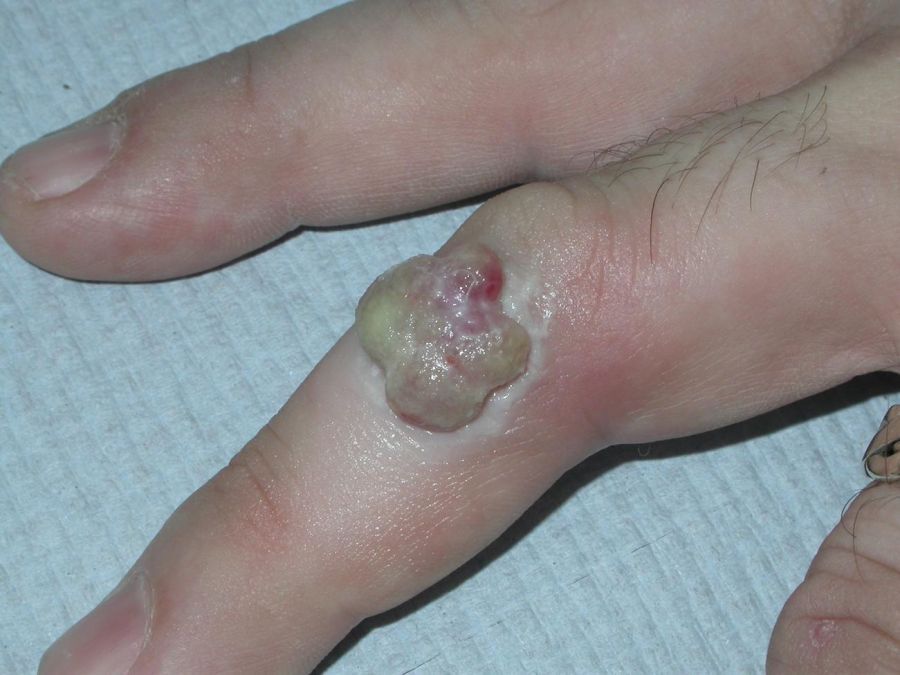
- Initially smooth, but often becomes eroded and bleeds significantly on contact
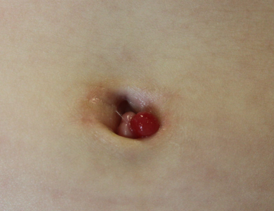
Clinical Images
Please refer to notes on image rights at bottom of the page with regards to individual image
ownership.
Management
- Typical lesions, small: advise the patient/carer as follows - apply Vaseline to the surrounding skin, then as much table salt as possible should then be applied to the whole lesion. The Vaseline protects the surrounding skin and helps hold the salt in place over the lesion. Once salt has been applied, it should then be covered (eg using surgical tape or Clingfilm). The process should be repeated every day until the lesion resolves. Advise the patient that if the lesion has not shrunk significantly within 2 weeks, they must contact the GP immediately to review the diagnosis, and the patient would need to be referred urgently to secondary care
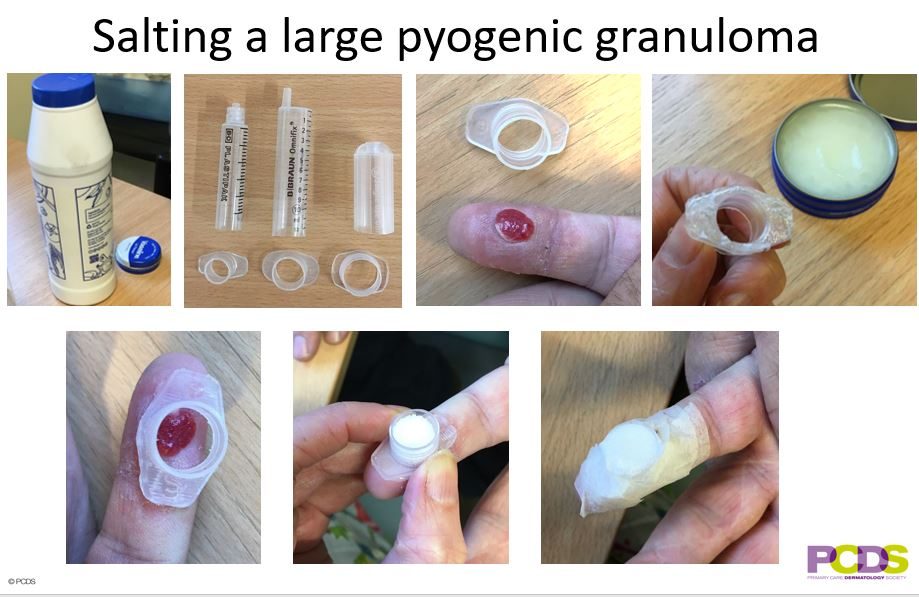
- Typical lesions, large: advise the patient/carer as follows - apply Vaseline to the surrounding skin, then as much table salt as possible should then be applied to the whole lesion, a plastic syringe can be used to hold the salt in place (see photo below). The dressing should be kept dry and the whole process repeated each day until the lesion resolves. It should not bleed after the first application of salt, and should go away completely (or almost completely) with 14 days of salting. Advise the patient that if the lesion bleeds after the first 24 hours of treatment or has not shrunk significantly within 2 weeks, they must contact the GP immediately to review the diagnosis, and the patient would need to be referred urgently to secondary care
-
Although the diagnosis is often straightforward the main differential diagnosis is that of a hypomelanotic melanoma, which tend to bleed less than pyogenic granuloma. Other features that may increase the level of suspicion include no clear history of trauma and an atypical site or age group. To this end, lesions needing diagnosis (as well as treatment) are best removed surgically (deep curettage and cautery, or excision) and sent for urgent histology - if this cannot be done in Primary Care within four weeks of presentation / or there is a concern about possible hypomelanotic melanoma, then the patient should be referred urgently to Secondary Care
-
It is also important to be aware that persistently discharging umbilical granulomas in neonates could signify deeper involvement
Additional images
1
Disclaimer - the author PCDS cannot accept responsibility for any misleading or incorrect statements, and the management of individual patients remains the direct responsibility of the individual doctor. We do however hope that visitors to this site can contact us regarding comments that are considered misleading or incorrect so that we can continue to improve the site.
Image Rights - The PCDS would like to thank Dermatoweb, DermQuest (Galderma), and others who have contributed images. All named individuals and organisations maintain copyright for the relevant images.