Vasculitis
LAST UPDATED: Jul 18, 2023
Introduction
Vasculitis refers to the inflammation and necrosis of blood vessels and may be localised or systemic. Many of the vasculitides (conditions associated with vasculitis) have a cutaneous component. In all cases a thorough work-up is required to investigate for an underlying cause and/or associated systemic features.
This chapter, which is set out as below, provides a logical approach to the patient presenting with cutaneous features of vasculitis (with particular detail in the section on investigations), in addition to management recommendations for cutaneous small vessel vasculitis.
Aetiology
Classification of vasculitis - as defined by the size of the blood vessels involved
The list below excludes a number of other skin conditions classified as vasculitis such as rheumatoid nodules, Behçet’s syndrome, erythema elevatum diutinum, granuloma faciale, and lymphomatoid granulomatosis, as these differ greatly in their presentation, investigation, and management. Vasculitis can also be found secondary to a number of other conditions such as inflammatory bowel disease, sarcoid and haematological malignancies.
Small vessel vasculitis
-
Cutaneous small vessel vasculitis - there are multiple causes (see later) including:
- Infection or drug-induced (sometimes referred to as hypersensitivity or allergic vasculitis). Examples of infections include streptococcus pyogenes, hepatitis, and HIV
- Those associated with connective tissue disorders - refer to the related chapter Connective tissue disorders for more information
- Primary cutaneous small vessel vasculitis (idiopathic) - the most common type; a diagnosis of exclusion
- Septic vasculitis - these are infections causing sepsis such as infective endocarditis, meningococcus, and Dengue haemorrhagic fever
- Henoch-Schönlein purpura (HSP)
-
Exercise-induced vasculitis
- Urticarial vasculitis
-
Mixed cryoglobulinaemia
- Waldenstrom’s hypergammaglobulinaemic purpura
Small vessel vasculitis associated with Anti-Neutrophilic Cytoplasmic Autoantibodies (ANCA)
- Eosinophilic Granulomatosis with Polyangiitis (EGPA) - formerly Churg Strauss Syndrome (CSS)
- Microscopic Polyangiitis (MPA)
- Granulomatosis with Polyangiitis (GPA) - formerly Wegener’s Granulomatosis
Medium vessel vasculitis
-
Polyarteritis nodosa (PAN), which is subdivided into:
-
Microscopic polyarteritis
-
Cutaneous PAN
-
Systemic PAN
- Kawasaki disease
- Nodular vasculitis (syn. erythema induratum of Bazin and of Whitfield)
Large vessel vasculitis
- Giant cell arteritis
- Takayasu's arteritis
Clinical findings
There can be significant overlap in the clinical findings of the various vasculitides. To try and help better understand this complex area of medicine this section first describes the various clinical features seen in vasculitis, and then further examines the individual conditions.
Clinical features of vasculitis
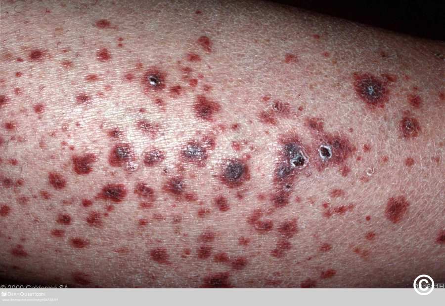
Palpable purpura
- Results from extravasation of erythrocytes into the dermis
- Is the most commonly identifiable clinical feature of the small vessel vasculitides. It can also be seen, although less commonly, in conditions where there is additional involvement of medium-sized vessels, such patients are usually constitutionally unwell with features of a systemic vasculitis
- Presents predominantly on dependent sites e.g. lower legs, with papules, plaques and sometimes bullae, and is commonly associated with post-inflammatory hyperpigmentation
- A retiform pattern (net-like) of purpura correlates with medium-large vessel vasculitis
- The differential diagnosis of purpura includes easy bruising, infection, thrombocytopenia, and scurvy
Ulceration and necrosis
- Most commonly seen on dependant areas of skin
- Small vessel disease - mainly associated with more superficial ulceration, although some ulcers can be more substantial
- Larger vessel disease - ulcers tend to be larger, and are sometimes sharply demarcated
- The differential diagnosis of tissue necrosis includes infection, physical factors (eg perniosis - chilblains), inflammatory dermatoses (eg pyoderma gangrenosum), emboli, warfarin-induced necrosis (and occasionally other adverse cutanenous drug reactions), calciphylaxis, and malignancy
Nodules
- Can be subcutaneous, tender, discoloured, and ulcerated
- The presence of nodules implies a larger vessel vasculitis
- In polyarteritis nodosa the nodules may run along the direction of the affected vessels
- In nodular vasculitis the affected skin is usually the bottom-third of the lower leg/ankle
Livedo reticularis
- Livedo reticularis is a livedoid discoloration of the skin in a reticular pattern
- Broadly speaking, livedo is divided into physiological and pathological livedo
- Physiological livedo (cutis marmorata) is commonly seen on the legs of infants and young women in cold weather and improves on rewarming
- Pathological livedo is a cutaneous manifestation of a number of systemic conditions including the Antiphospholipid syndrome, polyarteritis nodosa, cryoglobulinaemia, Sneddon's syndrome and occasionally the Anti-neutrophilic Cytoplasmic Antibody (ANCA)-positive vasculitides
- For more information refer to the related chapter on Livedo reticularis
Urticarial vasculitis
-
May be a vasculitis in its own right, or can be secondary to one of the other vasculitides
-
Patients may complain of burning symptoms, as opposed to itching
-
Clinically urticarial-like lesions persist greater than 24 hours, and often demonstrate purpuric foci and post-inflammatory hyperpigmentation
-
Is often associated with angioedema
-
Urticarial vasculitis is divided into two groups:
-
Normocomplementaemic ie normal levels of complement
-
Hypocomplementaemia, which is strongly associated with Sjögren's syndrome and systemic lupus erythematosus
-
For more information refer to the related chapter Urticarial vasculitis
Hands and feet
-
In addition to palpable purpura and ulceration, the hands and feet can provide other important clues as to the type of vasculitis and/or cause
-
Multiple sites of digital ischaemia/necrosis are usually associated with larger vessel vasculitis or a cryoglobulinaemia, but can also be seen in embolic events
-
Subungual (splinter) haemorrhages are commonly seen in subacute infective endocarditis, but may also be seen in patients with other types of vasculitis or secondary to trauma
-
Connective tissue conditions, which can be associated with vasculitis, will often have cutaneous changes on the extensor surfaces of the hands and nail folds
The vasculitides (conditions associated with vasculitis)
Identifying the type and/or cause of vasculitis requires a considerable work-up, with some cases requiring more urgency than others. The section on investigations provides a logical approach on how to approach cases presenting with cutaneous vasculitis.
Cutaneous small vessel vasculitis (CSVV)
-
The most common dermatological vasculitis
- Causes include infection (10-15%), autoimmune disorders such as systemic lupus erythematosus, dermatomyositis, and rheumatoid arthritis (15-20%), and 5% of cases are associated with malignancy
- Approximately 50% of cases are primary cutaneous small vessel vasculitis (idiopathic), which is a diagnosis of exclusion. It is important to highlight that some cases of what initially appear to be CSVV may instead represent the initial clinical presentation of an occult, pre-clinical, evolving systemic disease, hence the importance of a thorough work-up in all such patients - for more detail refer to the section on investigations (which also lists the most common drug causes)
-
Clinical features
-
Palpable purpura of dependant sites
-
Can cause ulceration, necrosis and post-inflammatory hyperpigmentation
-
Livedo reticularis is uncommon
-
Systemic involvement is uncommon
-
Histopathological examination shows leucocytoclastic vasculitis (ie vascular damage caused by nuclear debris from infiltrating neutrophils), which is a feature of several of the other vasculitides
-
Cutaneous small vessel vasculitis is usually self-limiting with lesions resolving within several weeks to months, although up to 10% of cases will have periodic flare-ups over a number of years
- Refer to the section on management for how to treat patients with CSVV
Henoch-Schönlein purpura (HSP)
- Defined as a small vessel vasculitis involving deposition of IgA immune complexes, that characteristically involves the skin, gastrointestinal system and glomeruli with or without arthritis or arthralgia
-
90% of cases arise under the age of 10. Occasionally older children and adults are affected
-
In the majority no trigger is found, although Group A beta-haemolytic streptococci is found in approximately 30% of cases
-
Cutaneous features
- Cutaneous lesions are classically symmetrical affecting extensor surfaces, particularly the elbow, knees and buttocks. The trunk and face can also be affected
-
The lesions are erythematous patches within which develop areas of haemorrhage and palpable purpura. Necrotic ulcers are sometimes seen
-
Crops of new lesions arise periodically until the cutaneous features subside, which is usually around 2-3 months
-
Systemic features
- The main features of HSP are usually preceded by 2–3 weeks of fever, headache, muscle/joint aches, or abdominal pain
-
75% of cases have joint involvement, which usually involves one to four joints, especially the ankles and knees. It may be transient and move between different joints
- Abdominal pain is present in half to three-quarters of patients and precedes the rash in up to one third. Abdominal pain may be associated with bloody diarrhoea. Orchitis and intussusception are possible complications
-
50% of cases have renal involvement, with 10% having serious involvement at the onset. Less than 3% of cases progress to end-stage renal failure
-
Histopathology shows a leucocytoclastic vasculitis
-
HSP is usually self-limiting, the main exception being those patients with moderate-severe renal involvement
Exercise-induced vasculitis
- A small vessel vasculitis that affects one or both lower legs following strenuous exercise such as long-distance running and hiking
-
Symptoms are worse in warm weather. It is more common in women, and patients aged over 50 years
-
Clinical features
-
Intense itching, stinging, or burning pain
-
Erythema, urticated lesions and palpable purpura
-
Oedema
-
The patient is otherwise well
-
Lesions take several weeks to fade and may leave post-inflammatory hyperpigmentation
-
Histopathology shows a leucocytoclastic vasculitis
-
The condition is self-limiting. Treatment is not usually required, although for more marked symptoms, leg elevation and NSAID may be of benefit
Septic vasculitis
- A small vessel vasculitis that is typically immune-complex negative and caused by infective endocarditis or septicaemia from gonococci, meningococci, pseudomonas, staphylococci, streptococci, certain rickettsial infections, and other microorganisms
-
Clinical features
-
Purpura (petechiae and ecchymoses), vesiculo-pustules (often with grey roofs signifying necrosis), haemorrhagic bullae, and, rarely, ulceration
-
Infective endocarditis may also have splinter haemorrhages, Osler nodes (painful, palpable, erythematous lesions most often involving the pads of the fingers and toes) and Janeway lesions (non-tender, macular lesions most commonly involving the palms and soles, occuring more frequently in endocarditis caused by Staphylococcus aureus)
Waldenstrom’s hypergammaglobulinaemic purpura
- A rare syndrome that is characterised by recurrent bouts of purpura occurring mainly on the lower extremities and dorsum of the feet. Lesions last for approximately one week. Prolonged walking, standing or sitting may precipitate attacks. Itching or a burning sensation may occur
-
There is a female preponderance
-
Investigations
-
The hallmark of this condition is polyclonal hypergammaglobulinaemia primarily composed of IgG
-
The ESR is often raised and there can be associated anaemia and leucopenia
-
Most patients have a positive ANA and anti-Ro/SSA or anti-La/SSB antibodies
-
Histopathology may show features of a small vessel vasculitis
-
Numerous associations have been shown with other systemic conditions such as Sjögren's syndrome, systemic lupus erythematosus, and rheumatoid arthritis
Cryoglobulinaemic vasculitis
- A small-medium sized vessel vasculitis
-
Cryoglobulins are immunoglobulins that precipitate or gel in the cold and dissolve on rewarming. Three types of cryoglobulins are distinguished based on whether the cryoglobulin is monoclonal and has rheumatoid factor activity. Knowing the type usually allows the physician to predict the clinical features; alternatively knowing the clinical features allows one to deduce the type of cryoglobulin. Essential cryoglobulinaemia is a term sometimes used when there is no identifiable cause for the cryoglobulinaemia
- Type I
- A monoclonal antibody that does not have rheumatoid factor activity. It is less common than types II and III
- Most commonly associated with lymphoproliferative disorders including lymphoma, Waldenström’s macroglobulinaemia (a low-grade form of lymphoma), and multiple myeloma
- Because type I cryoglobulins do not easily activate complement, patients with type I are asymptomatic until the level of cryoglobulinaemia is sufficiently high to cause a hyperviscosity syndrome
- Types II and III (mixed cryoglobulinaemia)
- Are rheumatoid factors
- In type II, the rheumatoid factor is monoclonal, whereas in type III it is polyclonal
- It is now evident that many patients diagnosed with type II or type III cryoglobulinaemia have the disease as an immune response to chronic hepatitis C infection. Other causes include connective tissue disorders and other chronic infection
- Type II and III cryoglobulinaemia frequently presents as vasculitis, most commonly with recurrent lower extremity purpura, glomerulonephritis, and peripheral neuropathy
- Histopathology initially shows a leukocytoclastic vasculitis, more chronic lesions show a mononuclear-predominant infiltrate
-
Cutaneous features
-
Are nearly always present in cryoglobulinaemia
-
Observed lesions have a predilection for dependent areas (particularly the lower extremities) and include erythematous macules and purpuric papules (90-95%), as well as ulcerations (10-25%), livedo reticularis and urticarial vasculitis
-
Lesions in non-dependent areas are more common in type I cryoglobulinaemia, as are livedo reticularis and severe Raynaud's phenomenon with ulceration and necrosis of digits. Nail fold capillary abnormalities are common
-
Making a diagnosis of cryoglobulinaemic vasculitis is important because systemic steroids, which may be needed in the short-term, will in the long-term worsen the underlying infection that is present in most cases. Clues to the diagnosis include:
-
Symptoms are initially intermittent
-
Worsening of lesions after prolonged standing, or in colder weather
-
Involvement of non-dependant sites, marked livedo reticularis and severe Raynaud's phenomenon with ulceration and necrosis of digits (mainly features of type I)
-
Low complement C4 levels are found in 90% of cases, and rheumatoid factor is positive in types II and III
-
The presence of Hepatitis C infection
-
Histopathological features of a leucocytoclastic vasculitis
-
Testing for cryoglobulins - cryoglobulins are very unlikely to be present if complement levels and rheumatoid factor are normal. If levels need to be checked they can only be tested in Secondary Care as stringent temperature control, maintaining the sample at 37 degrees Celsius, is needed from the time the sample is collected until separation of the serum
Microscopic polyangiitis
-
A systemic vasculitis affecting blood vessels ranging in size from small capillaries to medium-sized arteries. The cause is unknown
-
Cutaneous features
-
50% have skin changes, mainly with palpable purpura on dependent sites
-
Livedo reticularis, digital ischaemia and urticarial vasculitis are uncommon findings
-
Systemic features
-
Constitutional symptoms such as fever, weight loss, myalgia and arthralgia are common
-
70-90% have necrotising glomerulonephritis
-
25-50% have respiratory complications
-
Investigations
-
Autoantibodies: MPO-ANCA (p-ANCA pattern) and RF are often positive. PR3-ANCA (c-ANCA pattern) is often negative
-
Histopathology from purpuric lesions show a leukocytoclastic vasculitis
Wegener’s granulomatosis (syn. granulomatosis with polyangiitis)
-
A triad consisting systemic vasculitis of small and medium-sized vessels, necrotising granulomatous inflammation of the respiratory tract (upper and lower) and glomerulonephritis
-
The aetiology is thought to be an amplified immune response to an antigenic stimulus, such as an infection
-
Cutaneous features
-
Uncommon at onset, although 40% will eventually develop skin changes
-
Palpable purpura on the extremities is the most common presentation, followed by oral ulcers
-
Less common findings include subcutaneous nodules, ulceration, non-specific lesions and pyoderma gangrenosum-like lesions
-
Systemic features
-
In 80% of cases respiratory tract involvement is present at onset, and the majority of these will have nasal, sinus, tracheal or ear involvement
-
Eventually 77% of cases develop glomerulonephritis
-
Investigations
-
Autoantibodies - 90% are positive for PR3-ANCA (c-ANCA pattern), many are positive for RF. Less than 10 % have positive MPO-ANCA (p-ANCA pattern) antibodies
-
Histopathology - leucocytoclastic vasculitis and/or granulomatous inflammation can be found in up to 50% of skin biopsies
Churg-Strauss syndrome (syn. allergic granulomatous angiitis)
-
Is a systemic vasculitis characterised by asthma, blood eosinophilia, and necrotising vasculitis with extravascular granulomas. The vasculitis affects small to medium-sized arteries and veins. The cause is unknown
-
Three phases have been described
- 1st stage - allergic rhinitis, nasal polyps, asthma and peripheral blood eosinophilia may last for years
- 2nd stage - multiorgan vasculitis, cardiac involvement is the most severe complication
- 3rd stage - results from renal damage and peripheral neuropathy, with complete resolution of many of the other features of the condition
-
Cutaneous features
-
Affect 70% of cases at some stage
-
Palpable purpura of the extremities, and infiltrated nodules are the most common findings, although other features such as livedo reticularis can be seen
-
Investigations
-
Autoantibodies - 50-80% are positive for MPO-ANCA (p-ANCA pattern). PR3-ANCA (c-ANCA pattern) is rare
-
Histopathology shows vasculitis of arteries and veins, with eosinophilia and extravascular granulomas
Polyarteritis nodosa (PAN)
-
Necrotising inflammation of medium-sized or small arteries, without glomerulonephritis or vasculitis in arterioles, capillaries or venules. Vascular involvement occurs preferentially at vessel bifurcations, resulting in microaneurysm formation, aneurysmal rupture with haemorrhage, thrombosis, and consequently organ ischemia or infarction. Hepatitis B is one of the most common causes
-
Patients are constitutionally unwell, and have significant weight loss, along with serious systemic involvement of the renal system (hypertension and renal failure), cardiovascular system (ischaemic heart disease), gastrointestinal tract (infarction, perforation) and the nervous system (peripheral neuropathy, and sometimes CNS complications). PAN does not affect the lungs and it does not cause glomerulonephritis
-
Cutaneous features
-
Are very common, and 40% of patients manifest with skin lesions
-
The classical cutaneous feature is that of a subcutaneous nodule or group of nodules, often on the lower limbs and in proximity to the vessels. The nodules may be tender, or ulcerated
-
Livedo reticularis may occur, is usually palpable, and may ulcerate. Ulcers have a 'punched-out' appearance
-
Ulceration and necrosis of the digits or penis may be seen
-
Investigations
-
Autoantibodies - 20% are positive for MPO-ANCA (p-ANCA pattern)
-
Histopathology initially shows a leucocytoclastic vasculitis, more chronic lesions show a mononuclear-predominant infiltrate
Cutaneous polyarteritis nodosa
-
Is a ‘benign’ variant that is largely confined to the skin. Patients have fewer constitutional symptoms, but may have mild involvement of the muscles and nerves. Cutaneous features are similar, however, necrotic tissue damage to digits is uncommon and could actually represent systemic PAN
Nodular vasculitis (syn. erythema induratum of Bazin and Whitfield)
-
Is a chronic relapsing lobular panniculitis with septal vasculitis. It is typically seen in healthy, middle-aged, sometimes obese women with venous stasis
-
Cutaneous features
-
Tender, dusky nodules or plaques
-
Often affects the posterolateral aspects of the lower third of one or both legs
-
The differential diagnosis includes other causes of panniculitis, PAN, and Takayasu's arteritis
-
Although in most cases no cause will be identified, it is important to exclude tuberculosis, which is more likely if lesions are ulcerated. Deep incisional biopsies should be taken for both histopathological examination, and tissue culture to look for either mycobacterium tuberculosis, or atypical mycobacteria. The use of PCR (polymerase chain reaction) testing aids in diagnosis and significantly reduces the number of false negative results
Takayasu’s arteritis
-
Is a large vessel vasculitis with granulomatous inflammation of the aorta and its major branches, resulting in cardiac and renal complications
-
Skin lesions have been reported in up to one-third of patients and may comprise subcutaneous nodules, with or without ulcerations, and less commonly papulonecrotic rashes
Lymphocytic vasculitis
-
There is some controversy as to the acceptance of the concept of lymphocytic vasculitis
-
The problem that arises when a lymphocytic vasculitis is evident histopathologically is that many of these conditions only show lymphocytic vasculitis in a small minority of cases
-
Lymphocytic vasculitis is recognised in a number of scenarios, including:
-
It is thought, by some, to be an end-stage finding of some cases of leukocytoclastic vasculitis
-
Behçet’s syndrome
-
Certain drug reactions
-
Some cases of SLE
-
Lymphomatoid granulomatosis
Clinical Images
Please refer to notes on image rights at bottom of the page with regards to individual image
ownership.

1

2

3

4

5

6

7

8

9

10

11

12

13

14

15

16

17

18

19

20

21

22

23

24

25

26

27

28

29

30

31

32

33

34

35

36

37

38

39

40

41

42

43

44

45

46

47

48

49

50

51

52

53

54

55

56

57

58

59

60

61

62

63

64

65

66

67

68
Investigations
A logical approach to the patient presenting with possible vasculitis
Before going further it is important to highlight the common features of exercise-induced vasculitis, and capillaritis as early identification of the these two conditions means that further investigations are not required. A drug-related cause may also avoid the need for thorough investigations:
- Exercise-induced vasculitis follows intense exercise presenting with intense itching, stinging, or burning along with erythema, urticated lesions and palpable purpura on the lower legs. The patient is otherwise well. Lesions take several weeks to fade and may leave post-inflammatory hyperpigmentation
- Capillaritis, which is also self-limiting, presents most commonly with red-brown patches (comprised of many small 'cayenne pepper-like' spots) or occasionally non-palpable purpura. For more information refer to the chapter pigmented purpuric dermatoses (syn. capillaritis)
- Medications and other drugs
- Are more likely to be associated with small vessel vasculitis, but are occasionally associated with systemic vasculitis
- Blood eosinophilia is noted in up to 80% of cases with systemic involvement
- The most common drugs implicated are penicillin's, minocycline, quinolones, 'sulfa' drugs, NSAID and other analgesics, thiazides and other diuretics, anticonvulsants, allopurinol, vaccines, cocaine, and other illicit drugs
While most cases of cutaneous vasculitis are self-limiting (primary cutaneous small vessel vasculitis being the most common) and without identifiable systemic involvement, some cutaneous changes may be the initial clinical presentation of occult, preclinical, evolving systemic disease. As such the assessment of the patient presenting in Primary Care with cutaneous features of vasculitis is best approached as follows:
- Red flags suggesting a patient could have systemic vasculitis requiring urgent Secondary Care intervention, include:
- Constitutionally unwell
- Neuropathy
- Swollen and inflamed joints
- Tender nodules
- Fixed livedo reticularis
- Necrosis of digits
- Abnormal renal findings (new onset or worsening hypertension / urinalysis - haematuria, proteinuria, casts / elevated serum creatinine levels)
- All patients without red flags require a through medical history, examination, and investigations as laid out in the screening tests referred to in the SEPARATE CHAPTER Investigations. These tests include screening for infections such as streptococcus, hepatitis B and C (the latter is associated with cryoglobulinaemia), and HIV, in addition to screening for other conditions in which features of a systemic vasculitis may not be apparent at the onset such as ANCA positive vasculitis and connective tissue disorders
- Vasculitis can be secondary to inflammatory bowel disease, haematological and other malignancies
- Only if no cause is found can we then think about a diagnosis of primary cutaneous small vessel vasculitis, which is a diagnosis of exclusion
Skin biopsies
Two biopsies of skin lesions (as opposed to peri-lesional skin) should be sent - one for histology and the other for direct immunofluorescence.
Histology
-
The correct biopsy technique is important
-
A punch biopsy for small vessel disease
-
Deeper, elliptical incisions should be performed for larger vessel disease
-
To use the term vasculitis in a meaningful way in dermatopathology, it must reflect damage to vessel walls and not merely the proximity of inflammatory cells to them
-
The most common finding in vasculitis is a leucocytoclastic vasculitis, which refers to vascular damage caused by nuclear debris from infiltrating neutrophils. A lymphocytic vasculitis is a less common finding, and is sometimes poorly understood
-
The concept of primary versus secondary vasculitis is difficult to grasp and apply, but helps to keep clinicopathologic correlation accurate
-
In a primary vasculitis the insult to vessel walls is an essential part of the condition - leukocytoclastic vasculitis is a clear cut example
-
In secondary vasculitis the essential part of the condition resides outside vessel walls, but the inflammatory cells that respond to the insult in turn involve vessels The determination of whether a vasculitis is primary or secondary turns on whether a pathologist can recognise the changes of the first injury, which can be subtle
-
In the case of both large and small vessels, the surest sign of injury is necrosis of the vessel wall. The term "necrotising vasculitis" reflects this, but is wildly overused, as very few vasculitides result in substantial necrosis of vessels
Direct immunofluorescence (DIF)
The findings depend on the cause:
- Perivascular IgA is characteristic of Henoch-Schönlein purpura
- Perivascular IgM suggests cryoglobulinaemia or rheumatoid arthritis
- Interface immunoglobulins may be a sign of systemic lupus erythematosus
- DIF is often negative in cutaneous polyarteritis nodosa and ANCA-positive vasculitis
Management
Assessing the patient with vasculitis
- Many cases of vasculitis require support from Secondary Care:
- Refer to the section above on investigations as to which cases require urgent intervention
- In terms of the 'well' patient with apparent cutaneous vasculitis, those patients with necrotic lesions will require more urgent intervention compared to those without necrotic change
Management of cutaneous small vessel vasculitis (CSVV)
- Remove and/or treat any causal factors e.g. a drug reaction or infection
-
Conservative measures
- Leg elevation and compression (if not contraindicated)
- Potent to super-potent topical steroids
- NSAIDs can help improve symptoms
- Commonly used systemic treatments include:
- High-dose tetracycline antibiotics (eg doxycycline 100 mg BD), which have an anti-inflammatory affect
- Colchicine 0.5 mg BD
- Dapsone 50-100 mg OD
- If areas of skin appear to becoming necrotic consider a short course of systemic steroids - prednisone 0.5 mg/kg/day for 1–2 weeks, tapered over 3–6 weeks
- For more severe and recalcitrant disease other treatments considered include hydroxychloroquine, ciclosporin, methotrexate, mycophenolate, azathioprine, and the biologics
Disclaimer - the author PCDS cannot accept responsibility for any misleading or incorrect statements, and the management of individual patients remains the direct responsibility of the individual doctor. We do however hope that visitors to this site can contact us regarding comments that are considered misleading or incorrect so that we can continue to improve the site.
Image Rights - The PCDS would like to thank Dermatoweb, DermQuest (Galderma), and others who have contributed images. All named individuals and organisations maintain copyright for the relevant images.