Livedo reticularis and livedoid vasculopathy
LAST UPDATED: Jul 01, 2023
Introduction
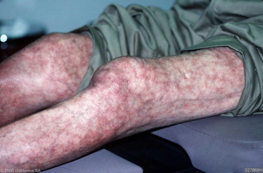
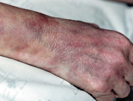
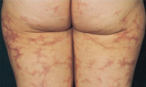
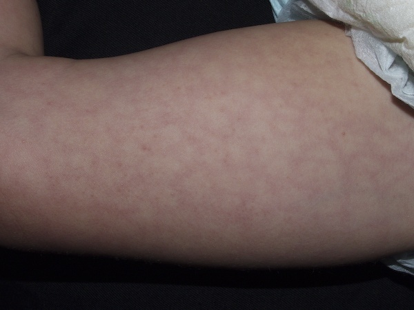
Livedo reticularis is a livedoid discolouration of the skin in a reticular pattern. Broadly speaking, livedo is divided into physiological and pathological livedo. Physiological livedo (cutis marmorata) is commonly seen on the legs of infants and young women in cold weather and improves on rewarming. Pathological livedo is a cutaneous manifestation of a number of systemic conditions, most notably the Antiphospholipid syndrome (APS), and is associated with arterial and venous thrombosis and increased pregnancy morbidity irrespective of the presence of antiphospholipid antibodies.
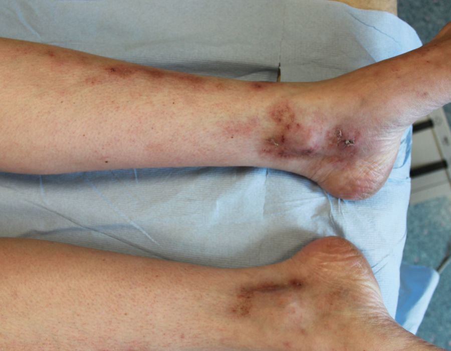
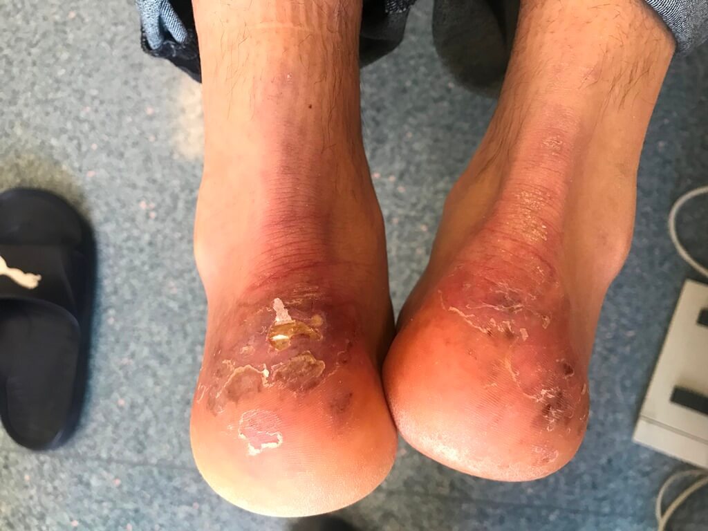
Livedoid vasculopathy is a rare, chronic vascular disorder characterised by persistent painful ulceration of the lower extremities. The condition occurs chiefly but not exclusively on the lower leg or foot. Occasionally, patients with livedoid vasculopathy will also have livedo reticularis, acrocyanosis, or Raynaud's phenomenon.
This chapter is set out as follows:
Aetiology
Pathological livedo reticularis may be a sign of vasculitis or of obstruction of the vessel by a circulating material. Causes include the following:
-
Livedo can be an initial manifestation in the primary APS, and up to 70% of patients with systemic lupus erythematosus associated with the APS
-
Polyarteritis nodosa and antineutrophil cytoplasmic antibody (ANCA)-positive vasculitis
-
Cryoglobulinaemia
-
Other connective tissue disorders such as dermatomyositis and rheumatoid arthritis
-
Thromboangiitis obliterans
-
Primary thrombocytopenia and polycythaemia vera
-
Pyoderma gangrenosum
-
Sneddon's syndrome is a form of idiopathic livedo reticularis with systemic involvement ie internal blood vessels are affected, most commonly in the brain, eye and heart
Clinical findings
Cutaneous features of livedo reticularis
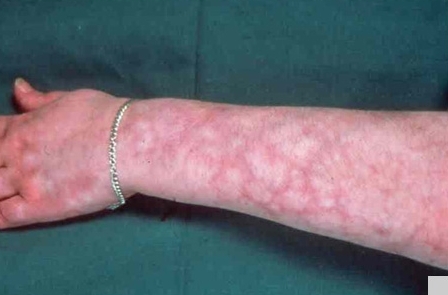
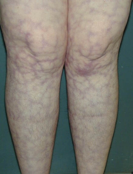
- A red-blue, reticular (net-like) discolouration surrounding pale central areas
-
The network may comprise regular unbroken circles or an irregular broken pattern
-
It occurs mostly on the legs, arms and trunk and is more pronounced in cold weather
-
In physiological livedo the signs improve as the skin warms, and the patient is otherwise well
-
In pathological livedo the rash is persistent, non-reversible with rewarming, and the patient may be unwell
-
The term livedo racemosa is sometimes used when the livedo is more generalised, and has an irregular-broken shape. While this pattern is recognised in Sneddon's syndrome, it can also be found in other causes of pathological livedo such as the APS and the ANCA-positive vasculitides
Systemic associations with pathological livedo reticularis
- Arterial and venous thrombosis has been observed in patients with livedo irrespective of the presence of antiphospholipid antibodies
- Pregnancy morbidity also appears to be higher in patients with SLE and livedo, even when negative for antiphospholipid antibodies
- Accelerated atherosclerosis may be a feature
Livedoid vasculopathy
-
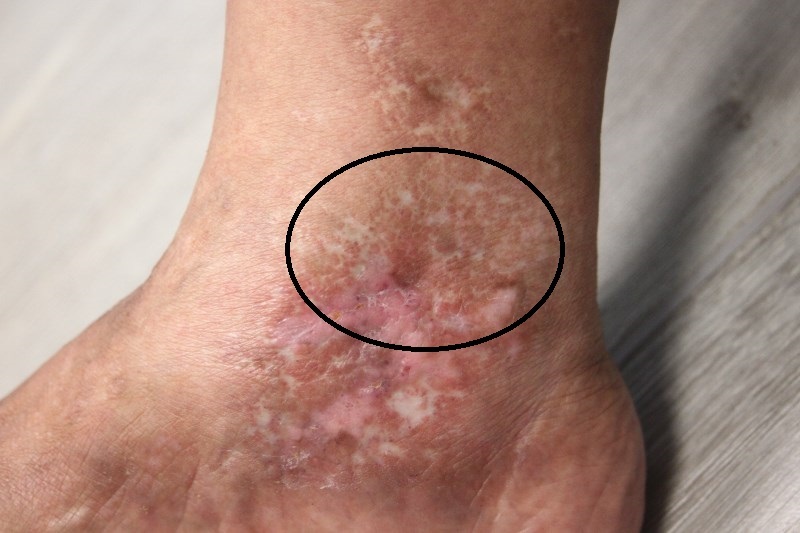
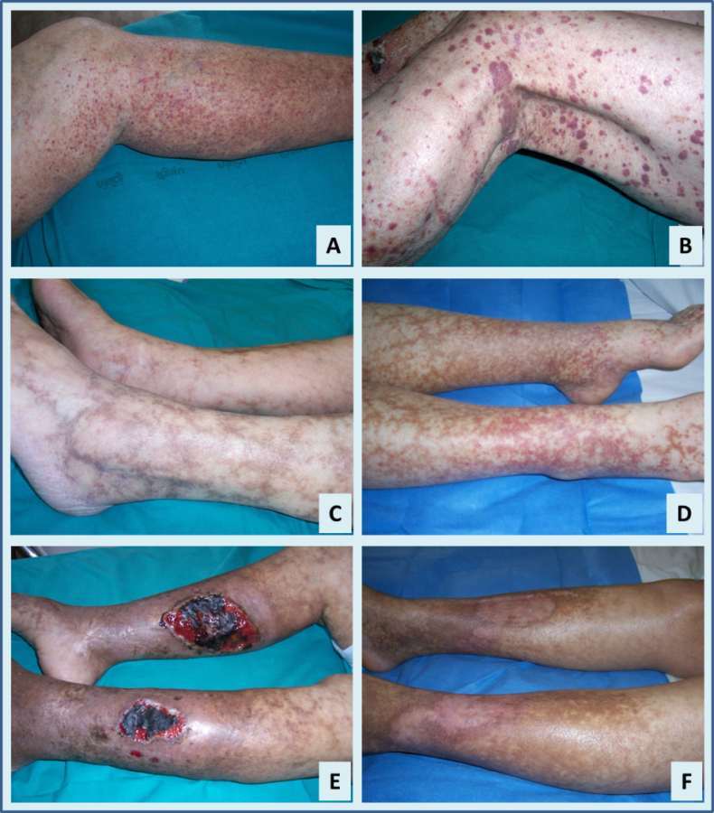
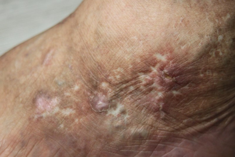
Livedo may present as a cutaneous ulcer (livedoid vasculopathy), and atrophie blanche may be a consequence
-
It usually affects the lower legs bilaterally and is more common in summer
-
It may affect all age groups but is more common in young females
-
Initially there is a painful papular rash that develops into small erosions/ulcers. Lesions subsequently heal leaving porcelain white scars (atrophie blanche) that are often star-shaped (stellate) or reticulated (net-like)
-
Livedoid vasculopathy was originally described as a clinical manifestation of vasculitis; however the present concept is that it is a vaso-occlusive phenomenon with thrombosis of intradermal venules. It may be primary, or secondary, for example to a coagulopathy such as the APS or cryoglobulinaemia. Livedoid vasculopathy is also a recognised complication of Covid-19 infection
Clinical Images
Please refer to notes on image rights at bottom of the page with regards to individual image
ownership.
Investigations
-
Physiological livedo - no investigations required
- Pathological livedo reticularis - requires an urgent and thorough work-up as per the chapter on on Investigations, including a thrombophilia screen to look for the Antiphospholipid syndrome
- Livedoid vasculopathy in the absence of livedo reticularis:
- FBC
- ANA, and other relevant autoantibodies if connective tissue disease suspected
- A thrombophilia screen to look for coagulation disorders such as the antiphospholipid syndrome, and fibrinolytic disorders such as protein C deficiency and factor V mutation (Leiden)
Management
Physiological livedo reticularis
- Treatment is not required
Pathological livedo reticularis
- Management depends on the underlying cause - refer to the section on investigations
Livedoid vasculopathy
- The main goal of therapy is to reduce pain, ulceration, and the development of atrophie blanche:
- Protect the area from trauma
- Provide standard ulcer management
- Stop smoking, if relevant
- Various drug therapies may be prescribed to enhance blood flow, although there is no good evidence for a reliably effective treatment
- Livedoid vasculopathy is a chronic disorder, with spontaneous remissions and exacerbations, the reported duration ranges from 3 months to 20 years
Disclaimer - the author PCDS cannot accept responsibility for any misleading or incorrect statements, and the management of individual patients remains the direct responsibility of the individual doctor. We do however hope that visitors to this site can contact us regarding comments that are considered misleading or incorrect so that we can continue to improve the site.
Image Rights - The PCDS would like to thank Dermatoweb, DermQuest (Galderma), and others who have contributed images. All named individuals and organisations maintain copyright for the relevant images.