
1. Referral pathways
Introduction
While there is no single dermatology model that works best across all localities, the gold standard of care is to make sure that patients are seen within agreed time frames, by the most appropriate health professionally, and ideally in a local setting. While this page (along with the conditions listed in the concise guidance section of the website) provides an insight into referral pathways, please refer to the website section on commissioning (including GPwERs) for more detailed advice that includes different models of care.
Skin lesion referrals: 2-week wait
There are a number of things to consider:
- Secondary Care only
- Diagnostics: should be undertaken by those trained and experienced in dermoscopy. Teledermoscopy should should be considered carefully as it has pros and cons. Teledermoscopy must come with education otherwise, even though the number of patients ending up in a two-week wait clinic may fall, large numbers of lesions may be referred unnecessarily to the teledermoscopy service (there is evidence across a number of specialties that improving access to a service increases referrals)
- Surgical treatment: should be undertaken by those competent in skin surgery as demonstrated by high levels of complete excision and good cosmetic outcomes
- Audit of Secondary Care skin cancer services: there are a number of entities that local commissioning groups can ask to be audited across different specialties including excision rates, levels of complete excision, and the number of patients diagnosed with any form of skin cancer (or pre-cancer) that receive a full skin examination
BCC and other skin lesions
There are a number of things to consider:
- Diagnostics: should be undertaken by those trained and experienced in dermoscopy. As above, consider teledermoscopy
- Surgical treatment: should be undertaken by those competent in skin surgery as demonstrated by audit showing high levels of complete excision and good cosmetic outcomes
- Primary or Secondary Care? Refer below for the role of GPwERs and GPs. In terms of Secondary Care a number of departments may be involved eg Dermatology, Plastic Surgery, Oculoplastics and Maxillofacial Surgery
- BCC and type of referral: usually routine, although if any of the following BCCs on the head cannot be seen as a soon appointment (within 6 weeks) then consider a 2-week wait referral:
- On or close to the nose, mouth, eyes or ears
- Large and/or poorly defined
- GPwERs: work as an integrated team with commissioned Secondary Care services. GPwERs have undertaken formal specialist training and assessment in the diagnosis (clinical and dermoscopic) of skin lesions, enabling the GPwER to be commissioned to provide skin lesion clinics (excluding 2-week waits). Groups 2 and 3 GPwER can also provide surgical services with a wider remit, which may include certain BCC on the head and neck. Formal accreditation is required
- GPs providing surgical services: manage benign skin lesions and small low-risk BCC beneath the clavicle. They are first and foremost surgical services. GPs would be expected to have an understanding of lesion recognition and management (such as surgical margins required) appropriate to their surgical remit but they have not undertaken specialist training and competency based assessment in skin lesion recognition and management. In essence, skin surgery services such as DES or LES only cover clinical remits that are within the normal scope of General Practice
- Audit: there are a number of entities that local commissioning groups can ask to be audited across different specialties including excision rates, levels of complete excision, and the number of patients diagnosed with any form of skin cancer (or pre-cancer) that receive a full skin examination
Dermatology emergencies
- Erythroderma: 90% or more of the body is erythematous, the skin feels hot and patient complains of feeling cold. Refer to the chapter Erythroderma
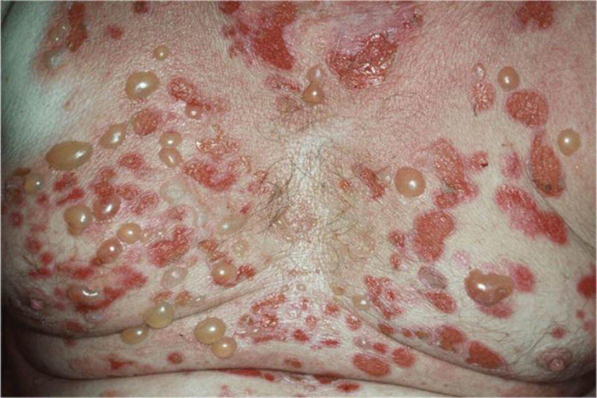
- Widespread blistering/erosions for example bullous pemphigoid (blisters) or pemphigus vulgaris (erosions)
- Suspected severe drug reactions for example toxic epidermal necrolysis (often associated with mucosal involvement) and the drug hypersensitivity syndrome
- Rash and systemically unwell: the cause may be infections (eg, cellulitis and necrotising fasciitis, disseminated herpetic infections) or inflammatory (eg pustular psoriasis)
- Vasculitis with rapid development of purpura / ulceration: if renal involvement suspected refer to nephrology otherwise dermatology. Refer to the chapter Vasculitis
For such emergencies phone the on-call dermatologist with a view to admission or urgent outpatient appointment. If there is no out-of-hours Dermatology service so you may need to speak to the on-call medical team for admission and inpatient Dermatology assessment.
'Soon' dermatology referrals (ideally to be seen within 6-12 weeks)
- Scarring acne: refer to the chapter Acne vulgaris
- Severe eczema: refer to the chapter Atopic eczema. Referral should be to the closest Secondary Care provider as multiple/frequent review appointments may be required
- Severe psoriasis: refer to the chapter Psoriasis. Referral should be to the closest Secondary Care provider as multiple/frequent review appointments may be required
- Scarring alopecia: refer to the chapter Alopecia
- Other conditions: this will be very much dependent on individual cases
Routine referrals
Most routine dermatology referrals should be given choice of providers. However, the conditions listed below require specific treatments that only some providers will be able to offer - direct referrals should be made to providers who offer such services:
- Leg ulcers: the place to refer depends on the cause and may include:
- Venous leg ulcers: tissue viability clinics / dermatology
- Arterial leg ulcers: vascular surgery
- Diabetic ulcers: diabetic clinics
- Paediatric dermatology: some Secondary Care Dermatology departments offer a paediatric service
- Allergy testing
- Patch testing: refer to Dermatology for suspected contact allergic dermatitis (eg moderate-severe hand or foot eczema, or facial/eyelid eczema). Patch testing is not appropriate for urticaria
- Food allergy: refer to the chapter Food allergy. If patients still need to be referred check with local policies, however, the usual route is to refer to Immunology, although some paediatric services also see children with moderate-severe food allergies
- Vulval conditions: refer to a specialist vulval clinic if available
- Alopecia areata: patients with worsening or unresponsive alopecia areata should be referred to Dermatology for intralesional steroids and more advanced options
- Hyperhidrosis (excess sweating): Hand and foot - refer to Dermatology for iontophoresis. Axillary - check to see if any local providers offer Botox on the NHS
- Laser: services offered depend on local policies but examples of conditions treated can be severe telangiectatic or rhinophymatous rosacea, some pigmentary disorders, and vascular lesions such as haemangiomas
2. Actinic (solar) keratosis
Key features
- Solitary-multiple asymptomatic macules on UV-damaged skin. Most skin-coloured/pink, can be red-brown
- Rough to touch and often develop a white-yellow surface scale
- Red flags for squamous cell carcinoma include any of - sudden growth in a pre-existing AK, pain, a palpable tender papule or nodule with or without surface scale, a non-healing ulcer of duration > 3 weeks on UV-damaged skin
- Differential diagnosis - refer to the Skin Lesion Diagnostic Tool
Management overview (including long-term self-management)
- For a simplied managment plan please refer to the PCDS AK Primary Care Treatment Pathway
- A full skin examination is best practice to look for signs of skin cancer elsewhere
- To treat or not treat? Up to 25% of lesions will resolve without treatment. While it often appropriate to treat persisting lesions, for those at low risk (eg older patients with a smaller number of lesions) treatment is probably unnecessary
- Refer to the bottom of the page for a patient information leaflet available through a QR code or printable PDF. The leaflet provides the patient with all necessary information including a place for the health professional to provide treatment notes, advice on UV-protection and vitamin D, self-management of actinic keratoses, and links regarding self-examination
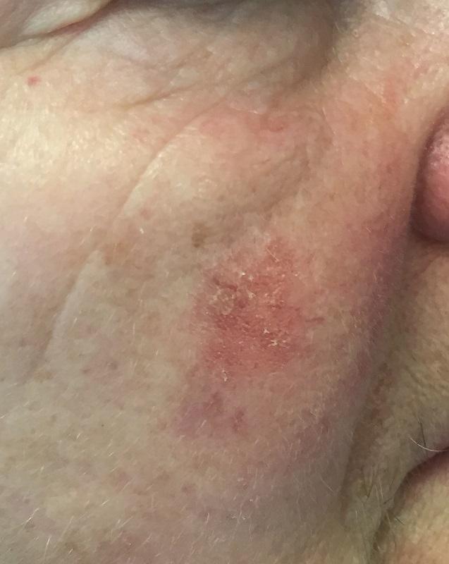
Treatment of grade 1 AKs - scale more palpable than visible, single or few lesions
- 5-Fluorouracil cream (5-FU) - 5% 5-FU cream or 4% 5-FU cream (Tolak): OD for 4 weeks, washing off after 8 hours. Provide a patient information leaflet, patients should be advised to expect erythema, crusting, and some discomfort during treatment; which is more effective than 3% diclofenac gel
- Klisyri ® (tirbanibulin) - apply OD for 5 days. Patients should be advised to expect erythema, crusting, and some discomfort during treatment
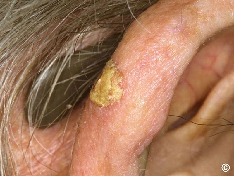
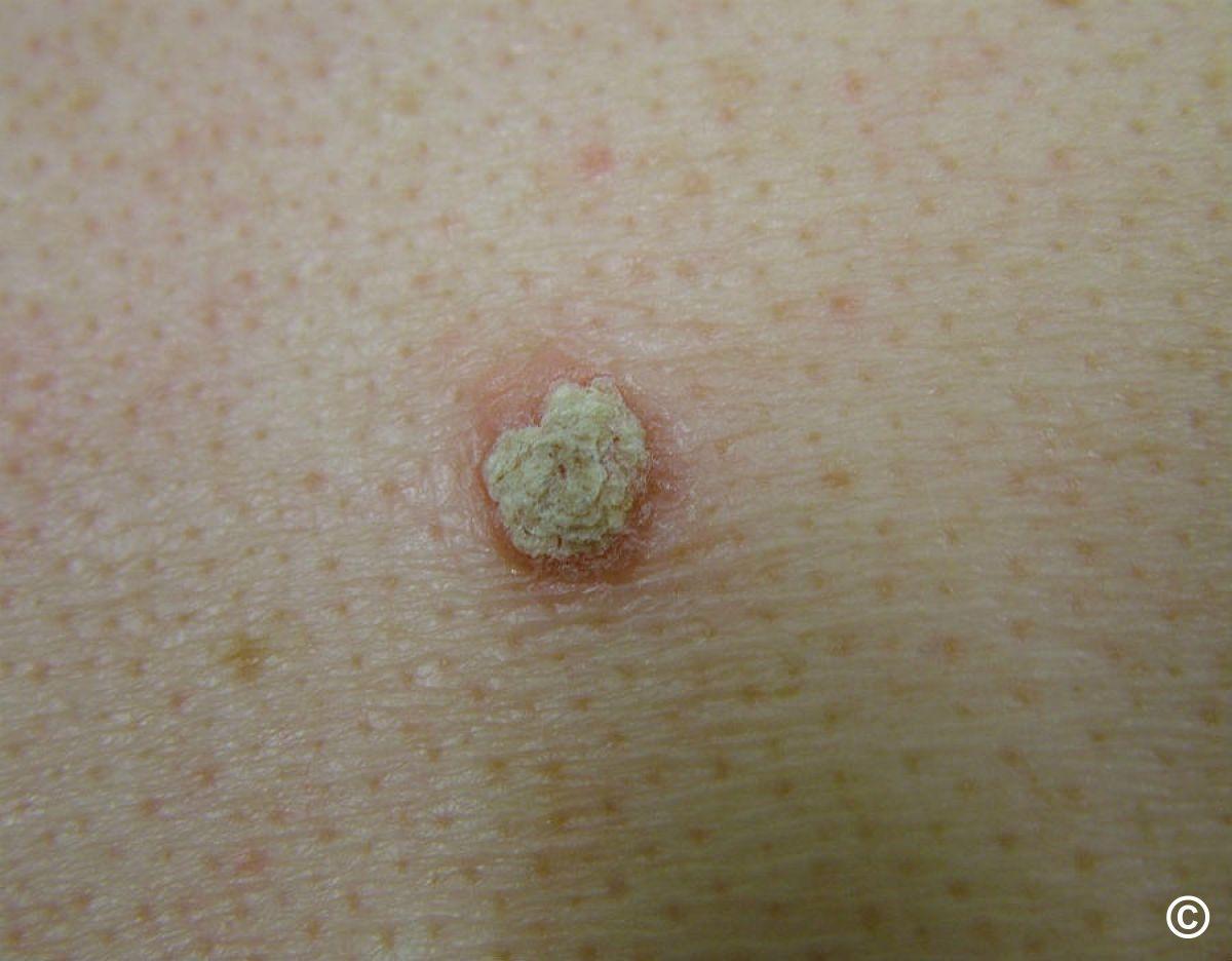
Grade 2 AKs - moderately thick scale, easily felt and seen, single or few lesions
- 5-Fluorouracil cream (5-FU) - 5% 5-FU cream or 4% 5-FU cream (Tolak): OD for 4 weeks, washing off after 8 hours. Patients should be advised to expect erythema, crusting, and some discomfort during treatment. Provide a patient information leaflet
- Actikerall ® solution (0.5% 5-FU+10% salicylic acid) - OD for 6-12 weeks. Actikerall ® tends to leave a film on the skin, which should be washed/peeled off before the next application. At the thicker end of the grade 2 spectrum, Actikerall ® may be preferable to 5-FU cream
- Cryosurgery - a singe 10-15 second freeze-thaw cycle with conventional liquid nitrogen. Other products, usually contained in a can, less likely to be effective. Cryosurgery can cause permanent hypopigmentation (eg on face), and avoid on gaiter area of legs
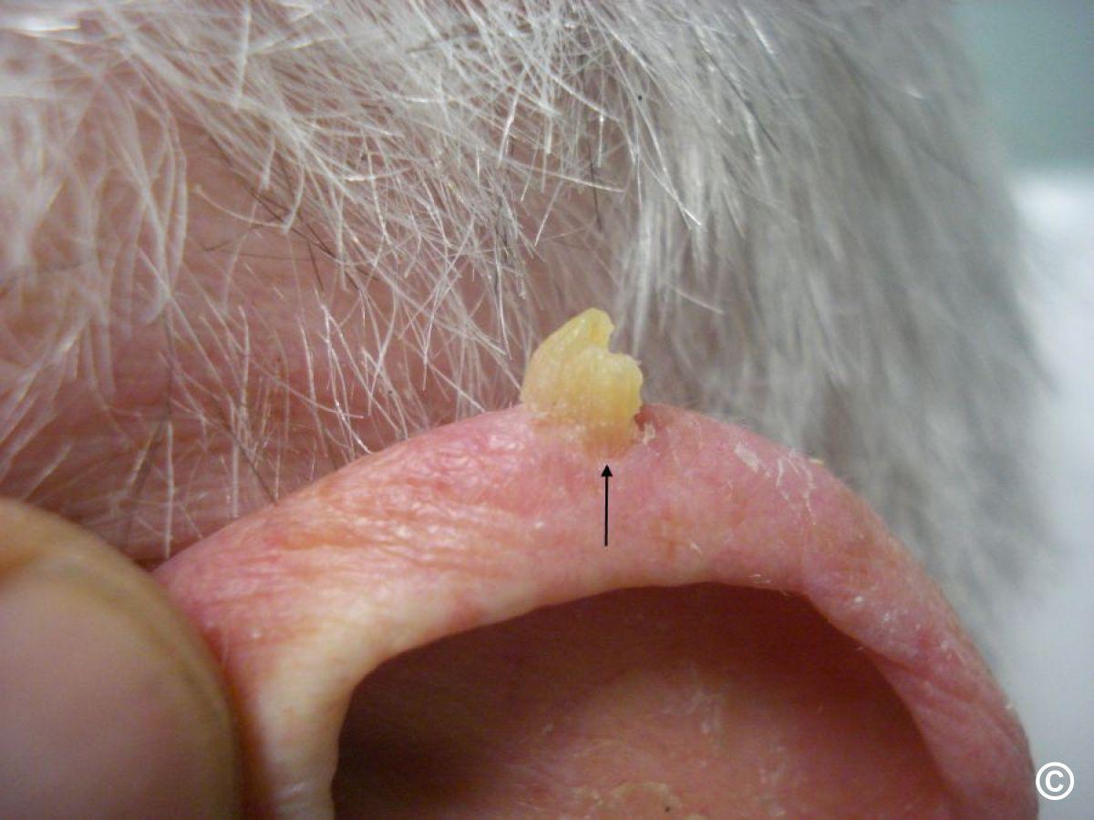
Grade 3 AKs - thick scale, single or few lesions
- Cryosurgery - with liquid nitrogen. Other types of cryosurgery unlikely to be effective. If no response consider a 2WW referral in case of early SCC change
- Curettage and cautery undertaken by a health professional with the correct training - important to include a few mms of skin depth under the scale, and send for urgent histology, to exclude early SCC transformation
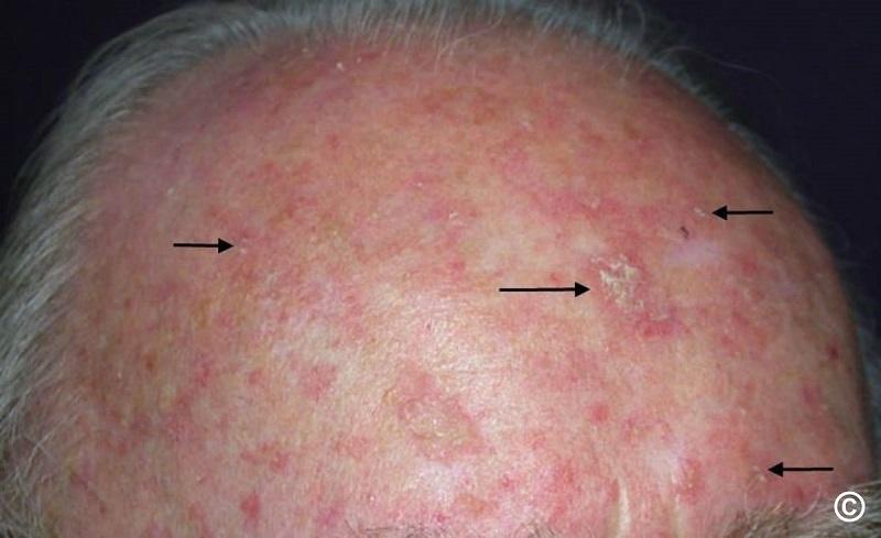
Field treatment - treating a larger area of skin in which there are several thin lesions
- First-line (best evidence for SCC reduction) - combination of 5% 5-FU cream and calcipotriol ointment BD for 4-6 days for face/scalp, and 6-10 days for arms/legs. Provide a PIL for this treatment
- In a case such as here, where the actinic keratoses are relatively thin, consider treating the whole area with 3% diclofenac gel BD for 8-12 weeks, and then review 4 months after treatment was commenced (improvements can continue for up to 4 weeks after treatment finished). At review treat remaining individual lesions with any of 5-FU cream, Actikerall ® solution or cryosurgery as above
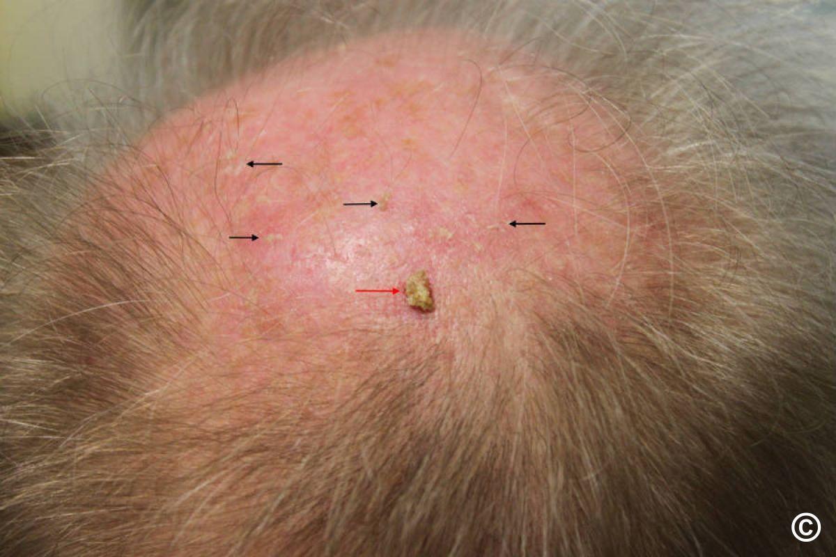
Field treatment - treating a larger area of skin in which there are several thicker lesions (eg grade 2 and above)
- Consider the combination therapy of 5% 5-FU cream and calcipotriol ointment BD for 4 to 6 days for the face, and 6 to 10 days for the arms and legs. Provide a PIL for this treatment
- Aldara ® cream (5% imiquimod cream) - 3 nights a week for 4 weeks (eg Mon, Wed, Fri). Adverse effects similar to above, and sometimes flu-like symptoms
- Share treatment images with patient (below)
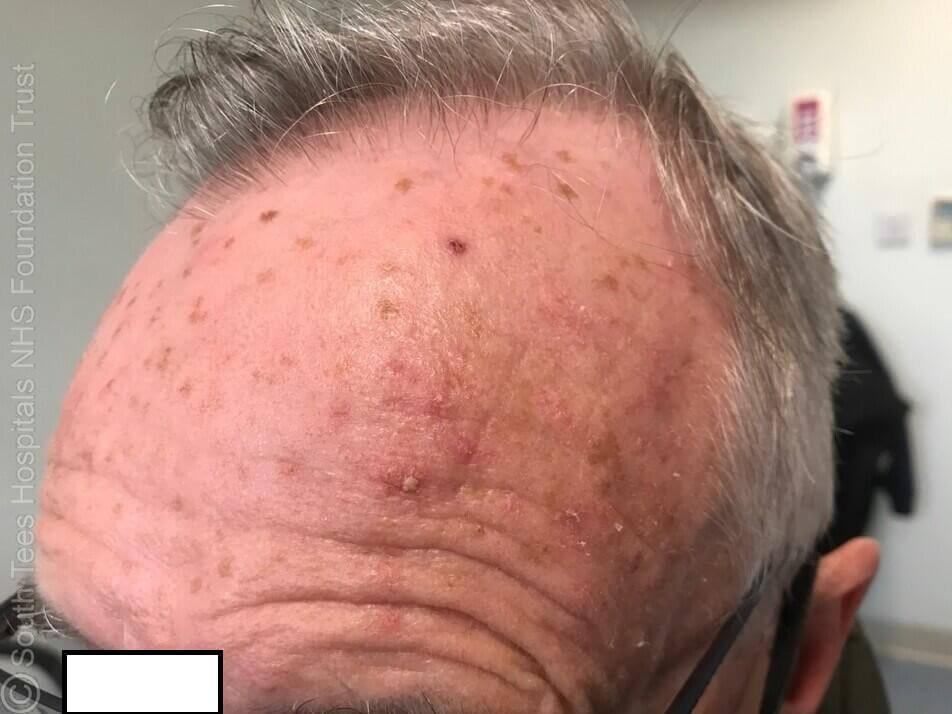
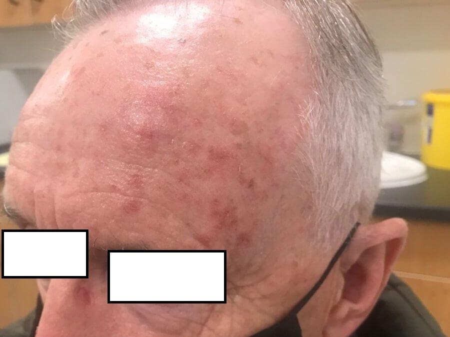
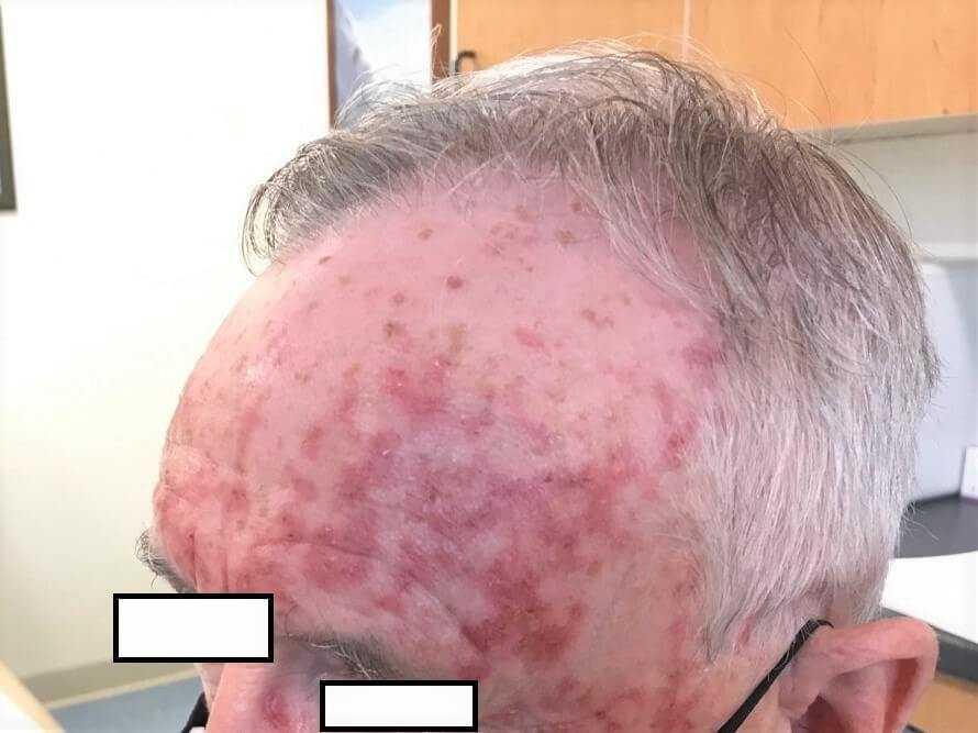
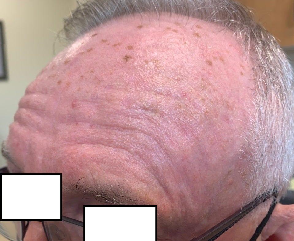
Treatment images - to share with the patient
When using one of the stronger treatments (eg 5-FU cream or Aldara ®) it is helpful to show the patient what to expect. The patient shown here had field damage to his left and right forehead, which were treated at the same time with Efudix cream OD for 4 weeks; in some patients the reaction will be less, in others it may be stronger. The pinkness that is still visible post-treatment will eventually fade. It is important to explain to patients that such treatments are more effective than milder treatments such as 3% diclofenac gel; this will help facilitate patient choice. When using such treatments think about timing; patients may wish to avoid important social dates / holidays.

Field treatment - more severe
- Such cases are best referred
- If any suggestion of SCC (beware of possible SCC under large areas of scale), refer urgently as a 2WW
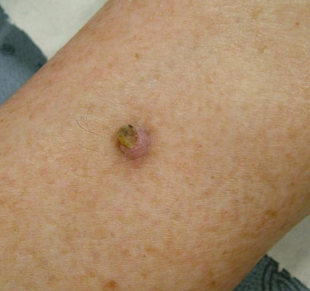
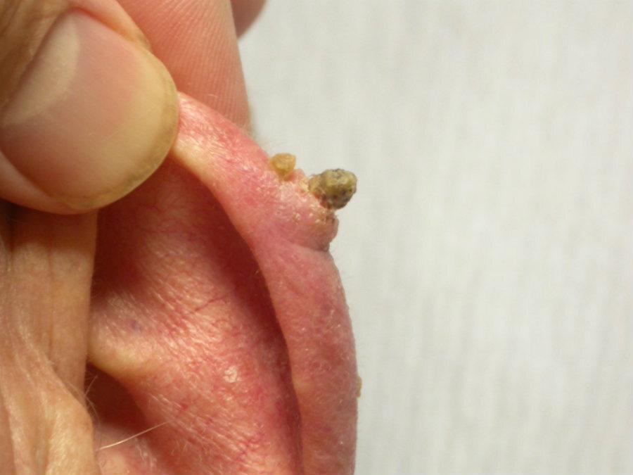
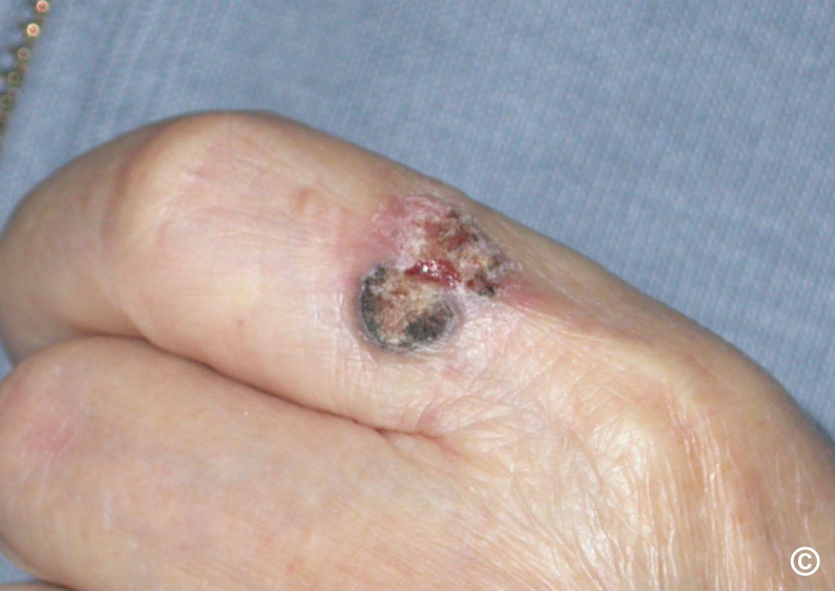
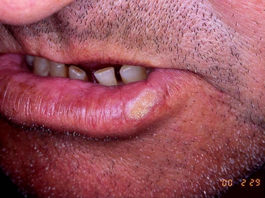
Red flags for squamous cell carcinoma
- Sudden growth in a pre-existing AK
- Pain
- Palpable tender papule or nodule with or without surface scale
- A non-healing ulcer of duration > 3 weeks on UV-damaged skin
- Have a low threshold for referring patients with significant immunosuppression (eg transplant patients)
Additional management resources
- The chapter Actinic keratoses
- A patient information leaflet on 5-FU cream and the combination therapy
- A Fast Facts learning course on actinic keratoses
Patient Information Leaflet
Link: Actinic (solar) keratosis

https://www.pcds.org.uk/patient-info-leaflets/actinic-solar-keratosis
Download the Patient Information Leaflet PDF: Actinic (solar) keratosis
3. Skin lesion algorithm - common benign lesions and skin cancer
This algorithm sets out a logical approach to the clinicodermoscopic evaluation of skin lesions. It does not replace the need for appropriate training when using dermoscopy. The main focus is on the clinical appearance of melanoma / SCC (considerable experience is required for the dermoscopic assessment of melanocytic lesions) and the dermoscopic appearance of common benign non-melanocytic lesions. For the best courses on dermoscopy please refer to our events section.
Follow this link for the algorithm.
4. Acne vulgaris
Key features
- Comedones (absent in rosacea), papules, pustules, nodules and scarring
- Differential diagnosis - refer to the General Dermatology Diagnostic Tool
Investigations
- The vast majority of patients do not require investigations
- If there is any suggestion of PCOS or other cause of hyperandrogenism refer to the relevant chapter
Management overview
- For a simplied managment plan please refer to the PCDS Acne Primary Care Treatment Pathway
- Refer to the bottom of the page for a patient information leaflet available through a QR code or printable PDF
- Various drugs can exacerbate acne - anabolic steroids, topical/oral corticosteroids, unopposed progestogens, lithium, ciclosporin, iodides taken orally (found in some homoeopathic therapies)
- When choosing treatment consider - type of lesions (comedones, papules and pustules, nodules), hyperpigmentation, and scarring
- Also assess the psychological impact - what is mild acne to a health professional may be causing severe distress to a patient

Step 1: mainly comedonal acne - a topical retinoid
- Choices include adapalene (Differin®), adapalene combined with benzoyl peroxide (Epiduo 0.1% or Epiduo Forte 0.3%®), or trifarotene (Aklief®) and clascoterone (Winlevi®). Topical retinoids are contraindicated in pregnancy
- Azelaic acid (20% cream or 15%) gel can be added to combat acne-induced hyperpigmentation
- Balance your prescribing choices and number of agents per what your patient can realistically do; adding multiple treatments can be burdensome
- To reduce excessive dryness/irritation, advise short-contract application e.g. leaving on for 30 minutes before washing off, then gradually increase duration of treatment as tolerated (eventually many patients can apply at bedtime and leave on overnight), or start one day a week and gradually increase daily use every 1-2 weeks (e.g. week 1 Monday, week 2 Monday and Friday, week 3 Monday, Wednesday, and Friday, week 4 every other day and week 5 daily)
- Review in 12 weeks


Step 2: mild to moderate papular / pustular acne
Read in conjunction with management notes below regarding hormonal treatments and so forth.
Use a fixed dose combination treatment, ideally containing benzoyl peroxide (BPO), which reduces bacterial resistance, with either a topical retinoid or topical antibiotic:
- First-line: Epiduo® 0.1% or 0.3% gel (adapalene + BPO) or Duac® gel (clindamycin + BPO) or Treclin® gel (clindamycin and tretinoin). Topical antibiotics are used for a maximum 12 weeks then switched to an alternative agent for maintenance
- Add-on treatments: Clascoterone (Winlevi®) to reduce seborrhoea; azelaic acid (20% cream or 15%) to treat inflammation and reduce acne-induced hyperpigmentation
- Advise as per step 1 - how to apply topical treatments and reduce adverse effects
- Review in 12 weeks, if no significant improvement go to step 3
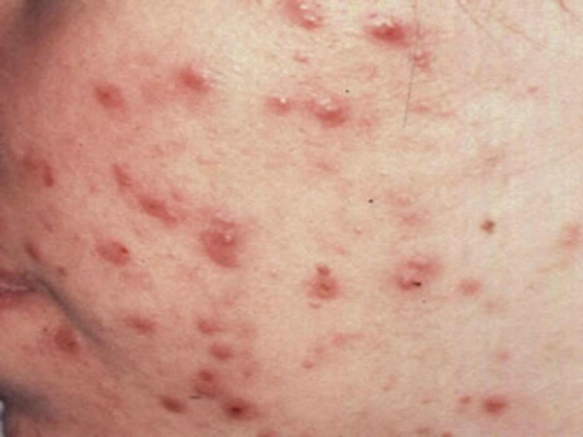
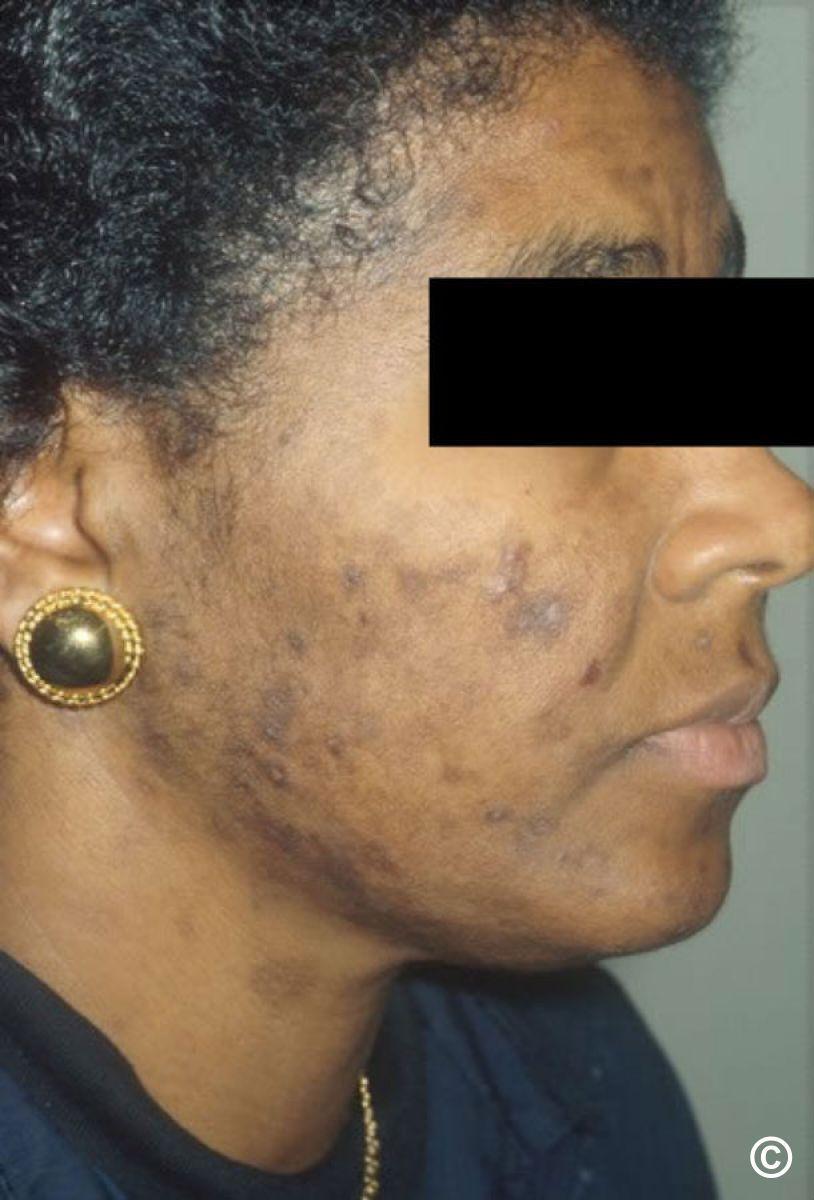
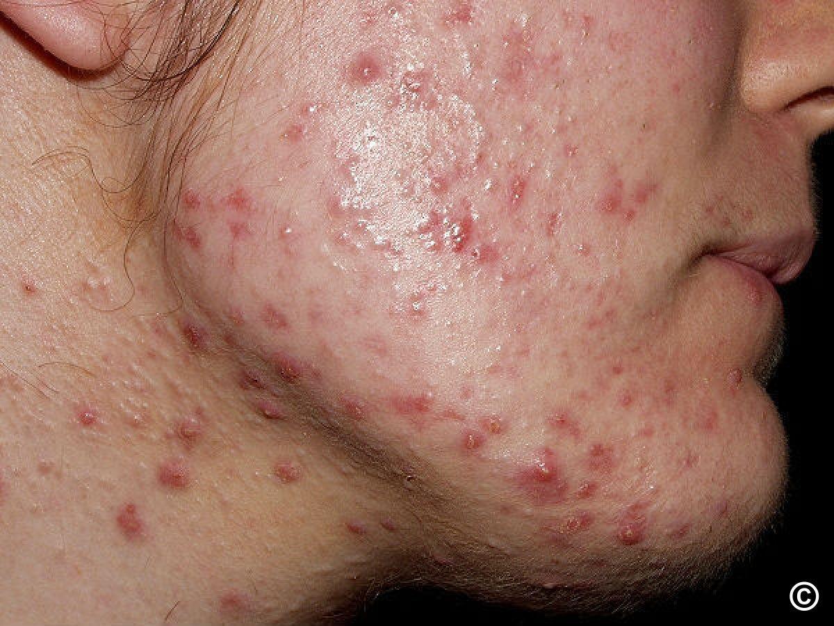
Step 3: not responding to the above and/or more widely distributed (without significant scaring)
Combine systemic antibiotics, or in some women COCP/antiandrogens (refer to additional notes below), with an appropriate topical agent, preferably Epiduo® 0.1% or 0.3% gel, or if not tolerated use BPO, adapalene, or trifarotene (Aklief ®) as single agents (advise as per step 1 - how to apply topical treatments and reduce adverse effects). Consider add-ons topical treatments where appropriate and adherence is possible. Choice of antibiotic:
- First-line: lymecycline 408 mg OD or doxycycline 100 mg OD. If partial response, consider increasing the dose of either to BD. Both are contraindicated in pregnancy and children under 12 years old. Lymecycline is better tolerated; doxycycline may be more efficacious
- If tetracyclines are contra-indicated:
- Erythromycin 500mg BD or Clarithromycin 500 mg OD - higher levels of bacterial resistance than tetracyclines. Clarithromycin may have less bacterial resistance than erythromycin
- Trimethoprim 300 mg BD - council patient as to very small risk of severe cutaneous adverse reactions and agranulocytosis (patients and carers should be told to seek immediate medical attention if case of fever, sore throat, rash, mouth ulcers, purpura/bruising or bleeding. The BNF also recommends regular FBC)
- Children under 12 years of age: erythromycin or clarithromycin (dose dependent on weight)
Duration of systemic antibiotic treatment
- Ideally 3 months, evidence suggests that for most patients there is little additional benefit in using antibiotics for more than 3 months in any given treatment period; however, patients relapsing quickly after stopping treatment may be better suited to 6-month courses, or consider alternative therapies e.g. spironolactone (where appropriate) or referral for oral isotretinoin (Roaccutane®)
- Once stopped, many patients will need to remain on their topical agent (step 2) as maintenance treatment
- The antibiotic course can be repeated in the future if needed, if remission (improved or clear skin > 4 weeks) is induced, e.g. a 3-month course once or twice a year
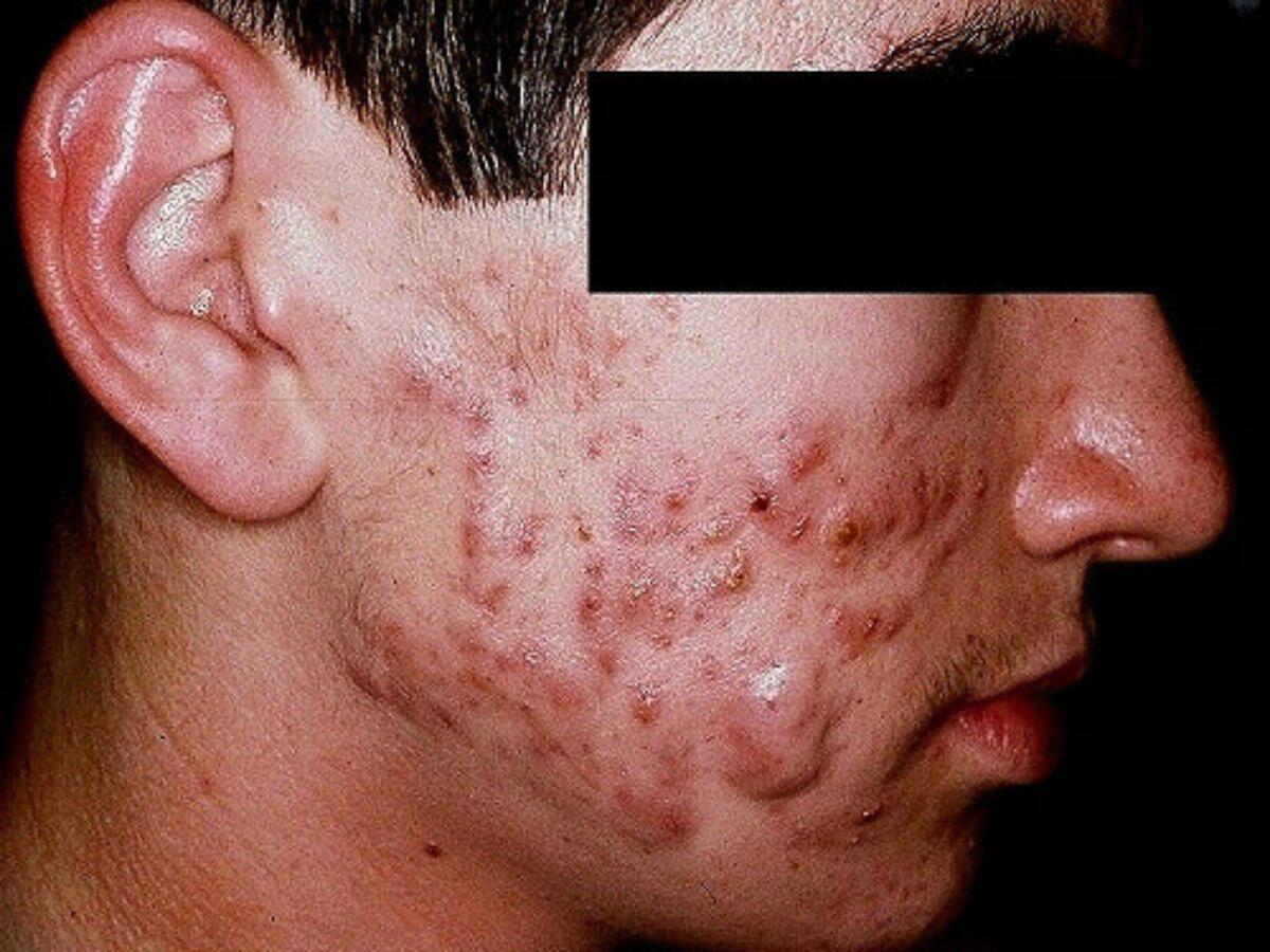
Step 4: Active scarring acne
- Patients with active acne and scarring should be started on treatment (as in step 3) and referred at the same time as urgent
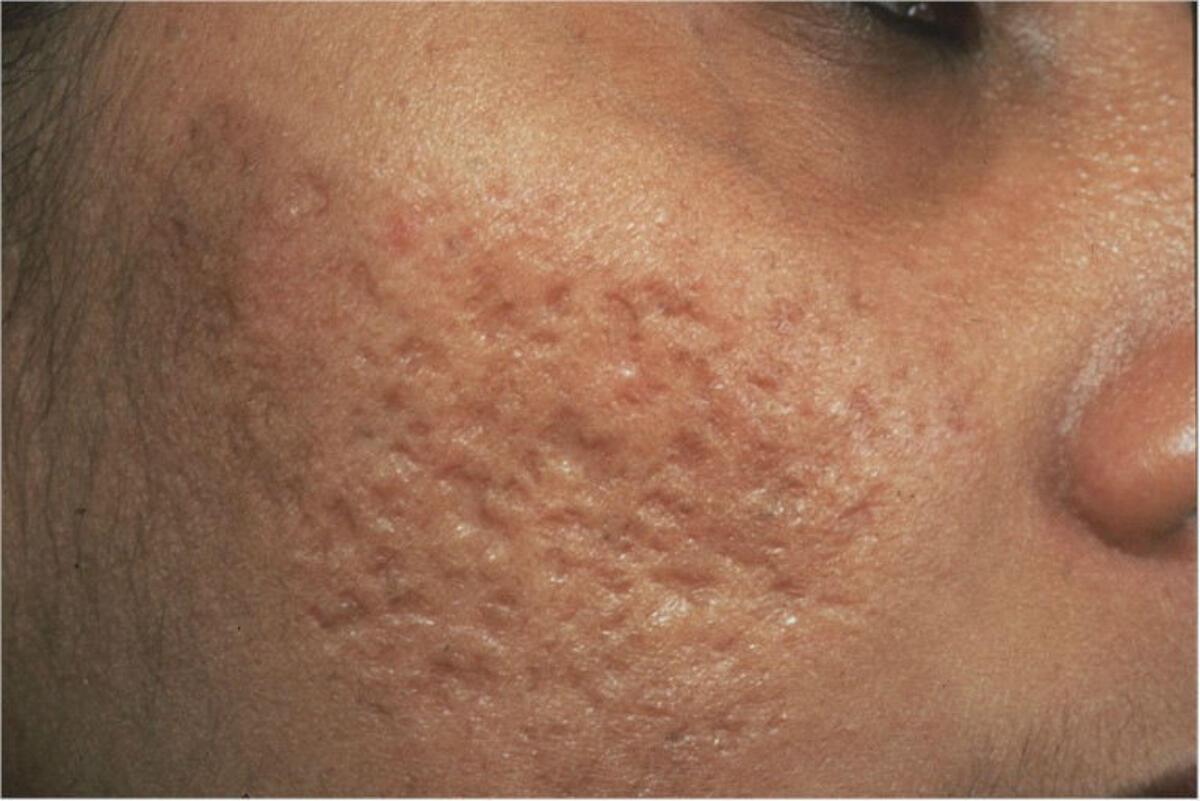
Scarring with no remaining active acne
- Depending on the type of scarring, physical treatments such as peels, subcision or punch excision, and laser therapy may help improve the appearance of the skin
- There is limited NHS availability - check local guidance
Additional notes
COCP / antiandrogens in women
- These can be used along with topical agents and in combination with a systemic antibiotic
- Clascoterone (Winlevi®) is a topical antiandrogen treatment that can improve seborrhoea and hence comedone formation subsequent acne progression. It is used form age 12 onwards in all genders and has negligible systemic absorption. It is a twice daily application and can be used as monotherapy or in combination with other topicals
- COCP - the effectiveness of oral contraceptives will depend on the content of ethinylestradiol and on the nature of the progestogen. COCP containing drospirenone such as Eloine ® and Yasmin ® are the logical choice, failing that Dianette could be considered if not contraindicated. The Faculty of Sexual and Reproductive Healthcare also promote tricycling of COCPs taking 9 consecutive weeks before having a 4–7-day break. It is also worth noting that the risk of venous thromboembolism is greater if Dianette/other COCPs are stopped and started as opposed to being kept on (if required). Can stop 3 months after acne is controlled unless needed for contraception. Drospirenone alone (Slynd®) is an alternative where the combined approach is contraindicated or not tolerated
- Spironolactone can be very effective in some patients with acne and is generally safe, it can take up to 6 months to work effectively. Refer here for more information
Skin of colour
- Acne-induced hyperpigmentation (previously known as post-inflammatory hyperpigmentation) can be significant and often persists for months to years. As such, timely and more aggressive treatment is advocated, including the use of topical retinoids and azelaic acid, and early referral for consideration of isotretinoin
Pregnancy
- The mainstay of treatment should be topical treatment, either benzoyl peroxide preparations or 2% topical erythromycin (topical retinoids are contraindicated in pregnancy, and should be avoided in women trying to get pregnant)
- The option of oral erythromycin 500 mg BD should be discussed with the patient if the potential benefits outweigh the possible risks
Referral
- Routine referral - mild-moderate acne that does not respond well to a 12-week course of antibiotic COMBINED with a topical retinoid or suitable topical, or relapse within 4 weeks of stopping the antibiotic
- Urgent referral - patients with acne that is scarring should be commenced on treatment and referred, as scarring is irreversible. Patient with moderate-severe body dysmorphia should be managed in the same way
- If referring for possible isotretinoin (usually the case) please make sure of the following:
- U&Es, LFTs and lipids including triglycerides are checked. Non-fasting sample is fine; some departments do insist on fasting tests, which is archaic
- Female patients during their reproductive years need to have appropriate contraception, where possible and where appropriate, but this is not mandatory. Check local advice
- Provide a patient information leaflet on isotretinoin
Additional resources
- Refer to the chapter Acne vulgaris for more detail
Patient Information Leaflet
Link: Acne vulgaris

Download the Patient Information Leaflet PDF: Acne vulgaris
5. Alopecia (hair loss) - an overview
Types of alopecia
- One of the most important aspects of care is to distinguish whether it is scarring or non-scarring (the vast majority)
- The clinical features of scarring alopecia may include: cicatricial lesions (circular to oval-shaped patches of scarring), an atrophic shiny appearance, inflammation (not always present), pustules and/or nodules. Under dermoscopy, features that may be seen include loss of follicular openings and tufting of hair
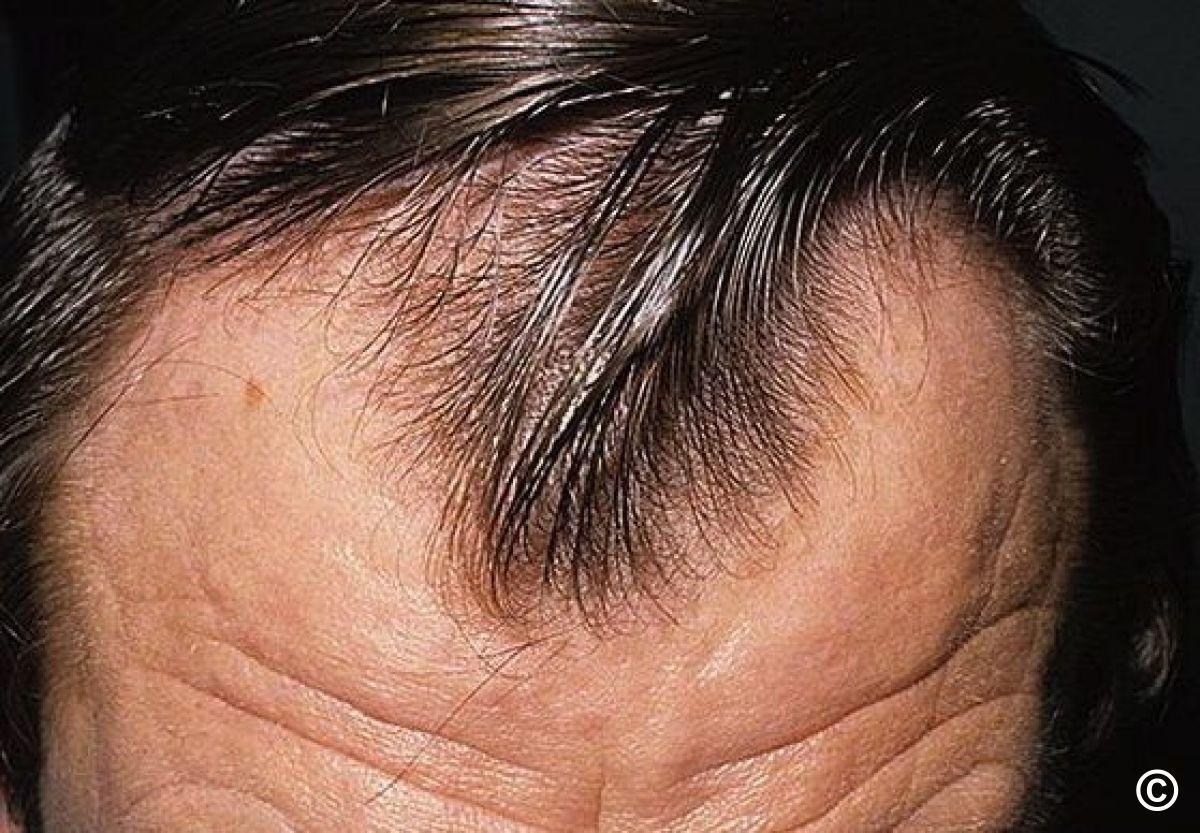
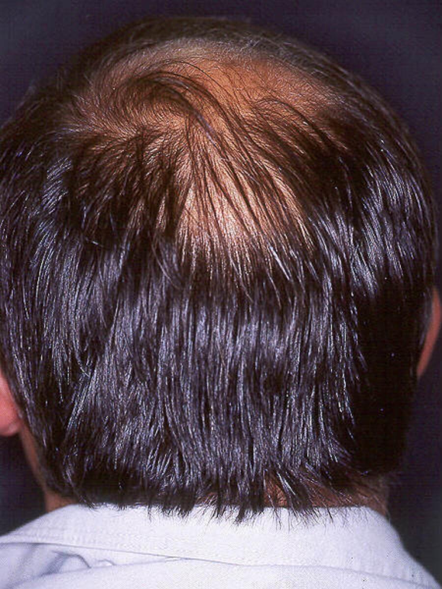
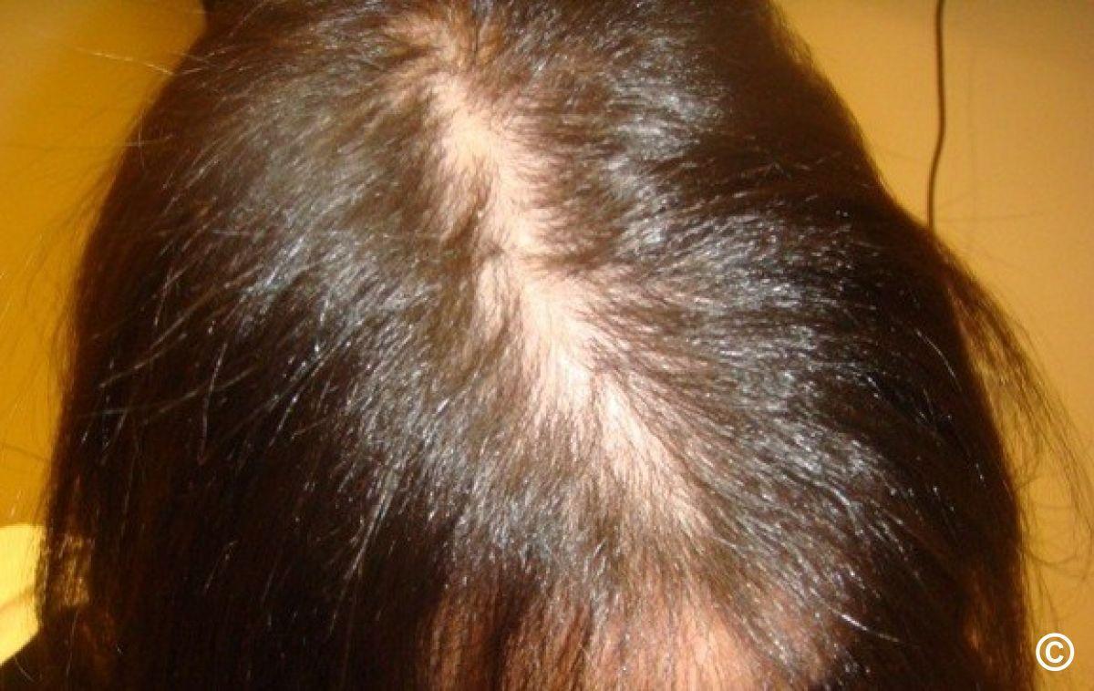
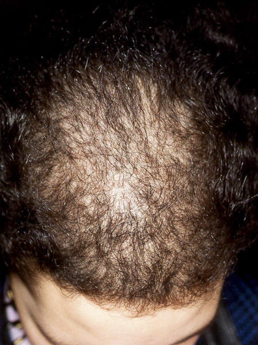
Non-scarring alopecia
- Male pattern alopecia (1&2) - recession of frontal hair line, mainly in a triangular pattern, followed by thinning of the vertex
- Female pattern alopecia (3&4) - a diffuse thinning of the centroparietal region, usually maintaining the frontal hair line
- Telogen effluvium - a more diffuse hair loss. Many causes including physical triggers such as pyrexia (such triggers tend to precede the alopecia by a few months), anaemia, thyroid disorders, and medications. Check FBC, ferritin and TFT

- Alopecia areata - one to several circumscribed, totally bald, smooth patch. The skin within the bald patch is normal or slightly reddened. Short broken hairs (exclamation mark hairs) are often seen around the margins of active expanding patches of alopecia. If scale present send both skin scrapings and plucked hair follicles for mycology in case of tinea
- There are many other causes of alopecia - refer to the chapter Alopecia: an overview for detailed information

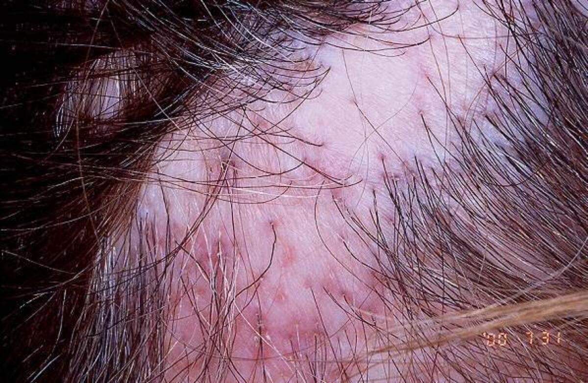
Scarring alopecia
There are several causes, including:
- Discoid lupus erythematosus - affected patches are erythematous and scaly (usually more so at the periphery) with follicular plugging
- Lichen planopilaris - erythema and scale centred on hair follicles, such changes are commonly located at the periphery of expanding areas of alopecia
- Folliculitis decalvans - pustules, crusting and sometimes erosions
Refer as semi-urgent (to be seen within 6-12 weeks) and commence treatment with Dermovate scalp ® application (making sure that the solution does not run on to the face). Untreated, scarring alopecia leads to irreversible hair loss. The only exception to the above is if tinea is suspected, which tends to present with fine scale and occasionally scarring alopecia - withhold topical steroids, send skin scrapings and plucked hair follicles for mycology, and refer to the chapter Tinea capitis.
For detailed information on scarring alopecia (including other causes) refer to the chapter Alopecia: an overview.
Additional resources
- Alopecia UK is a very good patient support group
6. Blistering (bullous) conditions
This section highlights common/important causes of blistering. There are many other causes of blisters - refer to the chapter Bullous disorders - an overview for detailed information.

- Leg oedema - bullae (blisters) commonly occur on dependent limbs due to an acute exacerbation or deterioration of peripheral oedema. Often contain clear fluid and can be tense

- Papular urticaria - a hypersensitive reaction to various insect bites. Bullae can be small or large and are often grouped together, sometimes in a circular or linear arrangement
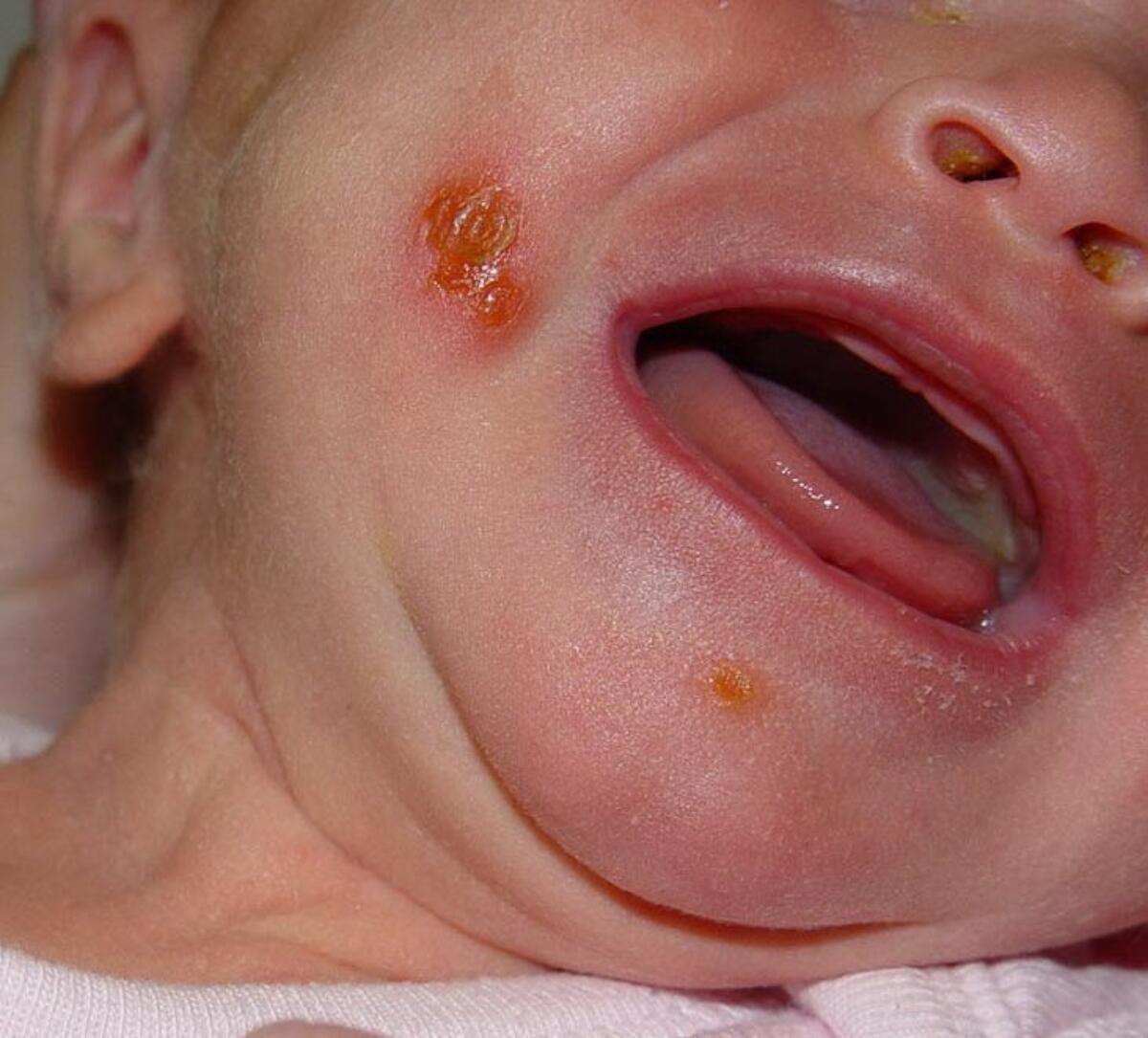
- Bullous impetigo - small or large bullae arise over a short period of time, usually spreading locally on the face, trunk, extremities, buttocks, or perineal regions and may reach distal areas. Bullae are fragile and burst spontaneously to leave a yellow crust that usually heals without scarring

- Child abuse, self-harm and domestic violence - look out for an inconsistent history, as well an unusual appearance / distribution of burns or scalds


Blisters on the hands
- Pompholyx (figure 1) - a type of eczema that usually presents periodically with intensely itchy vesicles, and occasionally large bullae, that predominate on the palms and sides of fingers
- Hand foot and mouth disease (figure 2) - initially macular lesions appear on the buccal mucosa, tongue, and / or hard palate, which rapidly progress to vesicles that erode and become surrounded by an erythematous halo
- Porphyria cutanea tarda and pseudoporphryia (figure 3) - skin fragility and bullae on the backs of hands. Pseudoporphryia is drug-related, being caused by a number of drugs, especially furosemide, nalidix acid and naproxen

- Phytophotodermatitis - caused by a reaction between naturally occuring plant psoralens, eg common hogweed, giant hogweed, cow parsnip, and UV exposure. The blistering rash, which is often streaky, arises some 24 hours later

- Herpes zoster - preceding pain. Closely grouped red papules, which rapidly becoming vesicular and then pustular, developing in a continuous or interrupted band, usually in one dermatome. Lesions can become bullous and necrotic


Adverse cutaneous drug reactions
- Stevens-Johnson syndrome / toxic epidermal necrolysis (figure 1) - these are life-threatening conditions. Painful / tender erythema with local erosions and blisters, which quickly progress to areas of confluent erythema with sheet-like skin loss. Mucosal involvement including the eyes and lips / mouth is common
- Fixed drug eruptions (figure 2) - a well-demarcated erythematous plaque that may blister. Tends to heal with post-inflammatory hyperpigmentation. Occurs at same site on re-exposure. Large numbers of drugs implicated. The most commonly associated drugs are tetracyclines, sulphonamides, NSAID and quinine
- Drugs can cause a number of other bullous reactions (figure 3)

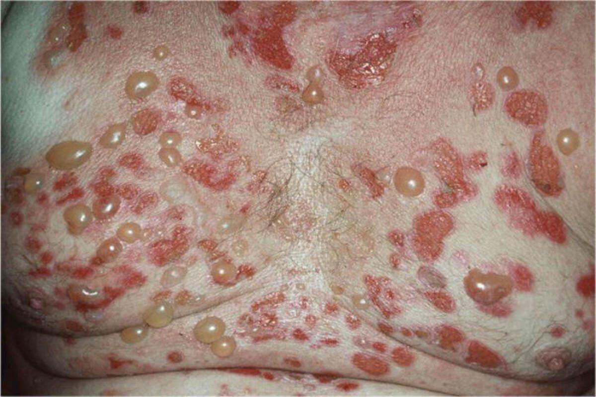

Immunobullous conditions
- There are several immunobullous conditions including bullous pemphigoid, a blistering condition of the elderly, which often starts with itch and fixed urticated and erythematous lesions (figure 1). Later, large tense bullae develop on both erythematous and on normal skin and there may be mucosal involvement with blisters and erosions (figure 2). Pemphigoid gestationis (figure 3) may arise at any time between four weeks gestation and five weeks postpartum. Initially there are itchy urticated lesions around the umbilicus, which then become more widespread evolving into large, tense bullae
7. Boils and folliculitis (including hidradenitis suppurativa): an overview
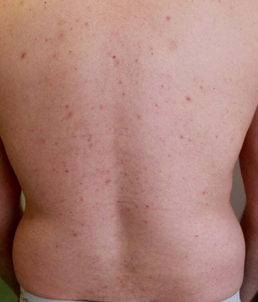
Folliculitis/pseudofolliculitis
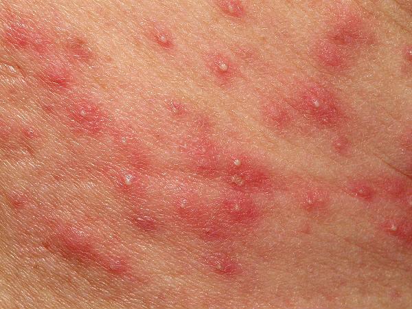
- Small papules and occasional pustules centred on hair follicles
- Many different types/causes
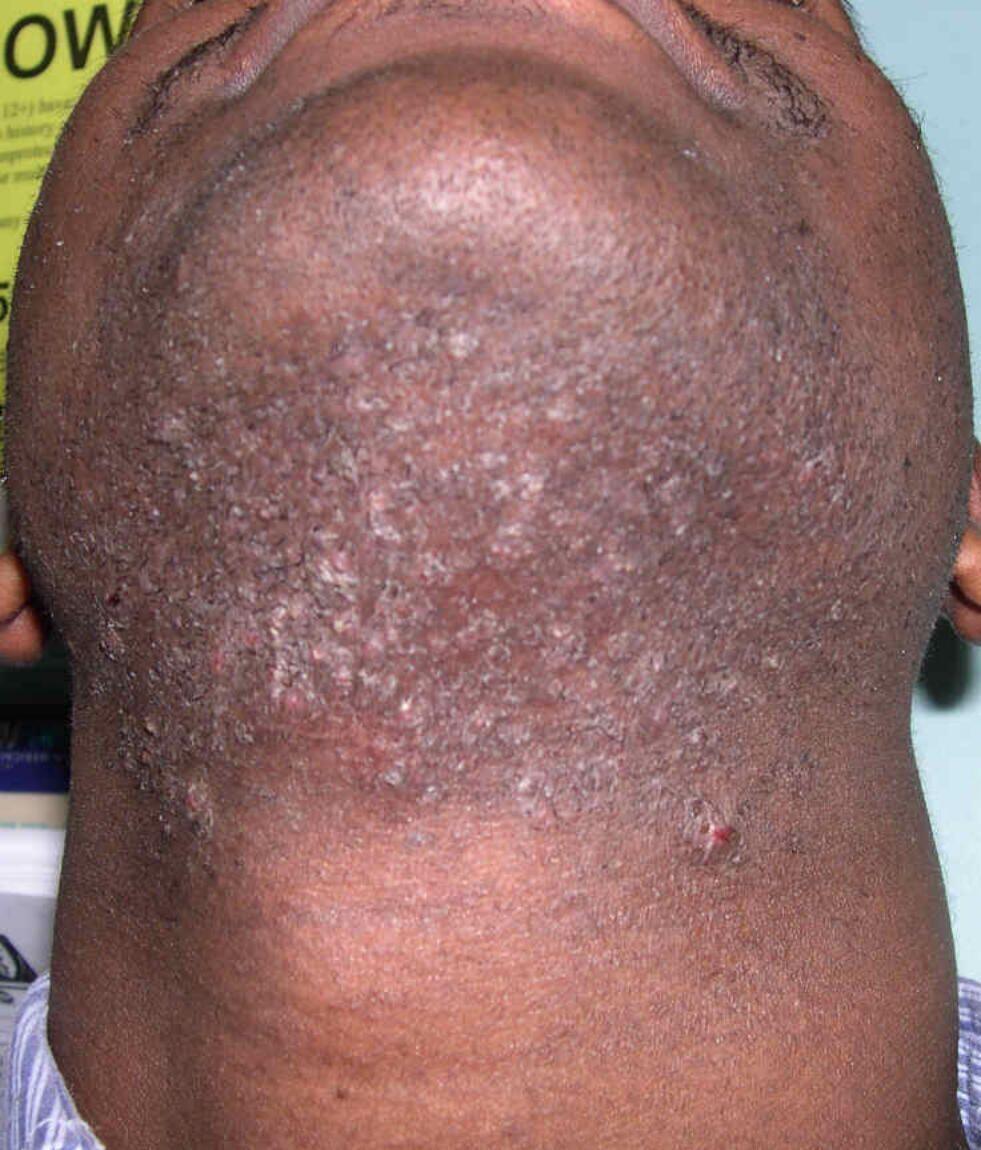
- Some cases related to shaving, eg, beard area, back of neck and legs, which can cause either a pseudofolliculitis resulting from ingrowing hairs and/or a folliculitis resulting from infection
- Take a skin swab of an active pustule to see if there is an infection (which can be primary or secondary)
- Refer to the chapter Folliculitis and Boils for diagnosis and treatment

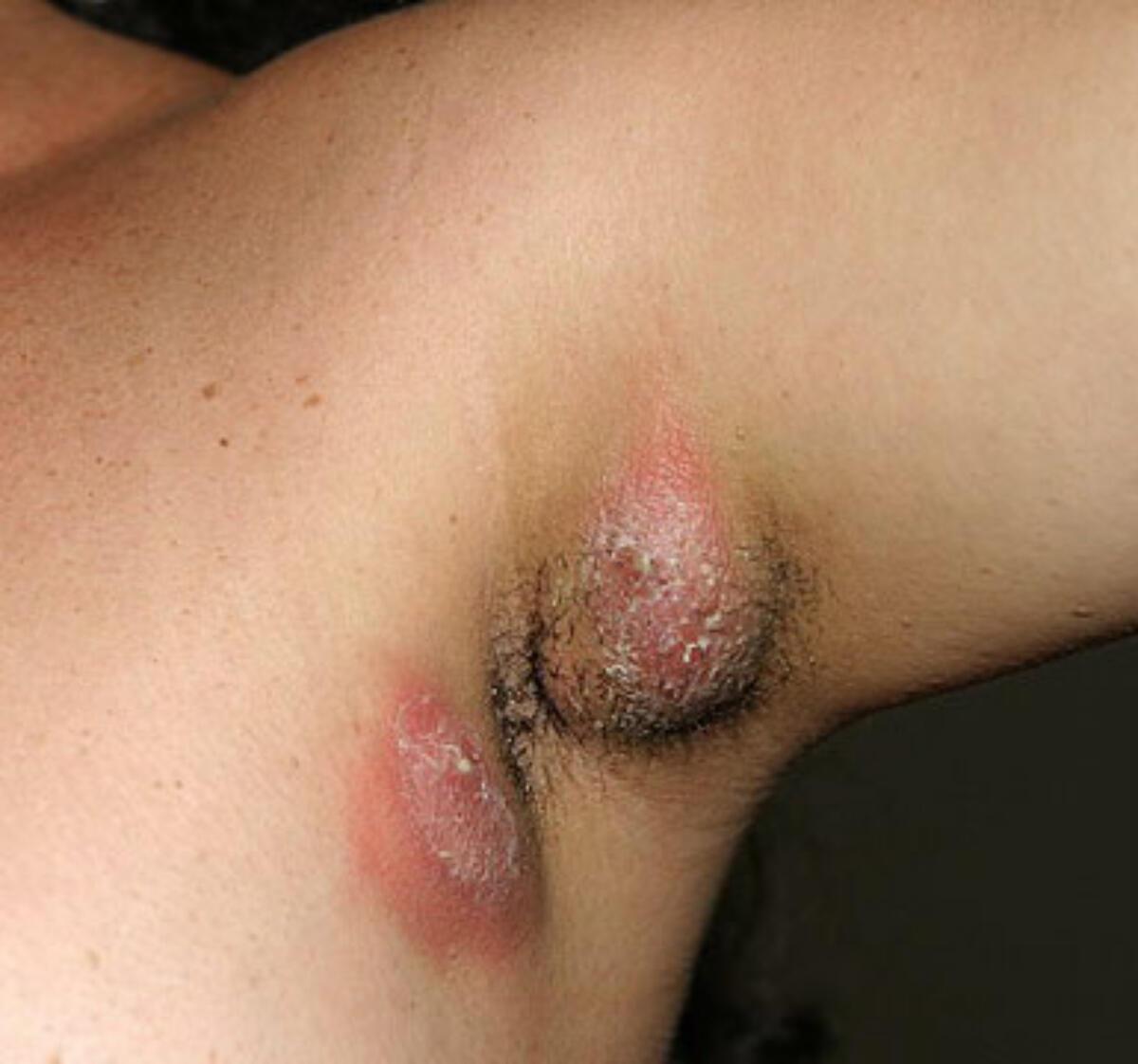
Boils (carbuncle/furuncle)
- If recurrent multiple boils swab for MC&S and check bloods for FBC, glucose
- If positive for staph. aureus ask lab to check for PVL (Panton-Valentine leucocidin) status - refer to the chapter Folliculitis and Boils for more information
- If multiple scarring boils in one or more of the following sites - axilla, groin, perineum, under breasts, then suspect hidradenitis suppurativa (HS). HS should receive prompt treatment as scar formation is irreversible and the longer the condition is left untreated the more difficult it becomes to manage. For detailed information refer to the chapter Hidradenitis suppurativa
8. Dermatological emergencies
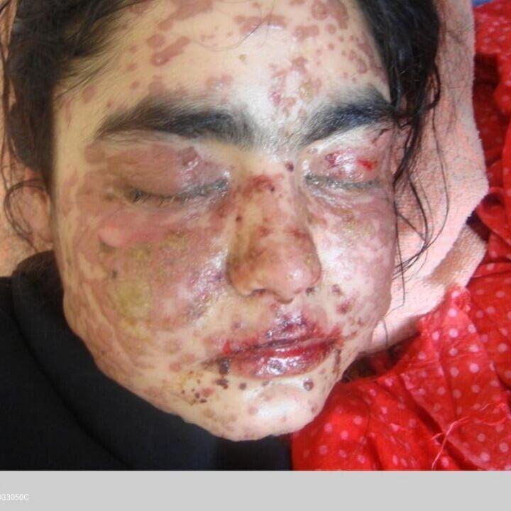

Requiring admission
Steven’s Johnson syndrome SJS (upper figure) / Toxic epidermal necrolysis TEN (bottom figure)
- These are drug reactions. Symptoms often start within a few days of the drug being commenced although it can take a little longer with anticonvulsants
- Symptoms - fever, malaise, myalgia and arthralgia
- Clinical features:
- Painful erythema with local erosions that quickly progresses to areas of confluent erythema with sheet-like skin loss
- Mucosal involvement includes the eyes, lips/mouth
- The term SJS is used when the disease involves less than 10% of the total body surface area. TEN is used when the disease involves more than 30% of the body surface area. Patients whose disease involves 10-30% of their body surface area are said to have SJS / TEN overlap
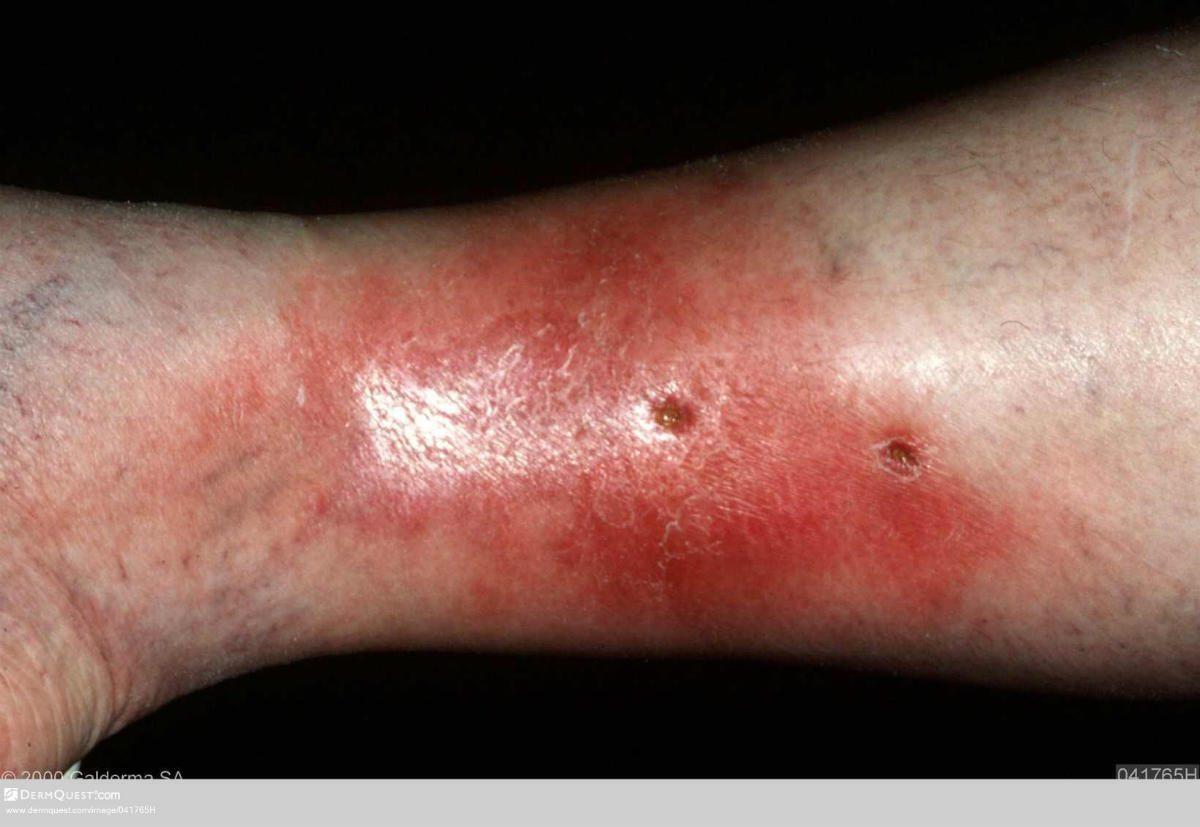
Requiring admission
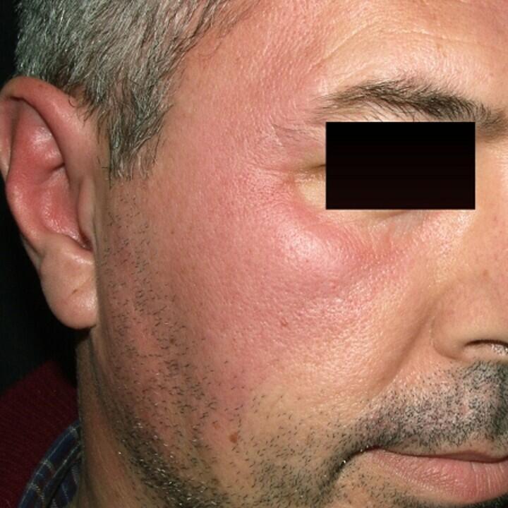
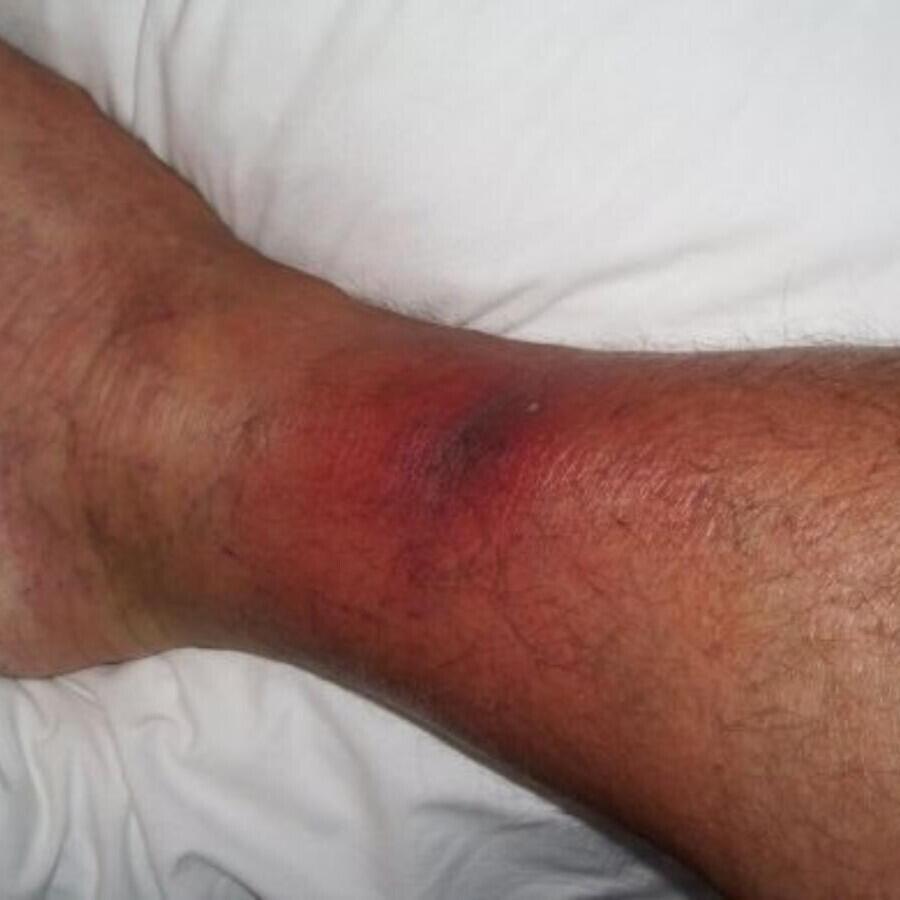
The following cases of cellulitis (upper figure) and erysipelas (bottom figure):
- Peri-orbital cellulitis
- Rapidly progressive cellulitis
- Patient constitutionally unwell eg vomiting, or temperature at or over 39 degrees C
- Major comorbidities
Requiring admission
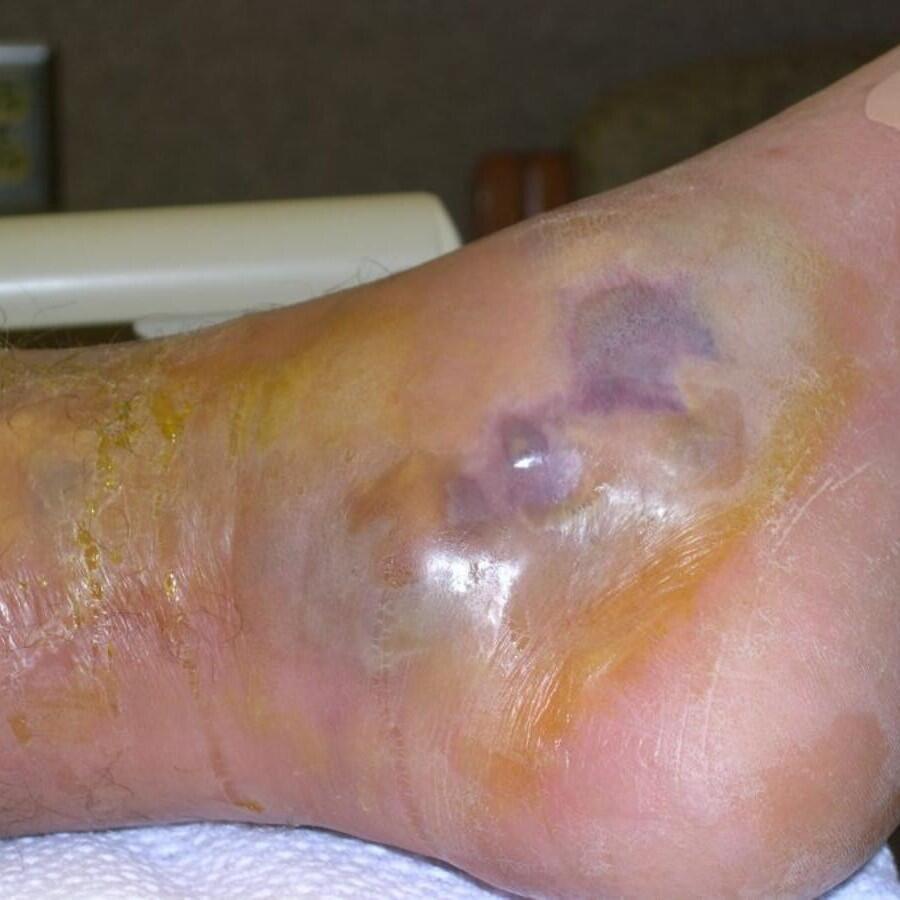
- Early features - erythema with moderate-severe pain/tenderness, or systemic upset, which is out of proportion to the physical signs
- As the condition progresses - dusky-violaceous areas (along with erythema) / crepitus / over a short period of time blisters develop, the affected area becomes necrotic, and the patient very toxic


Same day discussion with on-call dermatologist (with a view to urgent outpatient appointment or admission)
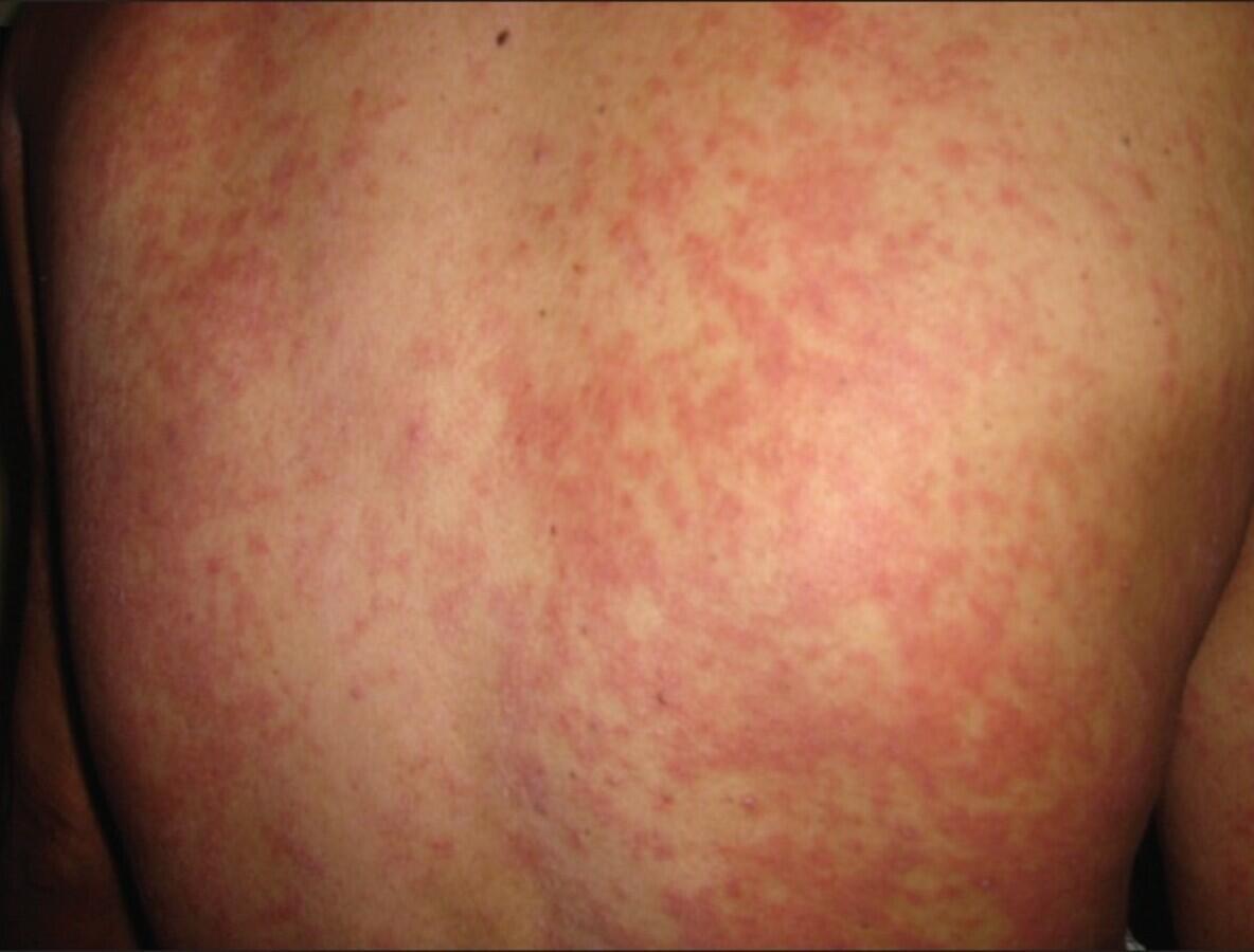
1. Erythroderma (top figure)
- 90% of more of body surface erythematous
- The skin feels hot but patient complains of feeling cold
2. Drug Hypersensitivity syndrome (second figure)
- Generally 3-6 weeks after medication started
- Rash, fever, facial oedema, lymphadenopathy
- Can have systemic features - hepatitis 50% and nephritis 10% of cases
3. Vasculitis (third figure)
- Urgent attention required if rapid development of purpura/ulceration
- If renal involvement discuss with nephrologists otherwise dermatology
4. Progressive / widespread erosions or blistering (bottom figure)
- Could suggest bullous pemphigoid or other immunobullous conditions
There are numerous other conditions that may be of concern, please follow the links below:
9. Drug rashes
Severe/life threatening
- Refer to dermatological emergencies in the section above
Mild-moderate
- Most drug rashes start at around 4-14 days after commencing the drug and settle within a few weeks of the drug being discontinued. However, some drugs occasionally take much longer
- Refer to the chapter Drug rashes: mild-moderate for more information on the most common clinical findings and associated drugs
10. Eczema - atopic (including facial eczema)
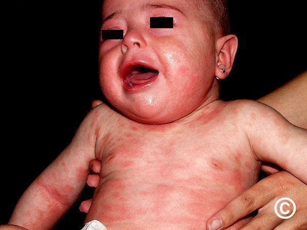
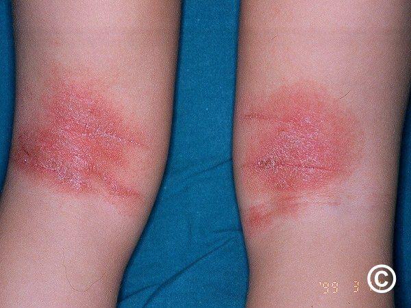
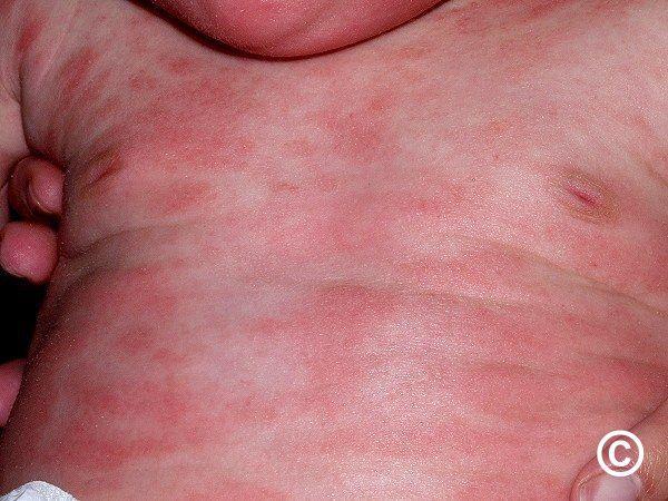
Infants & young children
Provide a management plan and refer to the bottom of the page for a patient information leaflet available through a QR code or printable PDF.
Emollients (creams, gels, ointments) - patient preference important. Prescribe large quantities (500-1000g a month minimum). If very dry may need ointment as well as cream/gel. Use at least twice a day, even if eczema is settled
Topical steroids - use on persistent areas of itchy erythema. Creams or ointments, consider the latter if skin very dry. Wait at least 30 minutes after using emollient before applying steroid. Strength determined by the age, site, and severity, eg:
- Face: mild potency eg 1% hydrocortisone once-twice daily. If requiring a regular topical steroid on the face, change to a topical calcineurin inhibitor - either pimecrolimus cream from the age of 3 months, or tacrolimus 0.03% ointment from the age of 2 years
- Trunk and limbs: moderate potency once-twice daily eg Eumovate ® (clobetasone butyrate 0.05%) or Betnovate-RD ® (betamethasone valerate 0.025%)
- Scalp: 0.1% betamethasone scalp application, or if stings too much Synalar ® scalp application
Diet relevant in a minority - consider if < 3 years of age with any of moderate-severe eczema, GI symptoms, wheezing, perioral flare when eating and/or perianal erythema. Refer below (additional management notes).
Other resources:
- For further advice refer to the A-Z clinical chapter on atopic eczema
- For a simplied managment plan please refer to the PCDS Primary Care Treatment Pathway for paediatric (0-12yrs) eczema
- The National Eczema Society - a patient support group
- Eczema Outreach Support - a support group helping children and young people with eczema to thrive
- Eczema Care Online - helps patients manage their eczema


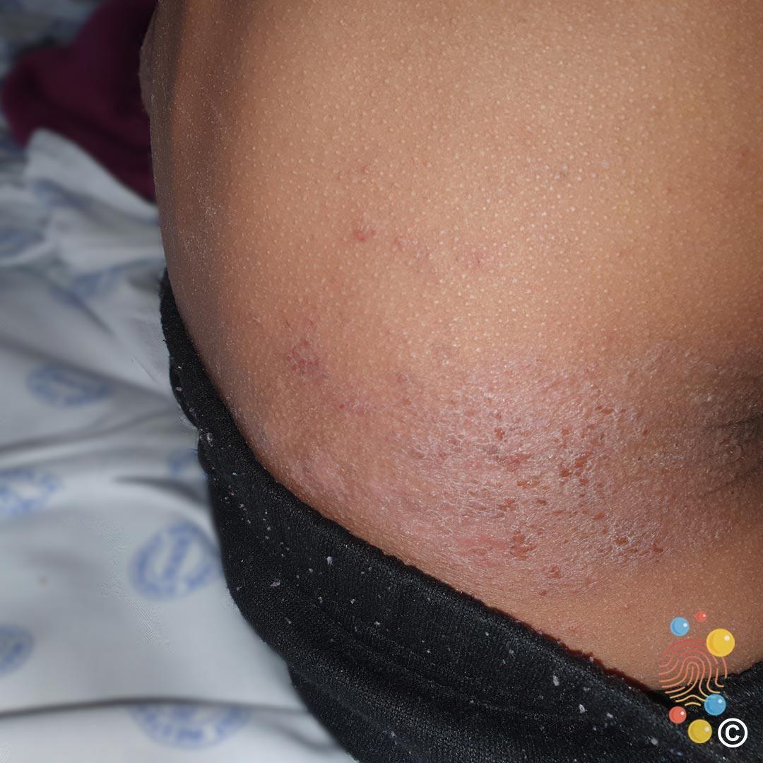
Older children / adults
Provide a management plan and refer to the bottom of the page for a patient information leaflet available through a QR code or printable PDF.
Emollients (creams, gels, ointments) - patient preference important. Prescribe large quantities (500g a week may be required for moderate-severe eczema). If skin very dry may need ointment as well as cream. Use at least twice a day, even if eczema settled, eg, morning and early evening (including after a shower/bath). In infants and very young children there is some evidence that washing hands before applying emollients may reduce the risk of peanut and various other food allergies.
Topical steroids (and calcineurin inhibitors) - use on persistent areas of itchy erythema. Creams or ointments, consider the latter if skin very dry. Use at bedtime, wait at least 30 minutes after applying emollient before applying steroid. Strength determined by the age of patient, site, and severity, eg:
- Face/flexures/genitalia - mild (1% hydrocortisone) or moderate potency eg Eumovate ® once-twice daily - if needed regularly refer to notes below (face)
- Trunk and limbs: potent once a day at night eg Betnovate ® (betamethasone valerate 0.1%), Elocon ® (mometasone) - it is safe to use up to 100g of a potent topical steroid per month
- Palms and soles: potent or very potent once a day at night eg Dermovate ® (clobetasol propionate 0.05%)
- Scalp: potent or very potent once a day at night eg 0.1% betamethasone scalp application or Dermovate ® scalp application. If stings too much consider Synalar ® scalp application
For frequent flares - consider the 'weekend' steroid regime, in which a potent topical steroid on 2 consecutive days a week as maintenance.
Additional notes on treating the face:
- The frequent use of topical steroids on the face should be avoided, instead consider topical calcineurin inhibitors - either pimecrolimus cream or tacrolimus 0.1% ointment. National guidance states these treatments can be initiated by any health professional experienced in treating eczema, which includes GPs
- Adults with predominantly facial eczema should be considered for patch testing
Other resources:
- For further advice refer to the A-Z clinical chapter on atopic eczema
- For a simplied managment plan please refer to the PCDS Primary Care Treatment Pathway for adult eczema
- The National Eczema Society - a patient support group
- Eczema Outreach Support - a support group helping children and young people with eczema to thrive
- Eczema Care Online - helps patients manage their eczema
Patient Information Leaflet
Link: Eczema - atopic eczema

Download the Patient Information Leaflet PDF: Eczema - atopic eczema
11. Eczema - discoid eczema
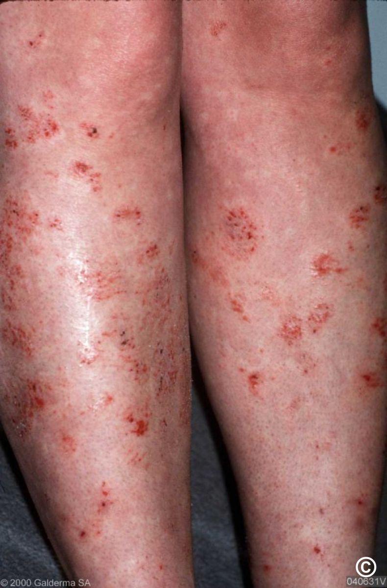

Key features
- Very itchy
- Can occur anywhere although limbs predominate
- Discrete round-oval patches - differs from psoriasis in that lesions tend to be lighter red, borders fade gradually at the periphery, and there is often surface crust (as opposed to white-silvery scale of psoriasis). Fungal infections such as ringworm have an advancing edge and central clearing
- Differential diagnosis - refer to the General Dermatology Diagnostic Tool
Management
- Refer below for a patient information leaflet
- Acute treatment - short-term use of a potent topical steroid cream/ointment, eg 0.1% betamethasone or mometasone furoate (both OD), until things settle (may take 2-6 weeks)
- Long-term treatment - regular use of a topical emollient at least twice a day (prescribe minimum of 500-2000 g per month, depending on how dry the skin is and size of patient). The same topical steroid can be reintroduced for a flare. It is safe for adults to use up to 100 g of a potent topical steroid per month, bearing in mind one needs to be more sparing on areas of thin skin, eg, face, lower legs
- Refer to the chapter Discoid eczema for more detail
Patient Information Leaflet
Link: Eczema - discoid eczema

https://www.pcds.org.uk/patient-info-leaflets/eczema-discoid-eczema
Download the Patient Information Leaflet PDF: Eczema - discoid eczema
12. Eczema - gravitational (venous)
Key features
- Itch and afebrile, although sometimes may just present as an erythematous asymptomatic rash
- Localised or diffuse involvement of the gaiter area - often bilateral, although one leg commonly affected more than the other
- Erythema, often with associated brown discoloration resulting from haemosiderin deposition
- Dry and scaly, or weepy
- Varicose veins may or may not be present
- Differential diagnosis - refer to the General Dermatology Diagnostic Tool. Many patients are referred to Dermatology with 'bilateral cellulitis' having had months of antibiotics - this is NOT cellulitis. Cellulitis is nearly always unilateral, presenting with hot, tender spreading erythema, and systemic symptoms
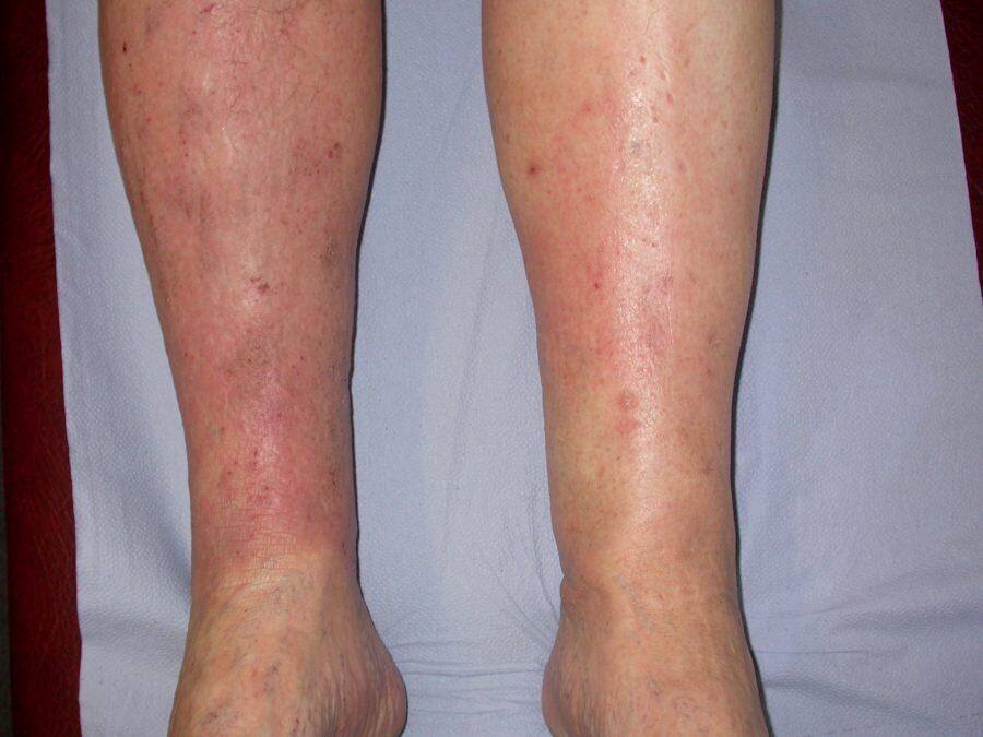
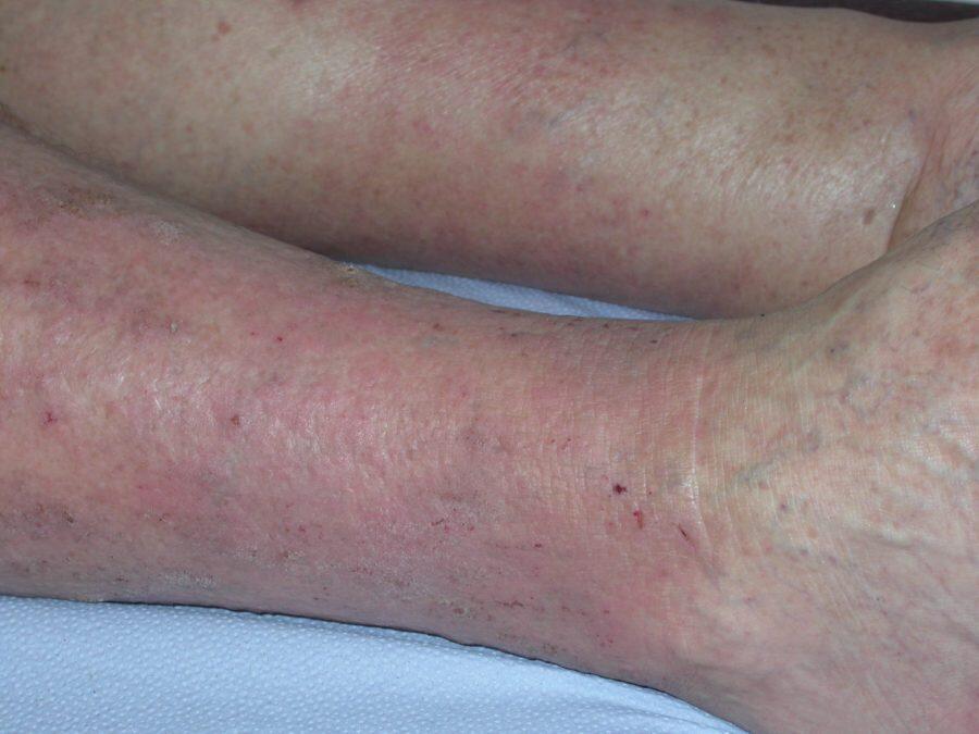
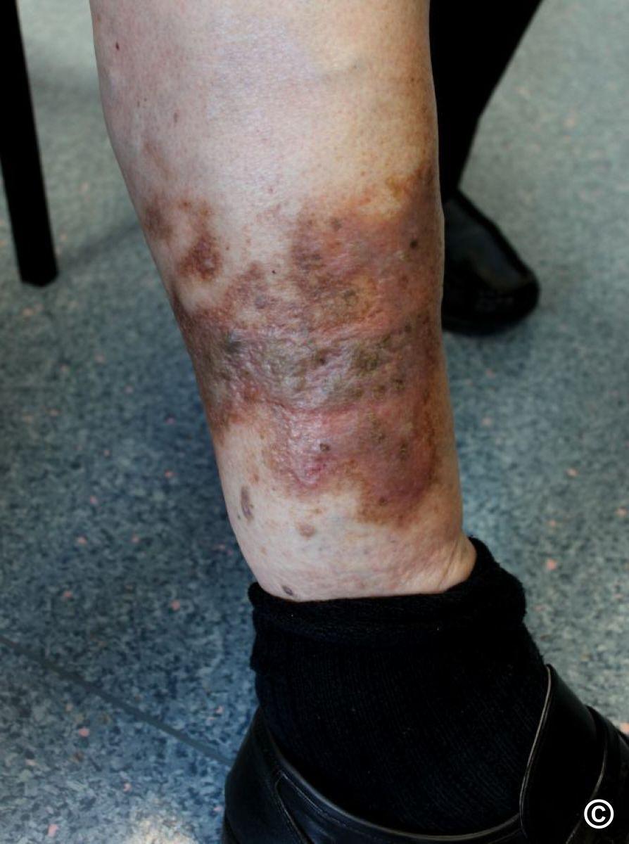
- Refer to the bottom of the page for a patient information leaflet available through a QR code or printable PDF
- Emollients - use copious amounts 2-3 times daily, creams often well-tolerated, ointments more effective if skin very dry (advise patients to scoop ointments out of tub using large spoon/other utensil, rather than hands). Patient preference is important
- Topical steroids - for persistent itchy erythema. Use the least potent preparation that controls symptoms, with steroid free periods to reduce risk of skin atrophy. Emollients must dry for at least 30 minutes before applying steroids
- Tacrolimus ointment - if topical steroids needed on a regular basis, settle eczema with the steroids, then replace with tacrolimus 0.1% ointment (Protopic ®) OD for 6-8 weeks, before gradually reducing to twice a week maintenance therapy. The topical steroid can be restarted during a flare
- Moderate-severe or persistent gravitational eczema
- Use a potent (eg Betnovate-C ® ointment) or super-potent topical steroid under Clingfilm occlusion at night, or failing that under medicated bandages eg ZIPZOC ® or Viscopaste ®. The bandages should be replaced once-twice a week. Continue treatment until things settle, which often takes 6-8 weeks. Click on the following link to see how to apply these bandages
- If symptoms persist consider contact allergic dermatitis to one of the treatments, more likely if eczematous spread to distant sites - refer to dermatology for patch tests
- Long-term - manage underlying venous conditions by the use of leg exercises and compression hosiery, normally prescribed from the pharmacy as 'made to measure' Class II below knee, open toe stockings. It is advisable for the practice nurse to check the ankle-brachial pressure index (ABPI) to make sure there is no significant arterial insufficiency that would exclude their use
- Provide patient with the attached document to help direct them to this page if they wish to view the videos
Patient Information Leaflet
Link: Eczema - gravitational eczema

https://www.pcds.org.uk/patient-info-leaflets/eczema-gravitational-eczema
Download the Patient Information Leaflet PDF: Eczema - gravitational eczema
13. Eczema - hand (and foot)
Key features
There are several different types of hand eczema including:
- Atopic eczema: can affect the hands and may be dry/scaly or wet/weepy
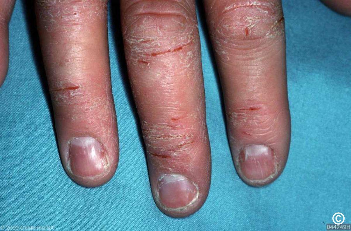
- Pompholyx: usually presents periodically with intensely itchy vesicles that predominate on the palms and sides of fingers. On occasions large blisters may develop
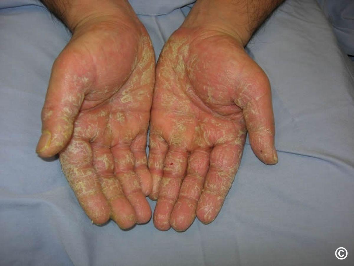
- Hyperkeratotic eczema: thick areas of scale on the palms and soles. Can be difficult to distinguish from psoriasis although the latter may be better demarcated
Investigations
- If dry and scaly, especially if unilateral and/or nail involvement send skin scrapings for mycology to look for tinea (fungal infection)
- If persistently weepy/crusted take swabs for bacteriology and treat relevant findings
- In cases of troublesome hand eczema (especially if no eczema elsewhere, or, out of proportion to eczema at other sites) refer to Dermatology for patch testing in case of contact allergic dermatitis (CAD)

Management overview - all patients
- Refer to the bottom of the page for a patient information leaflet available through a QR code or printable PDF
- Prescribe emollient cream to use regularly (patient preference important) and soap substitute, eg Dermol Wash Cutaneous Emulsion ®
- Potent/super-potent topical steroids may be needed, eg Betnovate 0.1% ® or Dermovate ®. If hands not weeping, treatment enhanced by using as ointments at night under occlusion with cotton gloves, or Clingfilm wrapped around troublesome areas
- Fissures - consider commercial superglue (patient must take great care when applying to a fissure and not have contact allergy to acrylates in glue ), or steroid impregnated treatment, which can be left on for up to 24 hours before reapplying, eg Haelan ® tape (fludroxycortide tape), or, Betesil ® medicated plaster (betamethasone valerate 2.25mg)
Wet/weeping eczema with or without large blisters (middle image)
- Manage as above in terms of information leaflets, emollients, soap substitutes and topical steroids
- Aspirate any large bullae
- In order to the dry the skin prescribe potassium permanganate solution (one Permitab ® dissolved in four litres of warm water, soak for 10-15 minutes, 2-4 times a day) - provide written instructions
Hyperkeratotic hand eczema (bottom image)
- Manage as above in terms of information leaflets, emollients and soap substitutes
- Topical steroid - use Diprosalic ® ointment OD-BD, treatment enhanced by using at night under occlusion with cotton gloves, or Clingfilm wrapped around troublesome area
Additional resources
- Who to refer - troublesome symptoms not responding to the treatments referred to above. If contact allergic dermatitis suspected refer to a Dermatology department with patch testing facilities
- Refer to the clinical chapter Eczema: hand (and foot) eczema for more detail
- Demonstrations on how to use medicated tapes - Haelan ® tape (fludroxycortide tape), Betesil ® medicated plaster
Patient Information Leaflet
Link: Eczema - hand (and foot)

https://www.pcds.org.uk/patient-info-leaflets/eczema-hand-and-foot
Download the Patient Information Leaflet PDF: Eczema - hand (and foot)
14. Fungal infections (tinea)

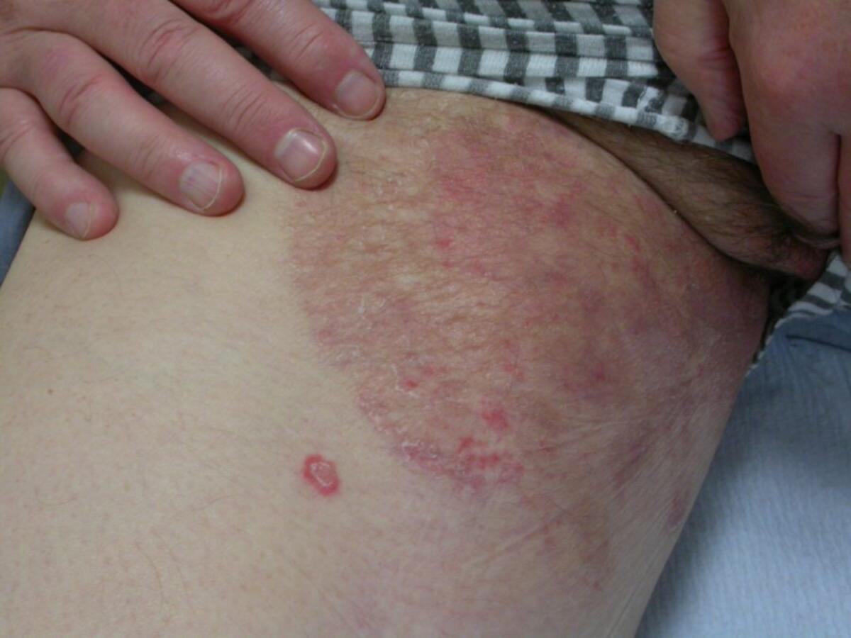
Key features of tinea corporis (body) and cruris (groins)
- Tinea, which can be itchy, should be part of the differential diagnosis of any rash that presents unilaterally and/or has a leading erythematous scaly edge. One of the main differentials is granuloma annulare - a smooth (non-scaly), annular plaque/s that enlarges centrifugally with a 'bumpy' edge. Refer to the diagnostic tools for other annular lesions
- Occasionally, tinea can have pustules, and is aggravated by topical steroids (tinea incognito), topical treatments may also disguise scale
Treatment
- Milder cases - a topical antifungal agent eg terbinafine (Lamisil ®) cream for two weeks (or longer if needed), or one of the imidazole creams eg miconazole (Daktarin ®) for up to four weeks. Terbinafine is more expensive but slightly more effective
- If the rash is extensive or inflammatory (pustules present) treat systemically with terbinafine 250mg OD for two weeks
- For more detail refer to the relevant clinical chapter
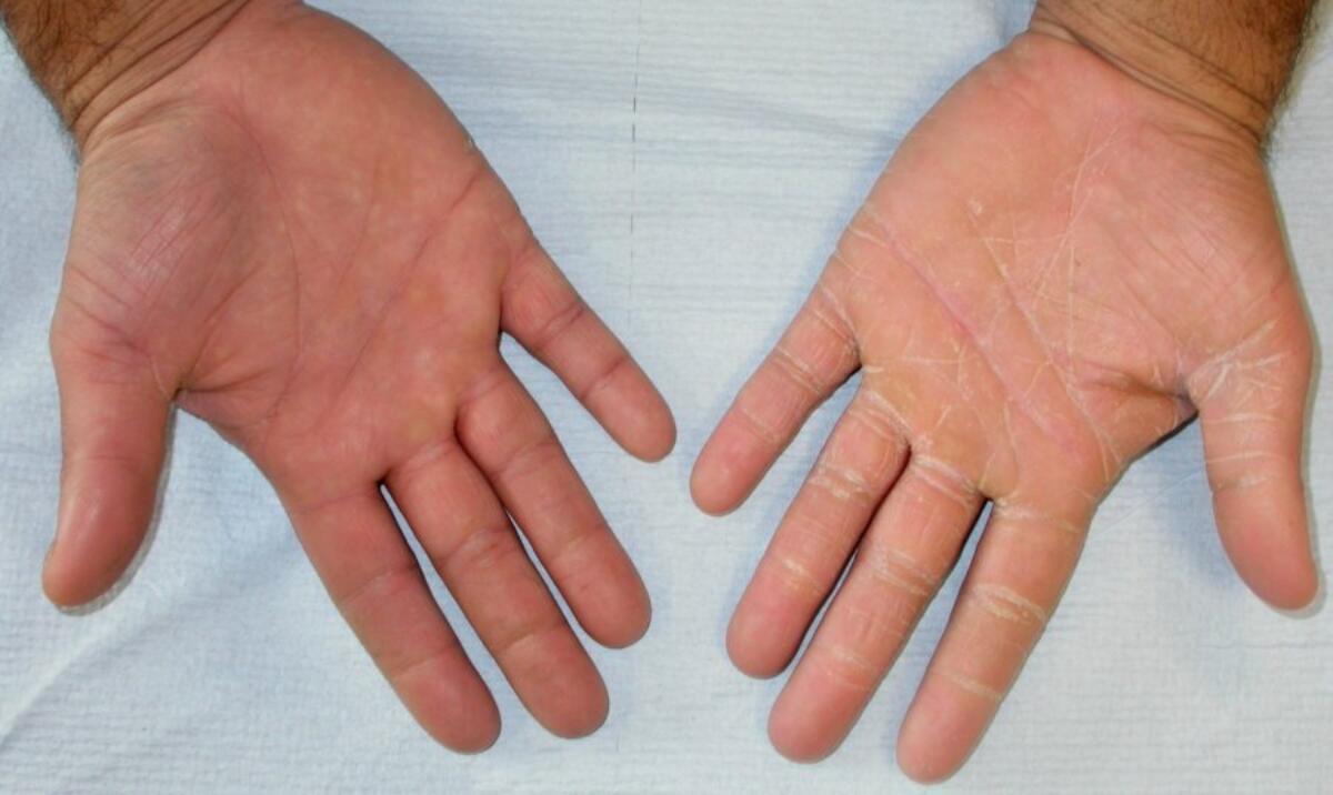
Key features of tinea manuum (hands) and pedis (feet)
- Fine scale especially in the skin creases, with or without erythema
Treatment
- Treat as above (tinea corporis), unless nail involvement, in which case refer below (tinea unguium)
- For more detail refer to the relevant chapter

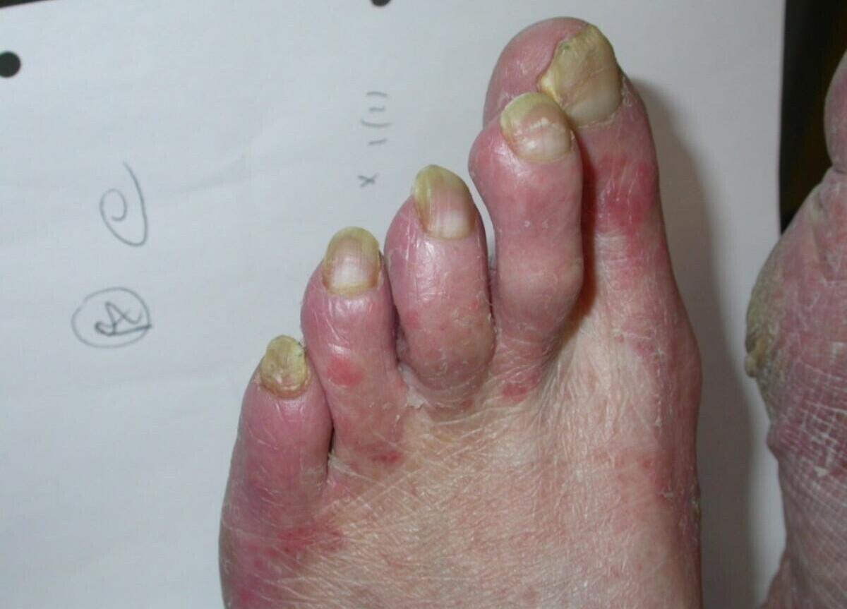
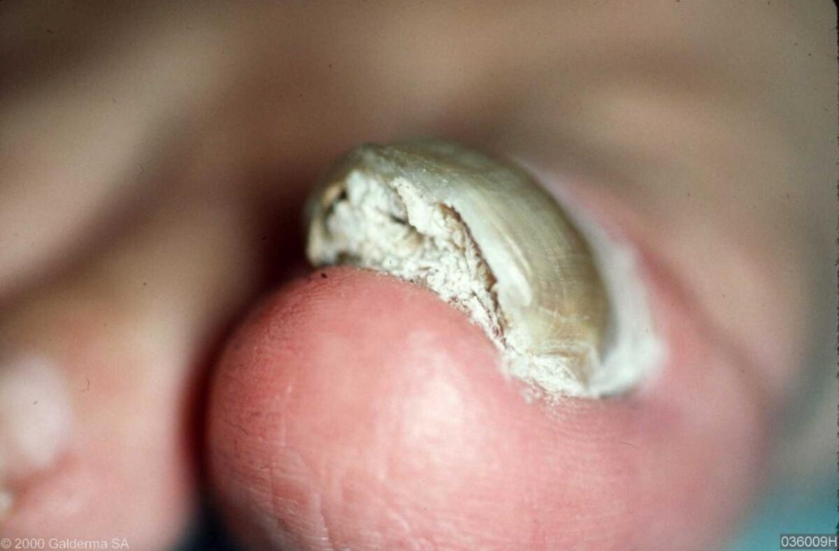
Key features of tinea unguium (nails)
- Often multiple nails. Superficial infection presents with white surface change (upper figure), otherwise affected nails are yellow, often with subungual debris as the distal nail plate starts to crumble (lower figures)
- To prevent high rates of false negative results, take clippings and scrapings as follows - cut nail back and remove subungual debris (where much of the fungus lives) by careful curettage / scalpel blade, sending the entire specimen for mycology. This must be done in the surgery, not by the patient
Treatment
- Topical treatments - have a low cure rate but may be suitable for treating distal nail infection (as opposed to involvement of the nail matrix) or superficial white infection. Options include Tioconazole ® nail solution BD for six to twelve months or Loceryl ® nail lacquer twice per week for three to six months for fingernails, and six to twelve months for toenails. Nails should be filed / cut back as much as possible prior to applying the treatment
- Systemic treatment with terbinafine (Lamisil ®) - should only be used is the nails are symptomatic, and preferably if mycology is positive (beware of the possibility of false negative results). The following regimes should be used:
- Adults: 250 mg OD for six weeks for fingernails and three to four months for toenails
- Children: six to twelve weeks (weight > 40 kg use 250 mg OD; weight 20-40 kg use 125 mg OD; weight up to 20 kg use 62.5 mg OD)
- For more detail, including on other treatment options such as itraconazole, refer to the relevant clinical chapter
- Refer to additional resources (below) for more information on terbinafine monitoring
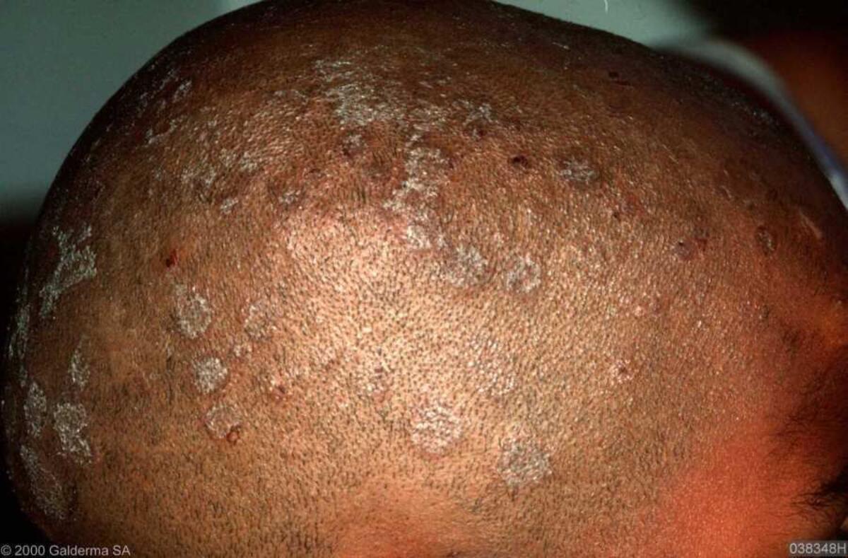
Key features of tinea capitis (scalp)
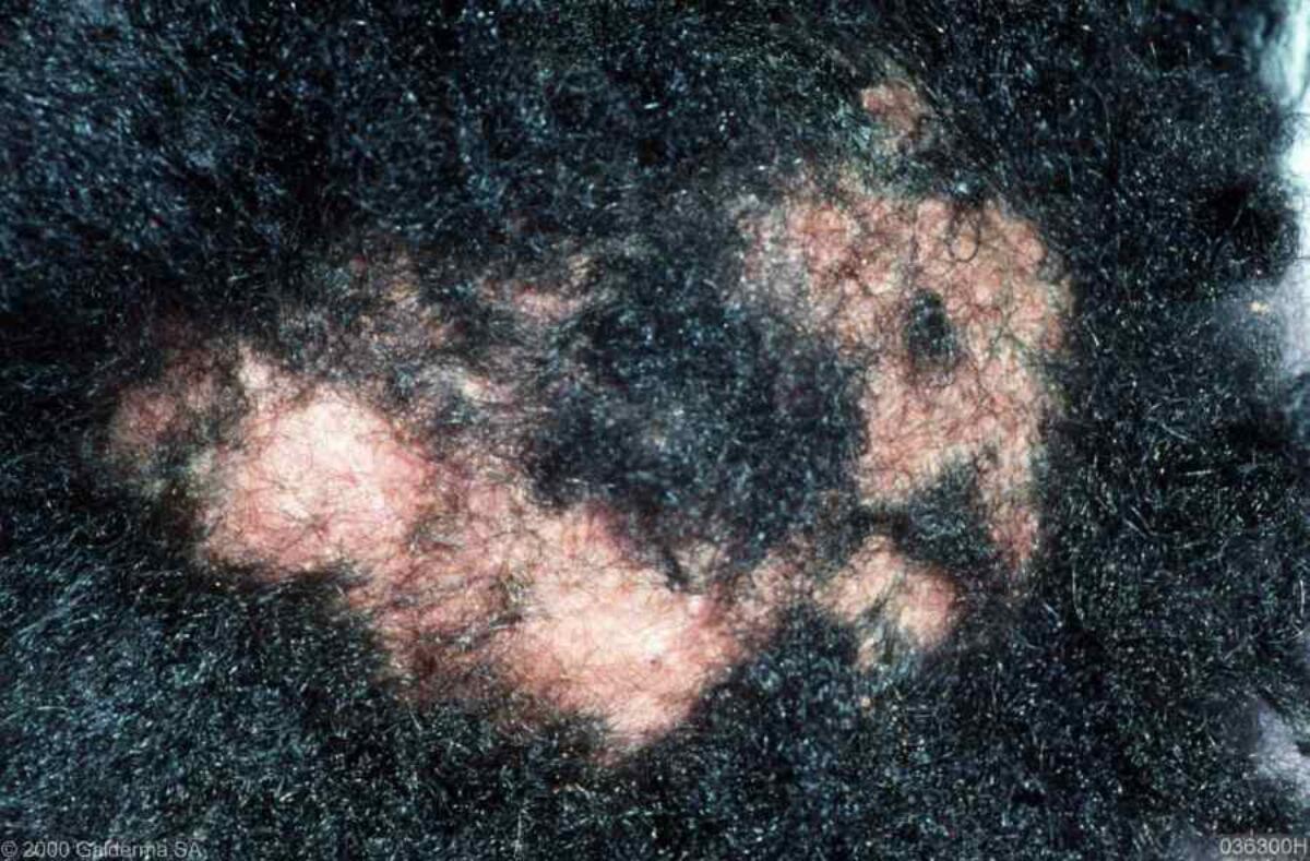
- Hair loss, often accompanied by scaling. Itch is variable. Dermoscopic features can include broken hairs with black dots, and comma-shaped hairs. Occasionally the hair loss can be scarring (bottom figure)
- Both skin scrapings and plucked hairs should be sent for mycology
Treatment
- Due to the risk of scarring alopecia treatment is usually with terbinafine 250 mg OD for two to four weeks in adults. For children refer to the dosages above (tinea unguium)
- The affected individual and other household members should also be treated with ketoconazole shampoo at least twice weekly for two weeks
- Refer to the relevant clinical chapter for more detail
Additional resources
- Clinical chapters - Tinea faciei (face) and barbae (beard)
- Guidance on monitoring with terbinafine: terbinafine is a safe and effective drug used drug to treat fungal infections, especially of the trichophyton species. While the BNF states that hepatic function should be monitored before treatment and then periodically after 4–6 weeks of treatment, the risk of liver damage is rare and as such many specialists agree that routine interval laboratory test result monitoring is unnecessary in the absence of underlying hepatic conditions. Individual review of the patient and clinical discretion should therefore be used when deciding on hepatic monitoring with regards to oral terbinafine usage. Patients should be counselled on recognising the symptoms of hepatotoxicity - jaundice, abdominal pain, generalised pruritus, dark urine, flu-like symptoms (and rarely a measles-like rash - the Drug Hypersensitivity Syndrome) and advised to discontinue treatment and seek urgent medical advice in the very unlikely event of such symptoms arising
15. Hyperhidrosis (excess sweating)
Management overview
- Provide a patient information leaflet
- Treatment depends on the site/s affected
Axillary hyperhidrosis
- Aluminium chloride antiperspirants eg Anhydrol Forte ® or Driclor ® should be used in the following way:
- Apply at night only
- Wash the area, dry before application and wipe on once only
- Wash off the following morning
- Due to the irritant nature of the treatment start twice a week, and gradually increase by one night a week
- If the skin becomes very irritable use a mild-moderate topical steroid (eg Eumovate cream) each morning
- Those not responding can be referred for Botox injections, providing these are available on the NHS. This is not curative and usually repeat treatment is required
- Very occasionally patients with more severe axillary hyperhidrosis may consider systemic anticholinergics (see below)
Palmar and plantar hyperhidrosis
- Antiperspirants can be tried as above
- Iontophoresis is offered by many dermatology departments, but treatment needs to be repeated and as such patients are generally encouraged in the long-term to buy their own machine for home use
- Very occasionally patients may consider systemic anticholinergics (see below)
General hyperhidrosis
- Generalised hyperhidrosis in a well patient with a classical history of sweating starting in late childhood and improving in middle age is seldom related to an underlying medical condition. In all other cases refer to the section on investigations in the chapter Hyperhidrosis
-
The mainstay of treatment are systemic anticholinergics:
- Propanthelene bromide is the only licensed systemic product for primary hyperhidrosis. Start at a low dose of 15 mg once to twice a day, increasing as tolerated to 30 mg tds
- Oxybutinin is an alternative - some patients are unable to tolerate standard release formulations, in which case consider a modified-release formula of oxybutynin eg Lyrinel XL 10 mg ®, the dose can be gradually increased up to a maximum of 30mg OD
- If dry mouth is a problem add in pilocarpine 10 mg tds
- Consideration needs to be given to prescribing long-term systemic anti-cholinergics as there is a small amount of evidence connecting them with dementia
Additional resources
- For more detailed information refer to the chapter Hyperhidrosis
- Hyperhidrosis UK is a very helpful patient support group
Patient Information Leaflet
Link: Hyperhidrosis (excess sweating)

https://www.pcds.org.uk/patient-info-leaflets/hyperhidrosis-excess-sweating
Download the Patient Information Leaflet PDF: Hyperhidrosis (excess sweating)
16. Intertrigo (inflammation in body folds/flexures)
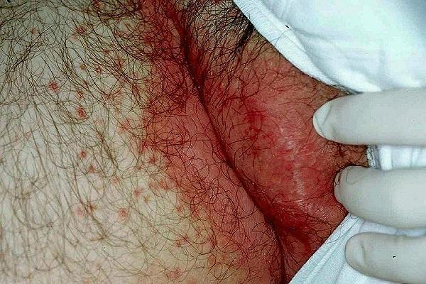
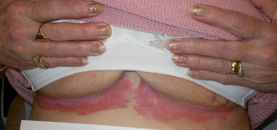


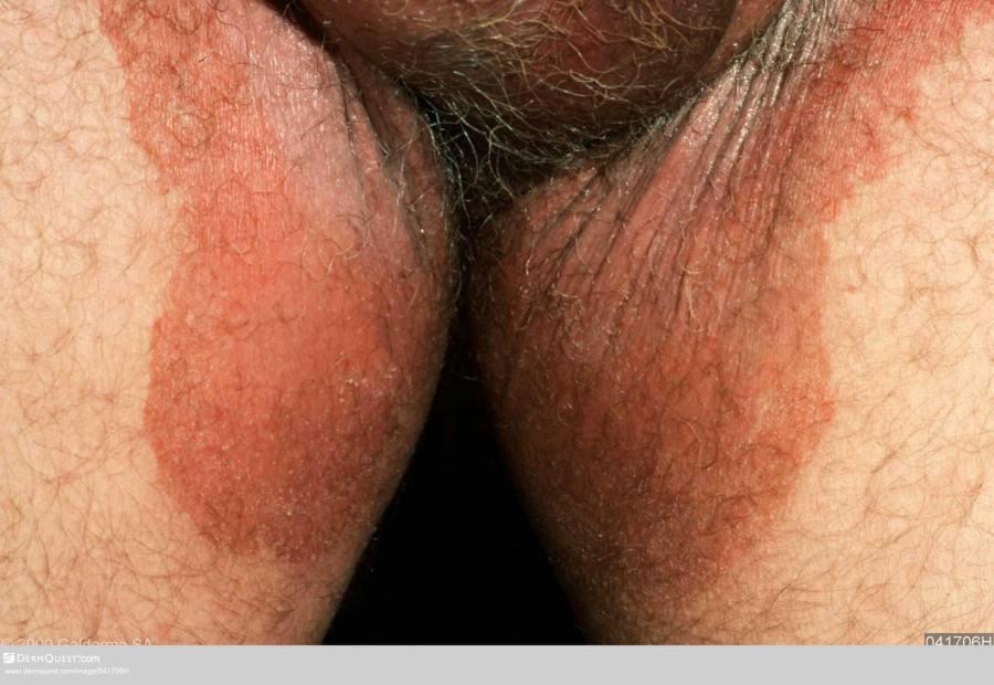
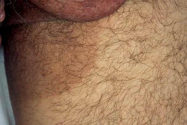
Differential diagnosis
In some cases inflammation is limited to body folds and simply reflects local skin changes (eg secondary to obesity), in others the inflammation is part of an underlying skin condition (eg psoriasis), as such patients with intertrigo such have a full skin examination. The most common diagnoses are:
- Secondary to obesity
- Candida (figure 1)
- Seborrhoeic eczema
- Psoriasis (figure 2)
- Tinea (figures 3&4)
- Erythrasma (figures 5-6)
Follow this link for the diagnostic guide to rashes affecting body flexures
Management overview
- Take swabs for bacteria and yeasts
- If dry or scaly take scrapings for fungus
- General measures: weight loss if appropriate, improve hygiene and use barrier creams
- If area is wet use astringent soak, eg, potassium permanganate and avoid talc
Treatment
- If suspected erythrasma (new lesions are smooth, and pink-red or brown, older lesions are brown with a wrinkled, scaly appearance. Woods light causes the erythrasma to fluoresce a coral-pink colour) the use of topical erythromycin, fusidic acid or miconazole will usually clear the condition. Occasionally a short course of oral erythromycin is needed
- If no erythrasma but significant inflammation use Daktacort ® or Trimovate ® cream BD
- If not responding to above:
- If findings suggestive of tinea (erythema with a leading scaly edge) treat with terbinafine cream
- If no suggestion of tinea manage with the short term use (4 weeks) of a potent combination preparation such as Lotriderm ® or Synalar C ® cream, and then review
Other management resources
- Refer to the chapter Intertigo for more detailed information
17. Itch (pruritus)
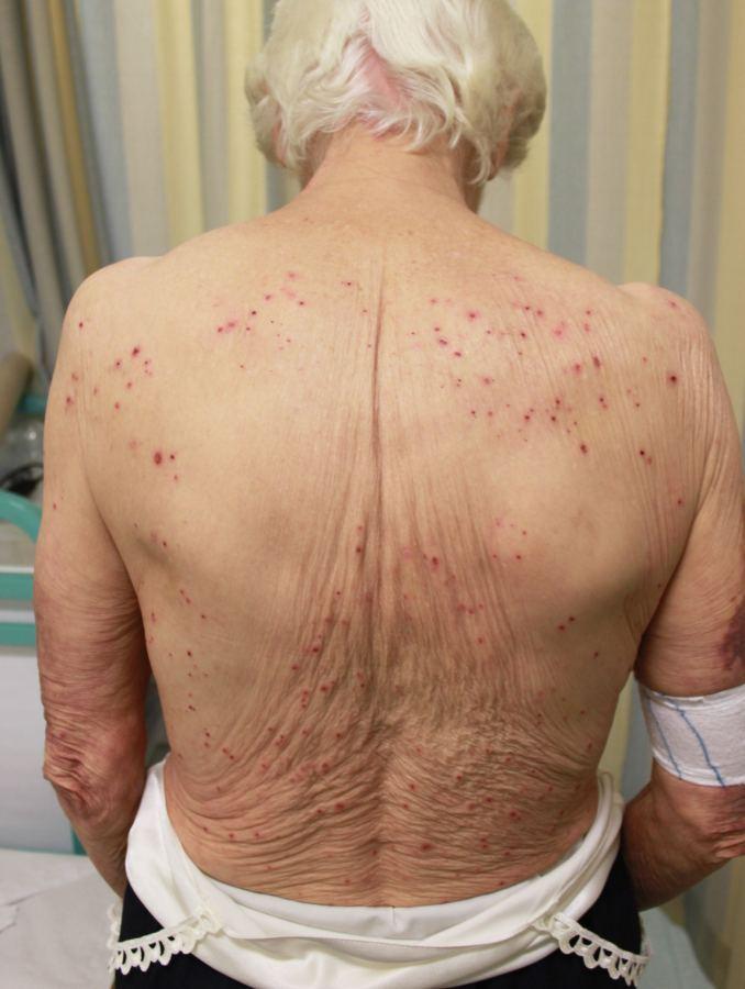
Key features
- This chapter focuses on pruritus (itch) with no or limited skin changes (other than excoriations)
- Where there are clinical features of a skin condition causing the itch follow the links below:
Investigations
- Many causes: systemic disease (including neoplastic), medications, psychogenic (including delusions of parasitosis), aquagenic (induced by water), although over 50% idiopathic
- Undertake a full history (including a systemic enquiry) and general examination (including lymph nodes and abdomen in case of lymphoma or other neoplastic disease) to try and identify a cause
- Standard screen: FBC and ferritin, CRP, routine biochemistry (U&E, creatinine, LFT, bone, TFT), antimitochondrial antibody (to rule out primary biliary cirrhosis), urinalysis, a CXR to help exclude lymphoma and bronchial carcinoma. Also consider hepatitis screen and HIV serology in at risk patients
- Patients with generalised pruritus and suspicion of haematological involvement also require a blood film, lactate dehydrogenase (can be a marker of lymphoma), and ESR (if available). Immunoglobulins and urinary paraproteins may also be requested, but will have a low yield, as myeloma is rarely associated with generalised pruritus
- Associated neoplastic/paraneoplastic conditions. If standard tests are normal then generally patients do not require further investigations, except the following two groups:
- Patients with relevant findings on systemic enquiry or physical examination require investigations tailored to their symptoms/signs
- Patients with new onset (within three months) severe and persistent pruritus should, in the absence of any obvious trigger or other symptoms or signs or abnormal basic investigations, be considered for a CT scan of the neck, chest, abdomen and pelvis. Patients with significant and unexplained weight loss should also have a CT scan
Management overview
- Refer to the bottom of the page for a patient information leaflet available through a QR code or printable PDF
- Tailor treatment according to the patient
Treatment
- Topical agents eg 1,2, or 5% menthol in aqueous cream are very cooling
- Anti-histamines:
- A four week trial of non-sedating antihistamines such as fexofenadine 180 mg or loratadine 10 mg, or a mildly sedating antihistamine such as cetirizine 10 mg. If there is no improvement consider increasing the dose, it is safe to take up to 4 a day if needed. While there is no clear rationale for non-sedating antihistamines in any condition other than urticaria, they are listed in the British Association of Dermatologist guidelines as a treatment for pruritus, however, if there is no significant improvement then the medication should be stopped
- Sedating antihistamines should only be used intermittently because of the risk of potentiating dementia
- A four week trial of non-sedating antihistamines such as fexofenadine 180 mg or loratadine 10 mg, or a mildly sedating antihistamine such as cetirizine 10 mg. If there is no improvement consider increasing the dose, it is safe to take up to 4 a day if needed. While there is no clear rationale for non-sedating antihistamines in any condition other than urticaria, they are listed in the British Association of Dermatologist guidelines as a treatment for pruritus, however, if there is no significant improvement then the medication should be stopped
- Other systemic treatments to consider:
- Gabapentin / pregabalin - a minimum of a six-week trial (if the patient is able to tolerate treatment):
- Gabapentin: small doses can be effective eg 100 mg OD to TDS, and gradually increasing to 300 mg TDS if needed. Gabapentin can be gradually increased up to 600 mg TDS if there is no effect at a smaller dose
- Pregabalin: start at 75 mg BD and increase to 150 mg BD if needed
- Antidepressants: SSRI's and mirtazapine have been used with success in some patients with generalised pruritus
- Gabapentin / pregabalin - a minimum of a six-week trial (if the patient is able to tolerate treatment):
Follow-up
- If no cause is found and the symptoms remain moderate-severe / recalcitrant then remain vigilant to the possibility of a late presenting paraneoplastic condition, especially lymphoma, in such cases monitor FBC and LDH (lactate dehydrogenase) as increasing LDH levels are a marker of lymphoma
Referral
- Patients not responding to treatment should be considered for referral to dermatology as some will respond to phototherapy
Additional resources
- Refer to the chapter Pruritus for more detail
Patient Information Leaflet
Link: Itch (pruritus) without a rash

https://www.pcds.org.uk/patient-info-leaflets/itch-pruritus-without-a-rash
Download the Patient Information Leaflet PDF: Itch (pruritus) without a rash
18. Molluscum contagiosum


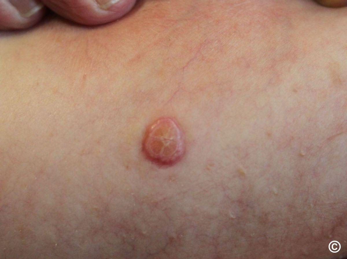
Key features
- Clusters of small shiny papules with an umbilicated centre
- Most 2-5 mm diameter, occasionally 1 cm or greater, especially in solitary lesions
- As lesions resolve they become inflamed and crusty
Management overview
- Refer to the bottom of the page for a patient information leaflet available through a QR code or printable PDF
- Most cases self-limiting with resolution over 6-9 months, can last up to 4 years
Treatment
- Most cases do not require treatment
- If treatment is required consider topical potassium hydroxide:
- MolluTinc ® (10% strength) - can be purchased over the counter
- Molludab ® (5% strength) - licensed from age of 2 years. Use BD until lesions become inflamed, usually around day 5, at which stage treatment can be stopped. If no inflammation by day 14 discontinue
- For stubborn lesions cryotherapy can be a very effective treatment in older children and adults
Additional resources
- Refer to the chapter Molluscum contagiosum
Patient Information Leaflet
Link: Molluscum contagiosum

https://www.pcds.org.uk/patient-info-leaflets/molluscum-contagiosum
Download the Patient Information Leaflet PDF: Molluscum contagiosum
19. Nail disorders
This section focuses on the most common and/or important nail conditions. Refer to the chapter Nail disorders for detailed information on a wide range of nail conditions.

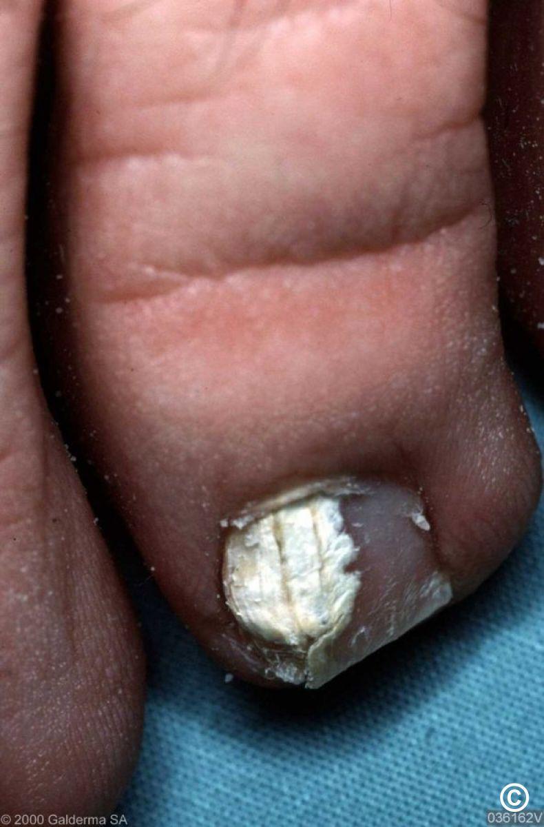
Tinea unguium (fungal infection)
- May be asymmetrical and can be associated with interdigital infection. In most cases the nail thickens and turns yellow, if the infection starts distally the nail plate crumbles. With superficial nail involvement the nail surface becomes white and powdery
- To help distinguish tinea from psoriasis and other nail conditions it is important to use the correct technique for nail samples in order to prevent high rates of false negative results. The nail must be cut back and the subungual debris (where most of the fungus lives) removed by careful curettage or by a scalpel blade, both the nail clippings and subungual debris should be sent mycology. This must be done in the surgery, not by the patient
- For more detail refer to the concise section on tinea and also the clinical chapter Tinea unguium
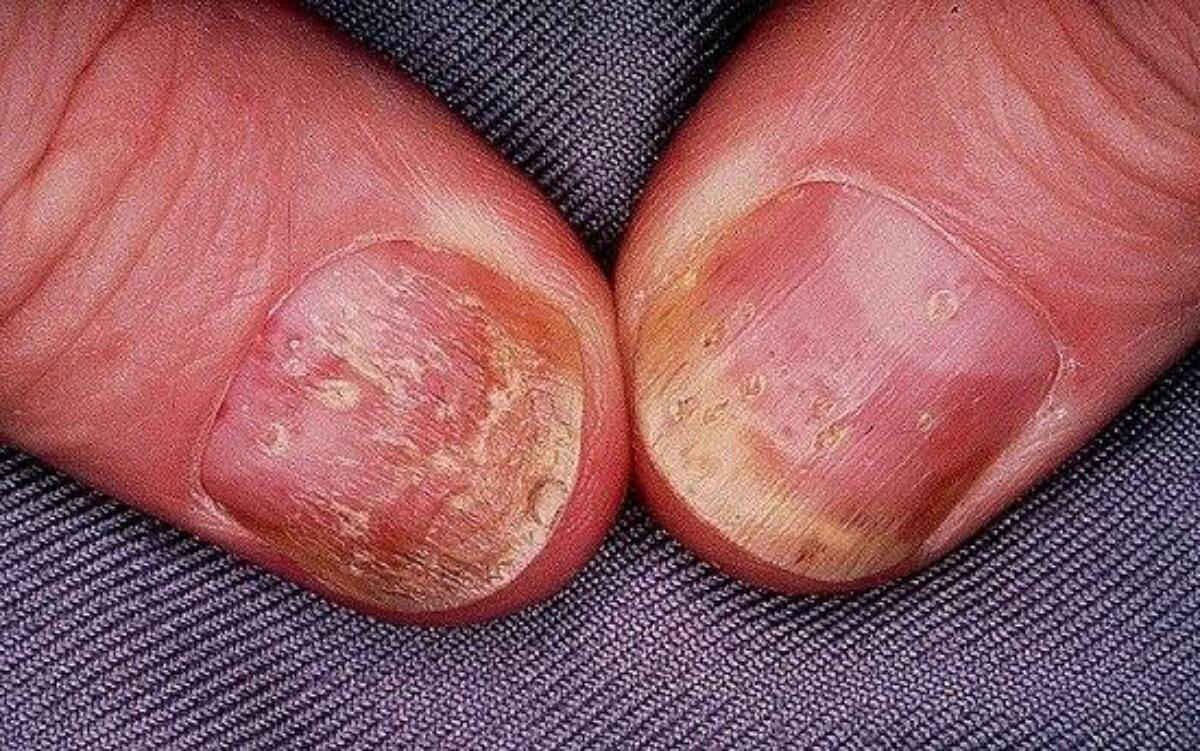
Nail psoriasis
- Can be more symmetrical than tinea
- Pitting
- Onycholysis - a yellow / white discoloration where the distal nail plate has separated from the nail bed
- Sometimes subungual hyperkeratosis - thickening of the nail bed
- Occasionally the oil drop or salmon patch sign - translucent yellow-red discolouration in the nail bed, resembles a drop of oil under nail plate
- For more detail refer to the chapter Nail psoriasis
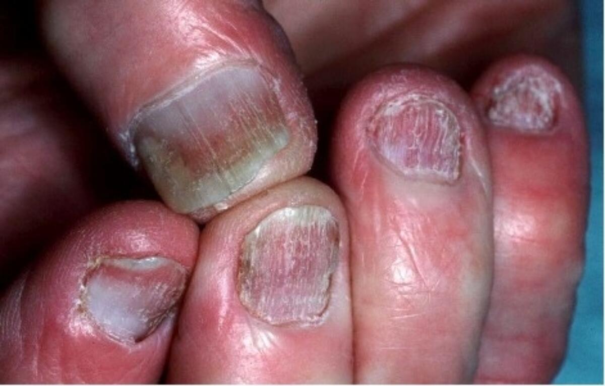
Lichen planus
- Longitudinal changes and thinning of the nail plate
- On occasions the nails can be severely affected causing permanent deformity - if nail change is significant refer as semi-urgent as treatment initiated early may prevent damage
- For more detail refer to the chapter Lichen planus

Chronic paronychia (nail fold inflammation)
- Predisposing factors: wet work, poor circulation, candidiasis
- Inflammation of nail folds and secondary nail changes
- Treatment: keep hands/feet dry, apply an imidazole antifungal cream to the nail folds 2-3 times per day until the cuticle reforms, or, in more persistent cases use Trimovate ® cream
- For more detail refer to the chapter Paronychia
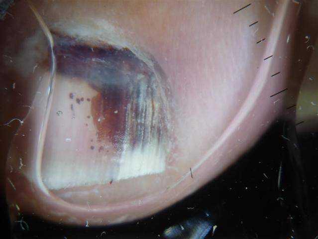
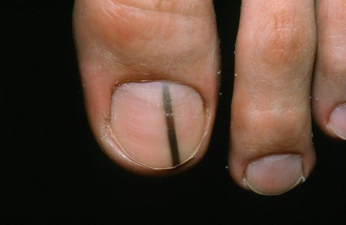
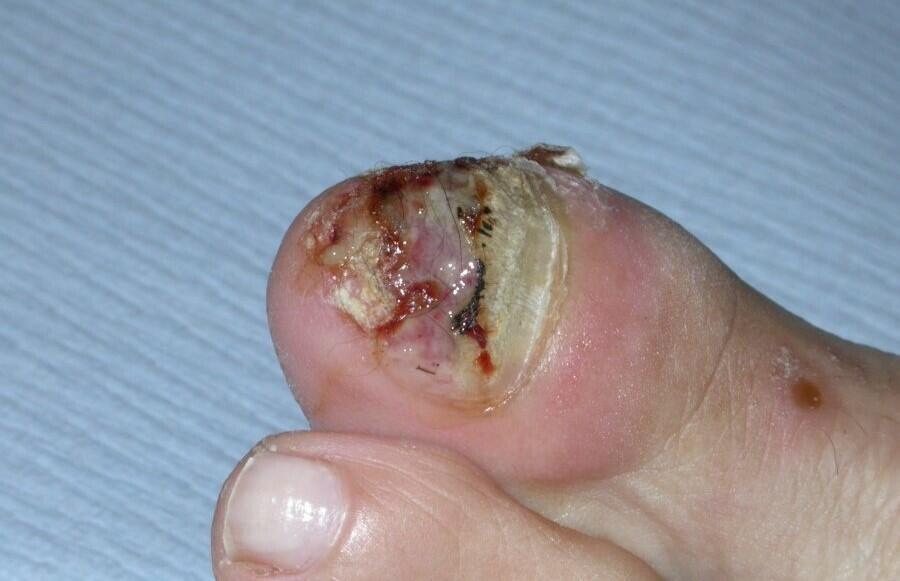
Subungual haematoma (figures 1&2)
- Not linear, can be curved
- Blood globules may be visible
- Over time, sparing of the proximal nail
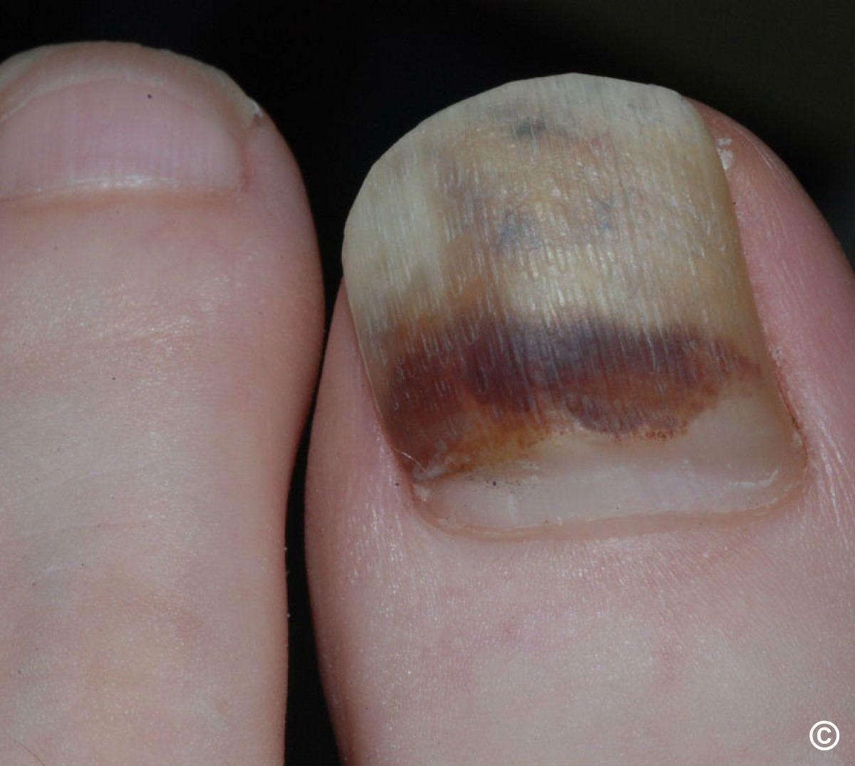
Melanoma (figures 3&4)
- Can present as a single line growing down the nail without proximal sparing, or, unexplained destruction of the nail
- For more detail refer to the chapter Subungual melanoma
20. Perioral dermatitis
Key features
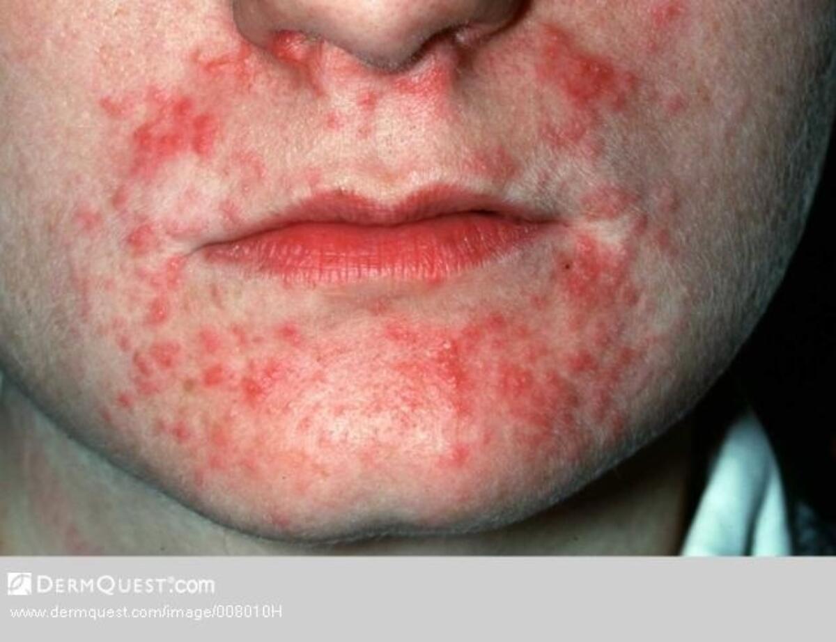
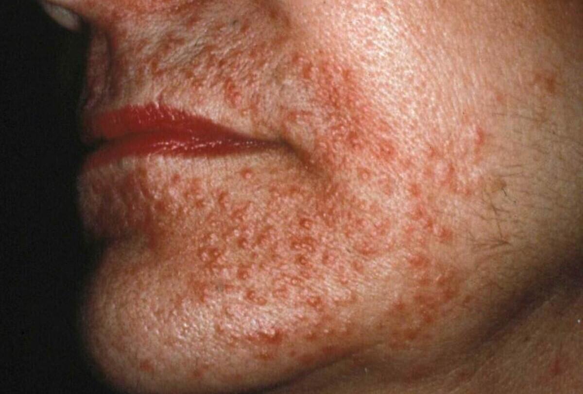
- Small monomorphic (of the same appearance) papules and occasional pustules in the perioral and occasionally periorbital areas
- Confluent erythema of nasolabial fold
- A poorly named condition as it is not a form of eczema, but is aggravated by topical steroids
- Differential diagnosis - refer to the General Dermatology Diagnostic Tool
Management
- Refer to the bottom of the page for a patient information leaflet available through a QR code or printable PDF
- Stop topical steroids if being used
- Milder cases - a topical antibiotic eg clindamycin, erythromycin or metronidazole
- Other patients - a systemic antibiotic for 6 weeks eg a tetracycline (lymecycline 408mg OD with food) or erythromycin/clarithromycin 500mg BD
Additional resources
- Refer to the chapter Perioral dermatitis for more detail
Patient Information Leaflet
Link: Perioral dermatitis

https://www.pcds.org.uk/patient-info-leaflets/perioral-dermatitis
Download the Patient Information Leaflet PDF: Perioral dermatitis
21. Photosensitive rashes (rashes made worse by UV light)
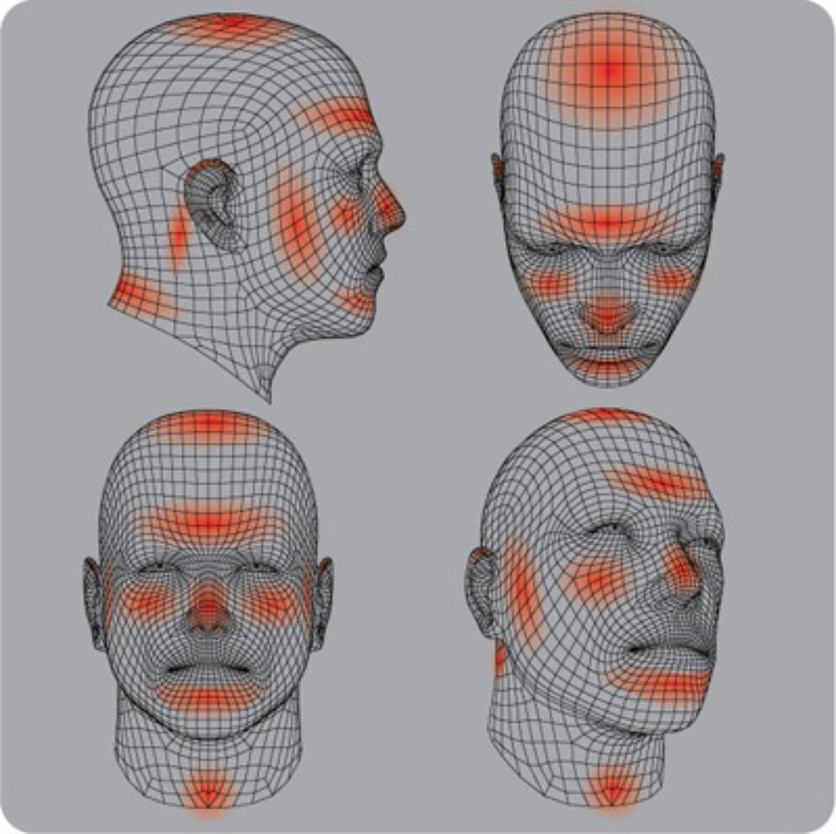
Photosensitivity refers to a wide range of skin conditions associated with an abnormal reaction to UV (ultraviolet) radiation. Photosensitivity is broadly split into two groups:
- Photodermatoses - skin conditions that only arise as a result of light
- Photoaggravated dermatoses - pre-existing skin conditions that can be made worse with light, eg lupus erythematosus and rosacea
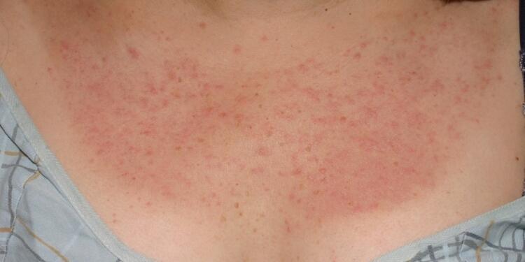
Polymorphic light eruption - a photodermatosis
- More common - teenagers / young adults
- Moderate itch. Symptoms commonly develop 24 hours after UV exposure (range 2 hours to 5 days) and settle within a week
- Affects exposed areas of skin, although less exposed sites can also be affected, and the face can be relatively spared due to natural tolerance
- Ill-defined papules, occasionally vesicles
- Refer to the chapter Polymorphic light eruption for more detail

Rosacea - a photoaggravated dermatosis
- Predominantly affects both middle-aged (age range 30-60) and fair-skinned people
- Erythema, usually described as burning/stinging, initially intermittent but becomes more permanent
- Central face (forehead, nose, cheeks and chin) with sparing of peri-oral and peri-orbital skin
- Associated with telangiectasia and/or papules and pustules
- Ocular involvement common
- Refer to the rosacea section on the concise guidance page for detail
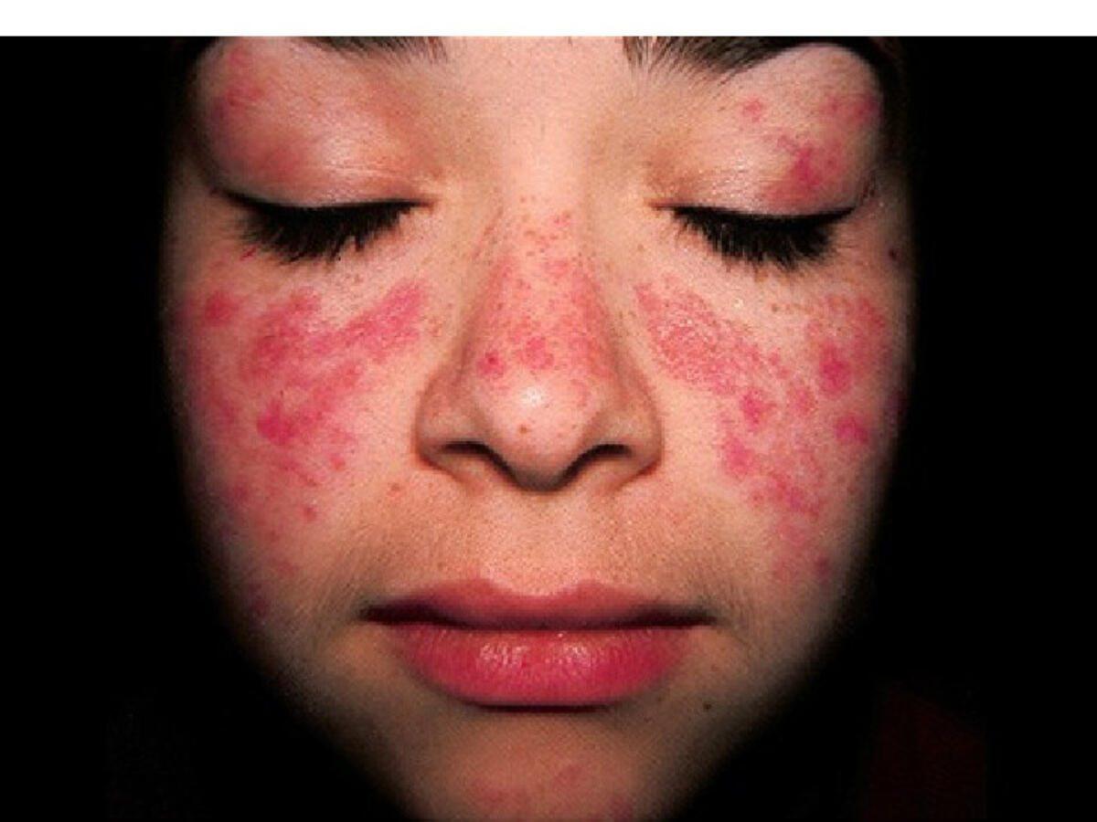
Systemic lupus erythematosus - a photoaggravated dermatosis
- More common in young to middle-aged adults, females, and in black skin
- Diffuse malar ‘butterfly’ rash and systemically unwell
- Other mucocutaneous features can include - Raynaud's phenomenon, persistent urticated lesions, a diffuse morbilliform eruption, mouth ulceration and non-scarring alopecia
- Non-erosive arthritis / multisystem disease
- Check bloods for both ANA & ENA - refer to the chapter Lupus erythematosus for more detail
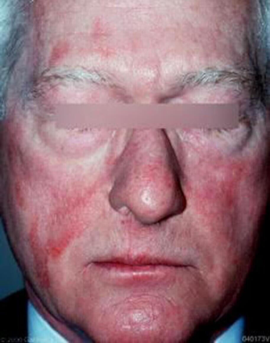
Drug / chemical induced - a photodermatosis
- The length of time between when the drug is first taken to the development of the rash is very variable. Symptoms can come on within weeks, although sometimes it can take years, eg quinine
- Various clinical features including one type that is painful / eczematous with erythema and scale of UV-exposed sites. There should be sparing of shaded areas (figure 6)
- Drugs include quinine, thiazides, carbamazepine, naproxen / other NSAID, and doxycycline
- Refer to the chapter Photodermatoses: drug / chemical-induced for detail
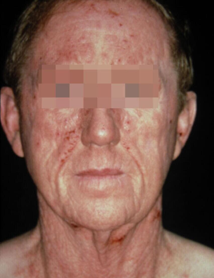
Airborne contact allergic dermatitis
- This is not a photodermatosis, but highlighted here as it can take on a similar appearance to the above
- Arises as a result of contact allergy to the Compositae plant group
- Mainly middle-aged and elderly adults during the summer months
- Often results in considerable erythema and swelling, sometimes with blistering, followed by scaling - unlike photodermatoses there should be no sparing of the photoprotected areas of the face
- Refer to the chapter Contact allergic dermatitis for detail
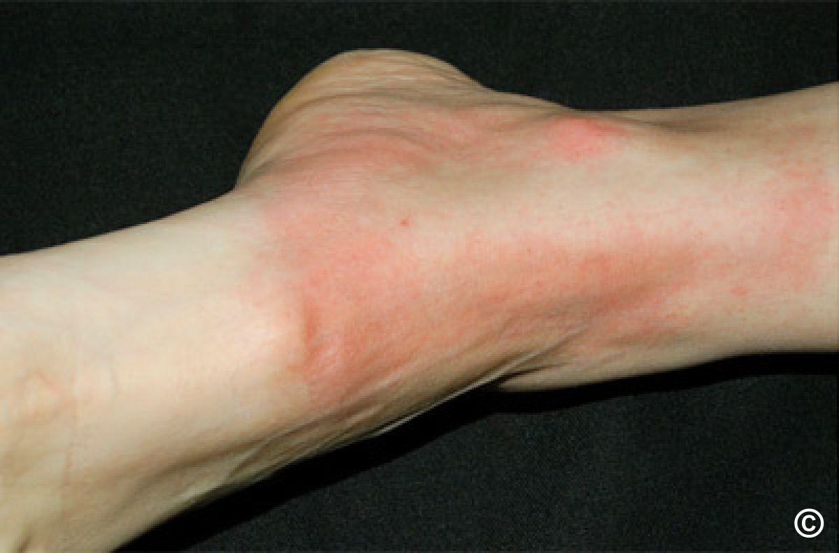
Additional resources
- This section highlights just a few common / important conditions, for an overview of all photodermatoses refer to the relevant clinical chapter
22. Pityriasis rosea
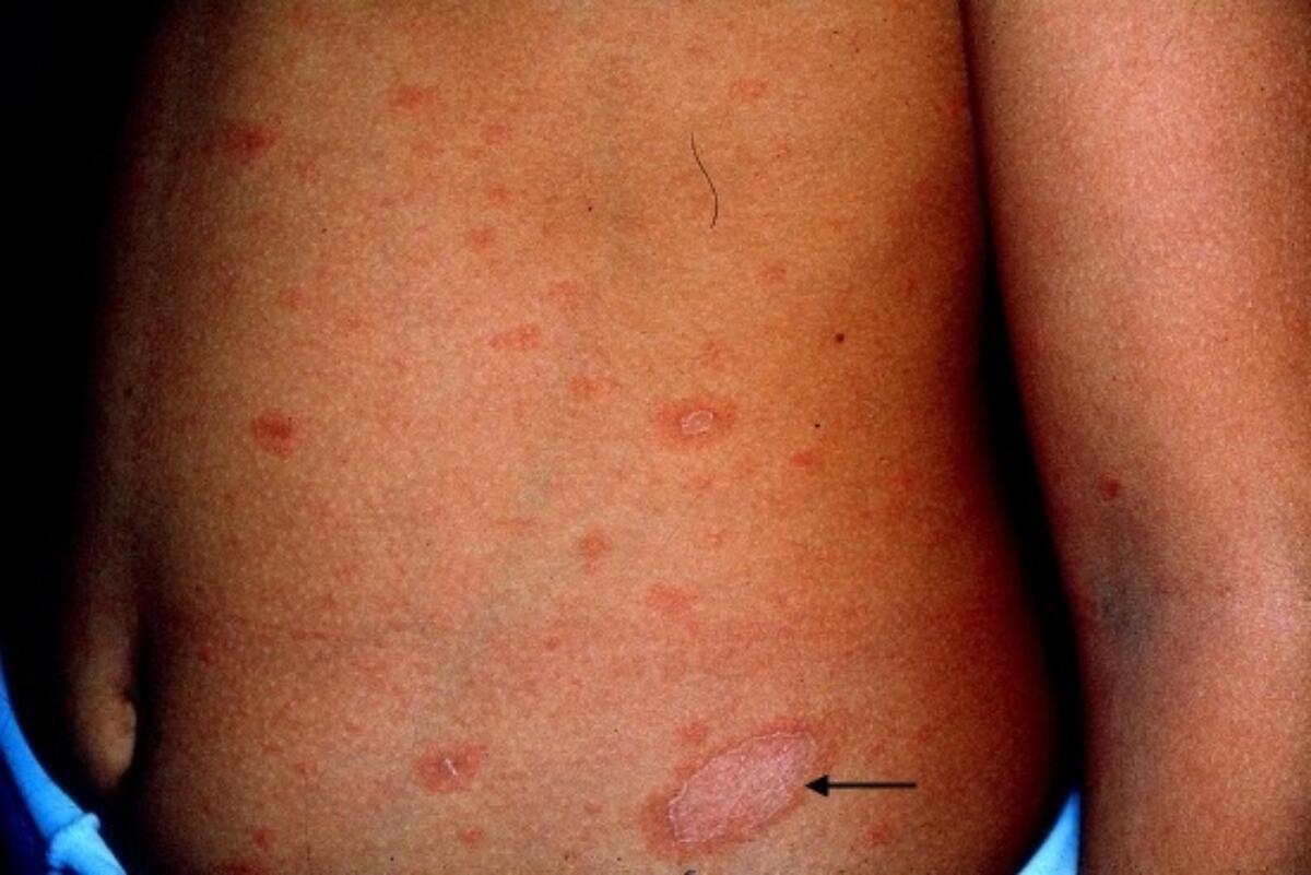
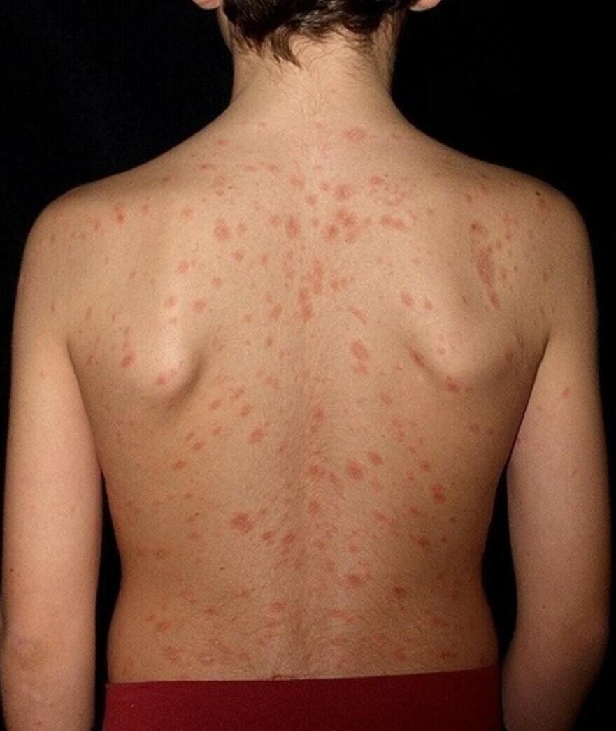
Key features
- Mainly young people
- A herald patch (top figure - arrow) precedes main rash by a few days - larger than subsequent lesions, 2-5 cm, bright-red with fine scale, sharply demarcated border
- Predominantly trunk, sometimes limbs, occasionally the face in children - lesions on trunk tend to run along the lines of cleavage parallel to ribs
- Oval dull-pink macules-patches, some of which have a fine peripheral collarette of scale
- Differential diagnosis - refer to the General Dermatology Diagnostic Tool. Syphilis is an uncommon but important differential with lesions that can be similar to those of pityriasis rosea, although additional sites such as palms and soles are frequently affected
Management
- Refer to the bottom of the page for a patient information leaflet available through a QR code or printable PDF
- The rash often resolves within 6-12 weeks
- An emollient, or mild to moderately potent topical steroid, eg, 1% hydrocortisone or Eumovate ® (clobetasone butyrate 0.05%) cream, can be used if itchy
- Refer to the chapter Pityriasis rosea for more detail
Patient Information Leaflet
Link: Pityriasis rosea

https://www.pcds.org.uk/patient-info-leaflets/pityriasis-rosea
Download the Patient Information Leaflet PDF: Pityriasis rosea
23. Pityriasis versicolor


Key features
- Upper trunk most common site, also neck, upper arms, abdomen. Sometimes additional sites
- Individual lesions often oval-shaped but large confluent areas frequently develop. Lesions fawn-colour in lighter skin types, and hypopigmented in darker skin types. Often have fine scale
- In Wood's light the lesions show yellow-green fluorescence
- Differential diagnosis - refer to the General Dermatology Diagnostic Tool
Management
- Refer to the bottom of the page for a patient information leaflet available through a QR code or printable PDF
- Ketoconazole shampoo (Nizoral ®) - make in to a lather and leave on affected areas of skin for 10 minutes before washing off, daily for 5 days
- In widespread/resistant cases add in itraconazole (Sporanox ®) 200 mg once a day for 7 days
- Inform the patient it will take several months for the skin colour to return to normal, on occasions the discoloration can be persistent
- Recurrent cases - repeat treatment, then consider ketoconazole shampoo once every 2-4 weeks to try and prevent recurrence
- Refer to the chapter Pityriasis versicolor for more detail
Patient Information Leaflet
Link: Pityriasis versicolor

https://www.pcds.org.uk/patient-info-leaflets/pityriasis-versicolor
Download the Patient Information Leaflet PDF: Pityriasis versicolor
24. Pruritis ani
Causes include:
- Hygiene
- Haemorrhoids
- Inflammatory eg eczema, psoriasis
- Threadworms
- Contact allergy to topical treatments (prescribed or over the counter)
- Serious bowel pathology should be considered if PR bleeding, change of bowel habit, discharge
Investigations
- Refer urgently to GI/colorectal departments if bowel pathology suspected
- If no red flags, take swabs for yeasts and bacteria - both candida and Strep. pyogenes can cause symptomatic infection
Management notes
- Refer to the bottom of the page for a patient information leaflet available through a QR code or printable PDF
- Discontinue current treatments eg topical anaesthetics used to treat haemorrhoids - they may be causing a contact allergic dermatitis
- Use an emollient cream (patient preference important) in the morning and post-defaecation
- If swabs reveal a significant infection treat accordingly
- If swabs show no relevant findings manage as follows:
- An antifungal/steroid application, eg Daktacort ® cream, once-twice daily
- If this does not help then treat with a potent topical steroid, eg 0.1% mometasone cream/ointment, every night until things improve and then 2 consecutive nights a week to keep symptoms at bay
- In resistant cases refer for patch testing in case of contact allergic dermatitis (including to topical treatments)
Additional resources
- Refer to the chapter Pruritus ani for more detail
Patient Information Leaflet
Link: Pruritus ani (itchy bottom)

https://www.pcds.org.uk/patient-info-leaflets/pruritus-ani-itchy-bottom
Download the Patient Information Leaflet PDF: Pruritus ani (itchy bottom)
25. Psoriasis
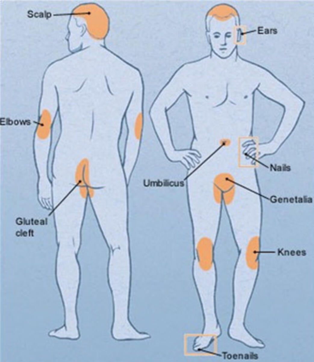
Management overview
- For a simplied managment plan please refer to the The PCDS Psoriasis Primary Care Treatment Pathway
- Refer to the bottom of the page for a patient information leaflet available through a QR code or printable PDF
- Direct to the Psoriasis Association
- Are any medications aggravating the psoriasis?
- Severe/recalcitrant psoriasis - test for HIV
- Manage CVD risk factors - psoriasis (especially moderate-severe) is associated with an increased risk of CVD
- Check for psoriatic arthropathy annually - refer to assessment tools. Patients with suspected PsA should be referred (usually to a rheumatologist) on an urgent basis as it is a destructive arthropathy
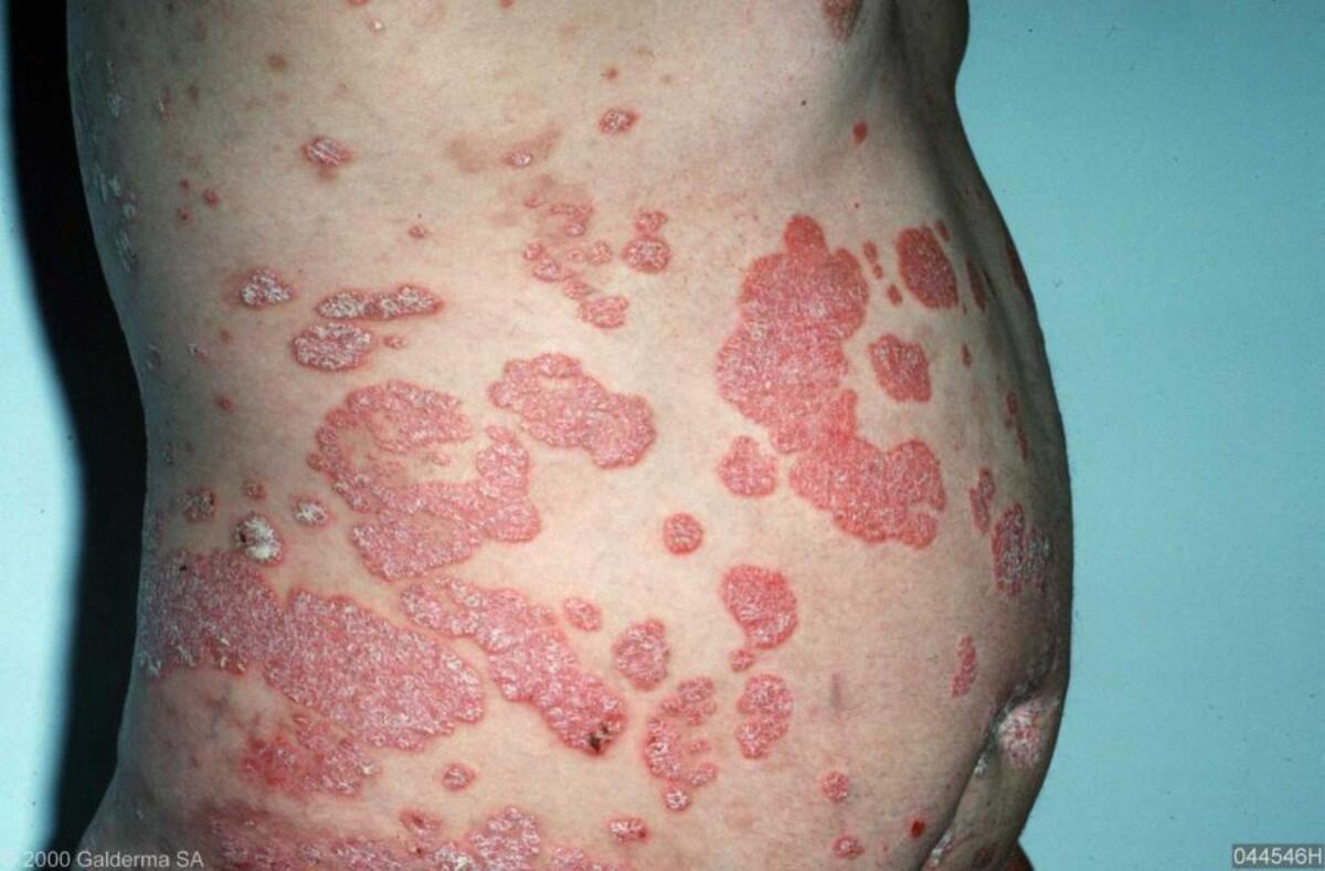
Chronic large plaque psoriasis
- Prescribe emollients to reduce scale and make the skin more comfortable
- Enstilar foam ® or Wynzora cream ® should be applied thinly OD until the plaques flatten and feel smooth. In terms of Enstilar ®, this can also be used twice a week as maintenance, and patients should shake the can containing the foam before use
- For large plaques on thin skin (eg shins) consider Exorex lotion ® BD-TDS, which does not contain a topical steroid
- Refer to dermatology if moderate-severe psoriasis with inadequate treatment response
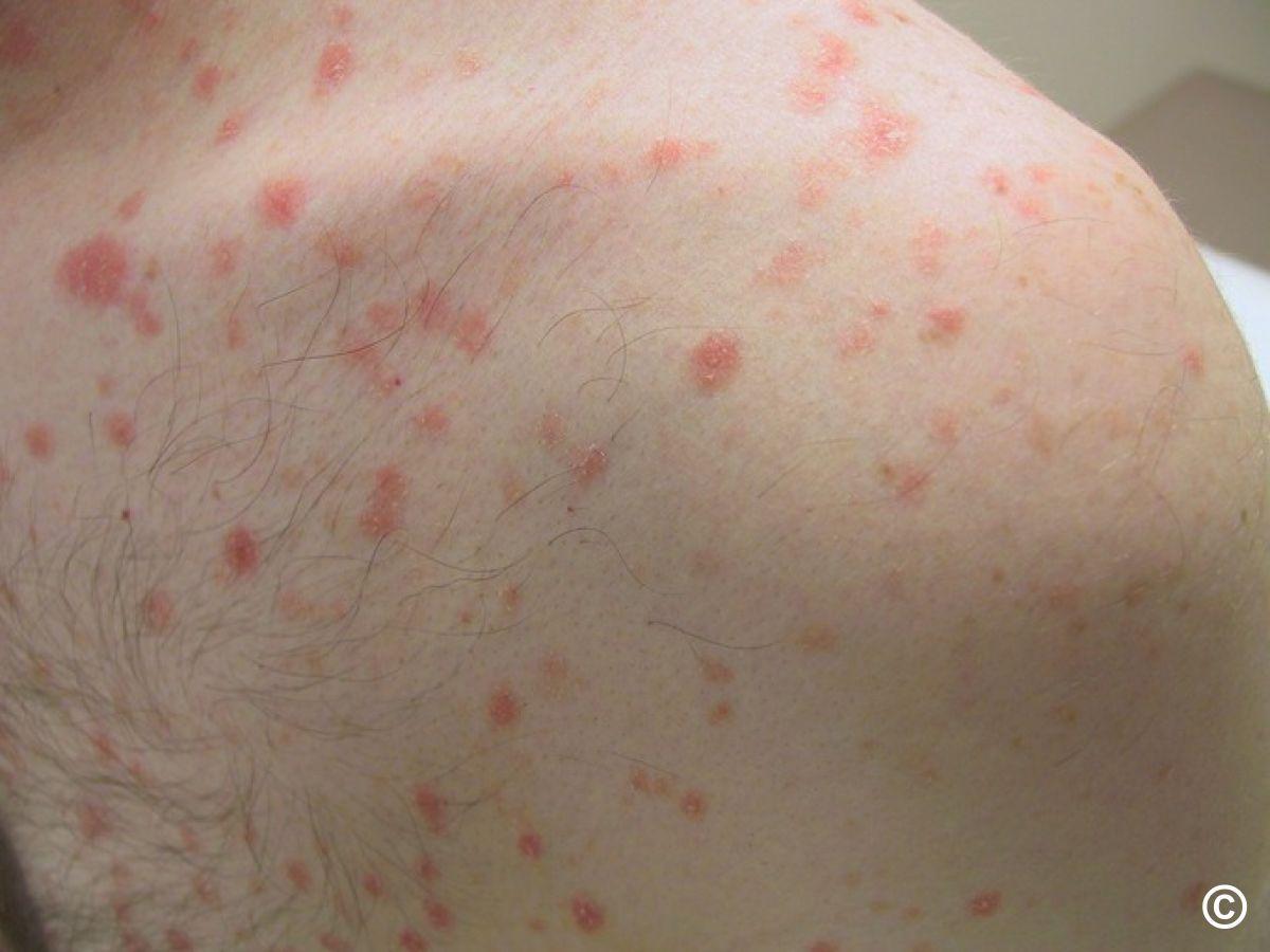
Chronic small plaque psoriasis
- Prescribe emollients to reduce scale and make the skin more comfortable
- For ease of use consider Exorex lotion ® BD-TDS, which does not contain a topical steroid, as such getting the lotion on to normal surrounding skin is not an issue
- Enstilar foam ® or Wynzora cream ® can be used OD but is likely to be very time consuming as care needs to be taken not to get it on normal skin
- Refer to dermatology if inadequate treatment response
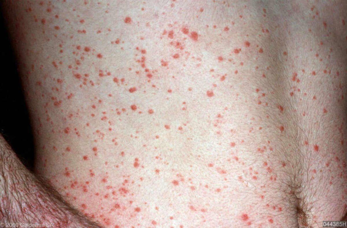
Guttate psoriasis
- Differs to the above - abrupt onset, often after an URTI. Multiple small 'tear drop' lesions that tend to affect most of the body
- Management:
- Prescribe emollients to reduce scale and make the skin more comfortable. Consider Exorex lotion ® BD-TDS, which does not contain a topical steroid
- While natural sunlight in moderation can improve guttate psoriasis, the use of sunbeds is discouraged
- Spontaneous resolution is common, but in persistent cases refer to dermatology
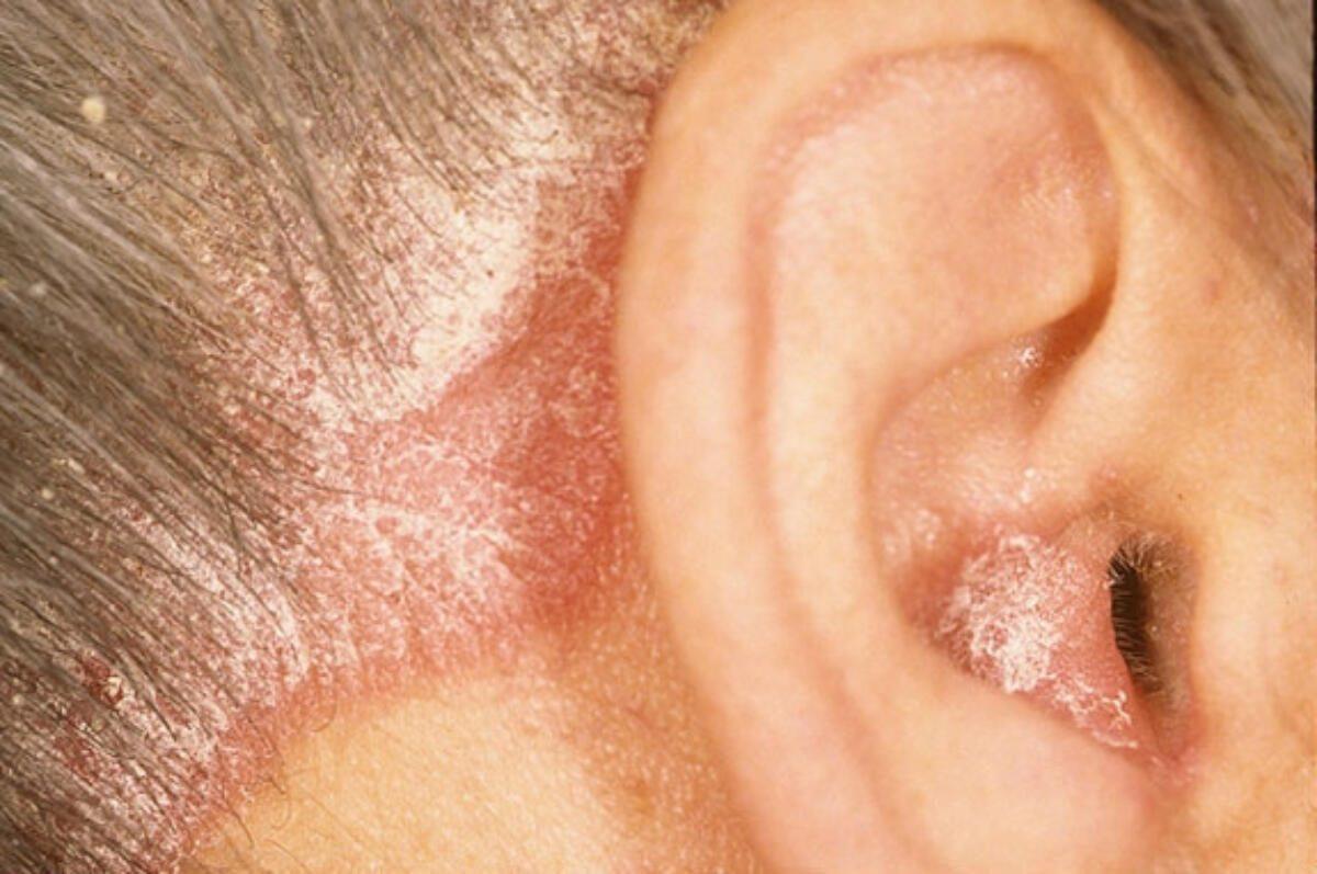
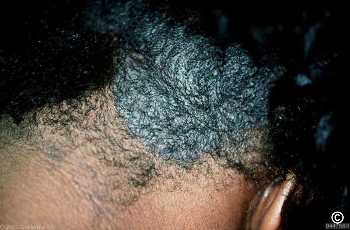
Scalp psoriasis
If main issues are scale and itch:
- Thick scale - massage Sebco ® scalp ointment in to affected areas for 5 minutes and leave on for at least 2 hours, but preferably overnight (treatment messy so use old pillowcase). Wash out in the morning with a tar-based shampoo, using a comb to lift scale - warn patients that hair loss may occur, but this will recover. Once scale much thinner, manage as below. Patients can follow this link for a video on how to apply scalp applications ('Treating scalp psoriasis' - at approximately 1 minute 40 seconds the nurse talks about applying an emulsifying ointment, the same can be done with Sebco)
- Thinner scale - Diprosalic ® scalp application OD and leave on. Use at other end of the day to shampoo. As things improve use less frequently. Use a tar-based shampoo twice a week, other shampoos/conditioners can be used in between. Patients can follow this link for a video on how to apply scalp applications ('Treating scalp psoriasis')
If main issue is itch with erythema - choose from a topical steroid lotion, foam, or shampoo.
Refer to dermatology if moderate-severe scalp psoriasis with inadequate treatment response.
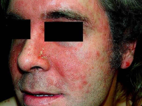
Psoriasis of the face, hairline, and ears
- Face - well-demarcated erythema and scale especially of central face. Prescribe emollients to reduce scale and make the skin more comfortable. The most effective treatment is tacrolimus (Protopic ®) 0.1% ointment OD-BD
- If mainly the hairline, Eumovate ® cream OD-BD is relatively easy to apply
- Ears - Eumovate ® cream OD-BD to the visible aspects. Use topical steroid drops for itchy ear canals (eg 0.1% betamethasone)
- Refer to dermatology if moderate-severe facial psoriasis with inadequate treatment response
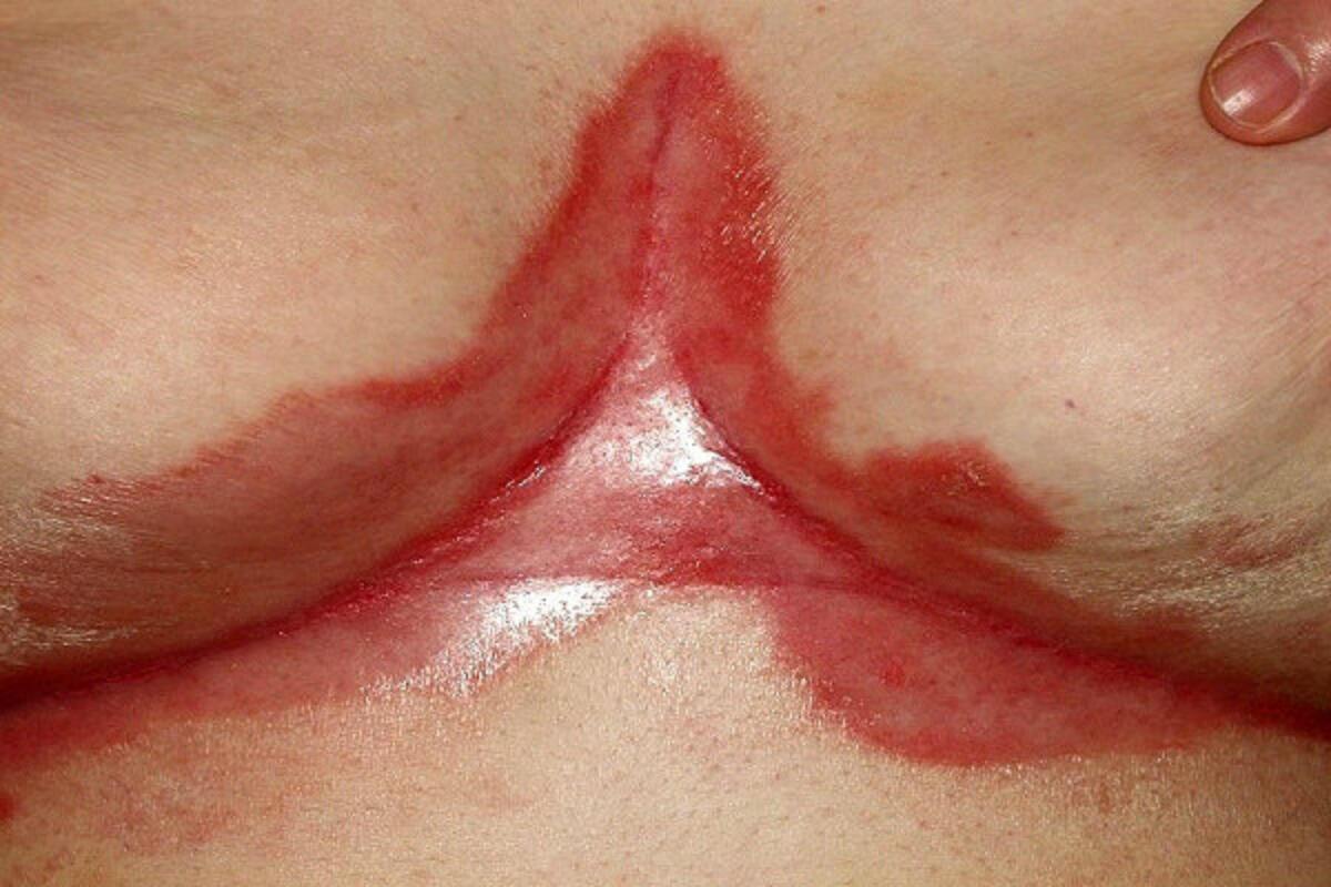
Flexural psoriasis
- Clinical features - shiny erythema without scale
- Treatment - emollient creams. Eumovate ® cream OD-BD if needed. If secondary infection likely, treat instead with Trimovate cream ® OD-BD
- Refer to dermatology if moderate-severe flexural psoriasis with inadequate treatment response
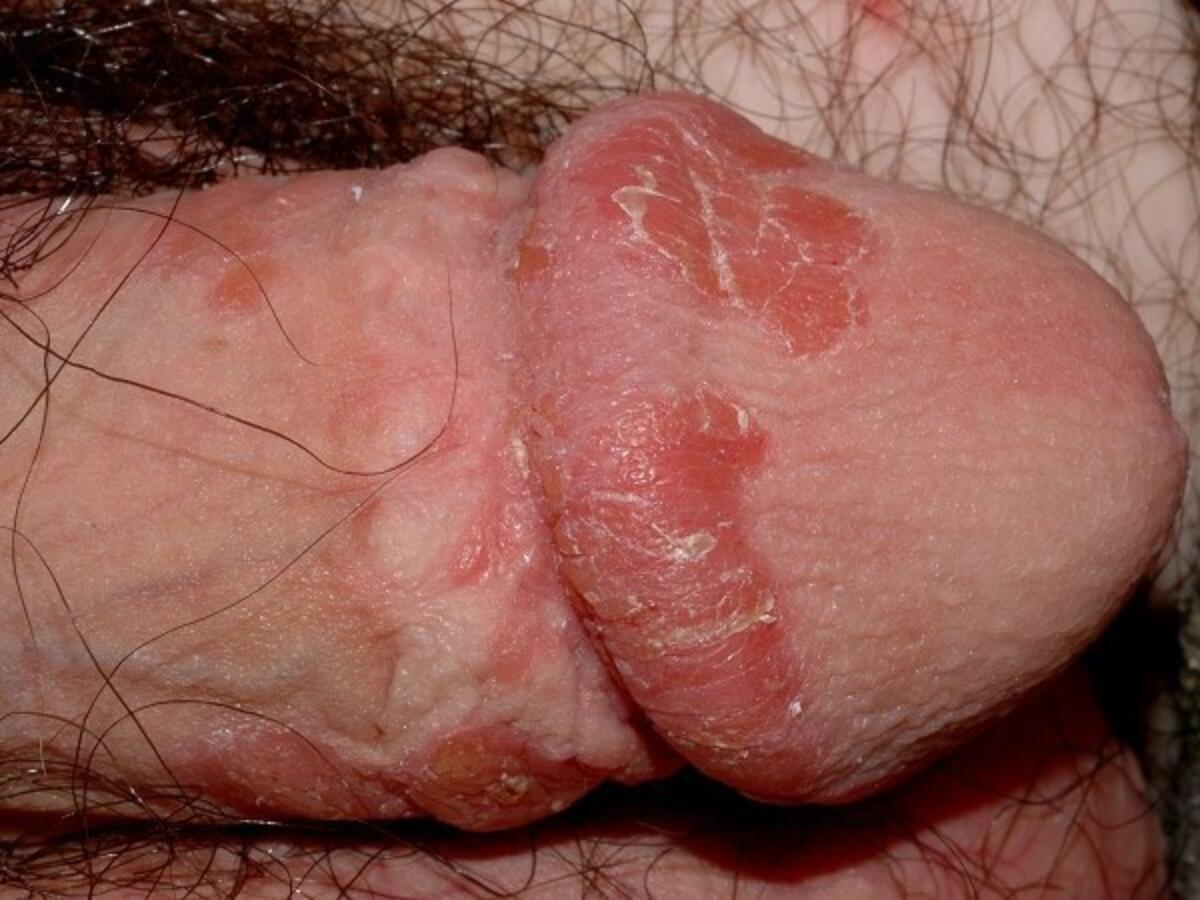
Genital psoriasis
- Clinical features - well-demarcated erythematous macules / small patches, often with scale
- Treatment - emollient creams. Eumovate ® cream OD-BD if needed. Other treatment options include Calcitriol (Silkis ®) or Tacrolimus (Protopic ®) 0.1% ointment
- Refer to dermatology if moderate-severe genital psoriasis with inadequate treatment response
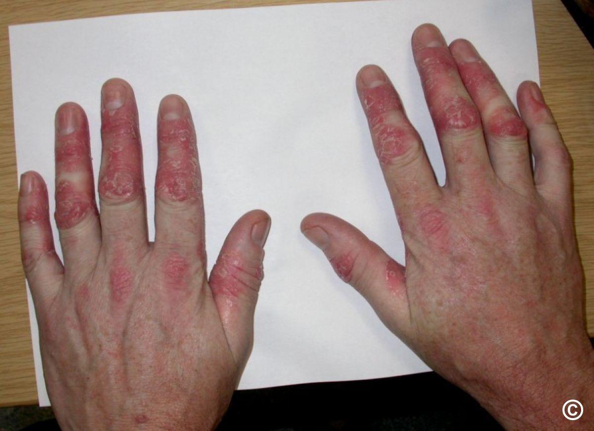
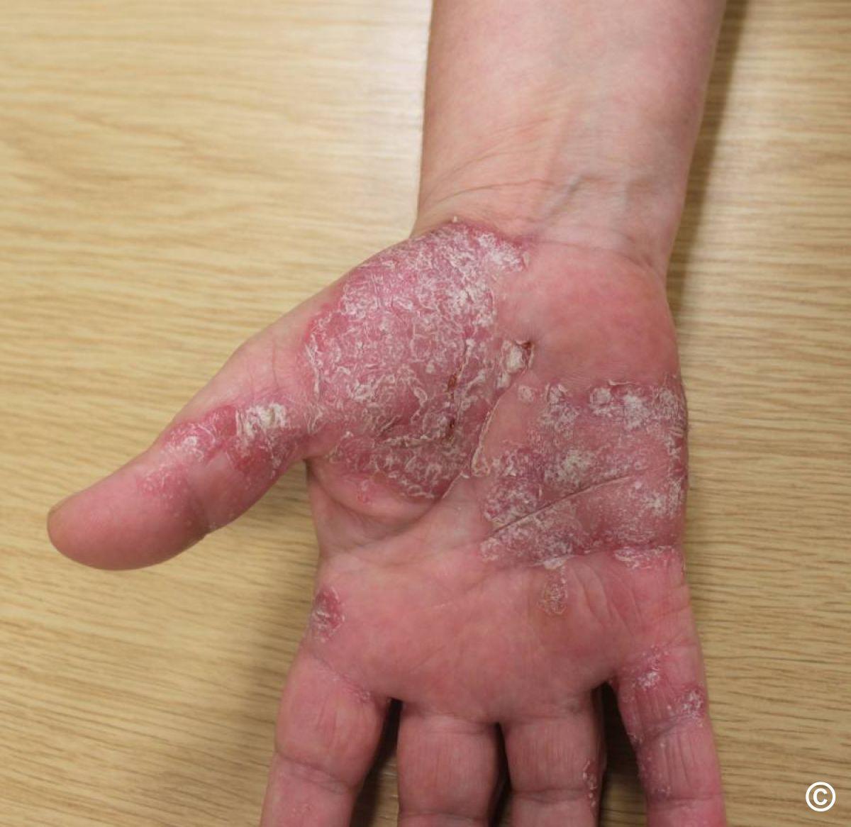
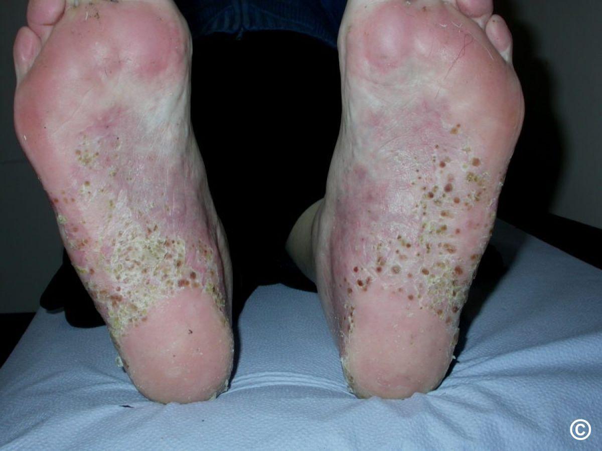
Hand/foot psoriasis
- Part of chronic plaque psoriasis (1) - manage in the same way
- Hyperkeratotic psoriasis (2) - well-demarcated erythema and scale. Treat with emollients daytime, and Diprosalic ointment ® OD at night, preferably under occlusion, eg cotton gloves, or Clingfilm for more isolated areas. Refer to dermatology if inadequate response to treatment
- Palmoplantar pustulosis (3) - grouped yellow pustules and brown macules on soles (and sometimes palms). Treat with super-potent topical steroids, eg Dermovate ointment ®, although often resistant to treatment, requiring referral
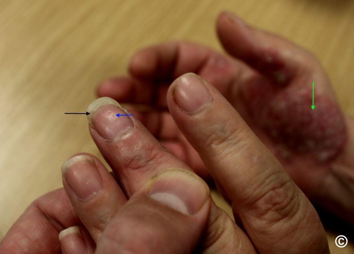
Nail psoriasis
- Presents with irregular pits, onycholysis, subungual hyperkeratosis
- Keep nails short. Refer to dermatology if very troublesome with multiple nails affected
Additional resources
- Patient support groups - the Psoriasis Association
- Psoriasis decision aid - this project was initiated and funded by Bristol Myers Squibb. This shared decision aid is for patients who have been diagnosed with psoriasis, when talking to their healthcare professionals. The decision on what to prescribe lies with the relevant healthcare professional at all times. This shared decision aid has been endorsed by The Primary Care Dermatology Society (PCDS)

Download the Patient Information Leaflet PDF: Psoriasis
26. Rosacea
Key features
The main features of rosacea include one or more the following:
- Inflammation: erythema, papules, pustules (and sometime nodules). Unlike acne, there are no comedones
- Vascular changes: telangiectases, and erythema that is initially intermittent but becomes more permanent, sparing peri-oral and peri-orbital skin
- Blepharitis
- Rhinophyma with marked thickening of the nasal skin
Differential diagnosis - refer to the General Dermatology Diagnostic Tool.
Management overview
- For a simplied managment plan please refer to the PCDS Rosacea Primary Care Treatment Pathway
- Refer to the bottom of the page for a patient information leaflet available through a QR code or printable PDF
- Do NOT use topical steroids, which aggravate rosacea
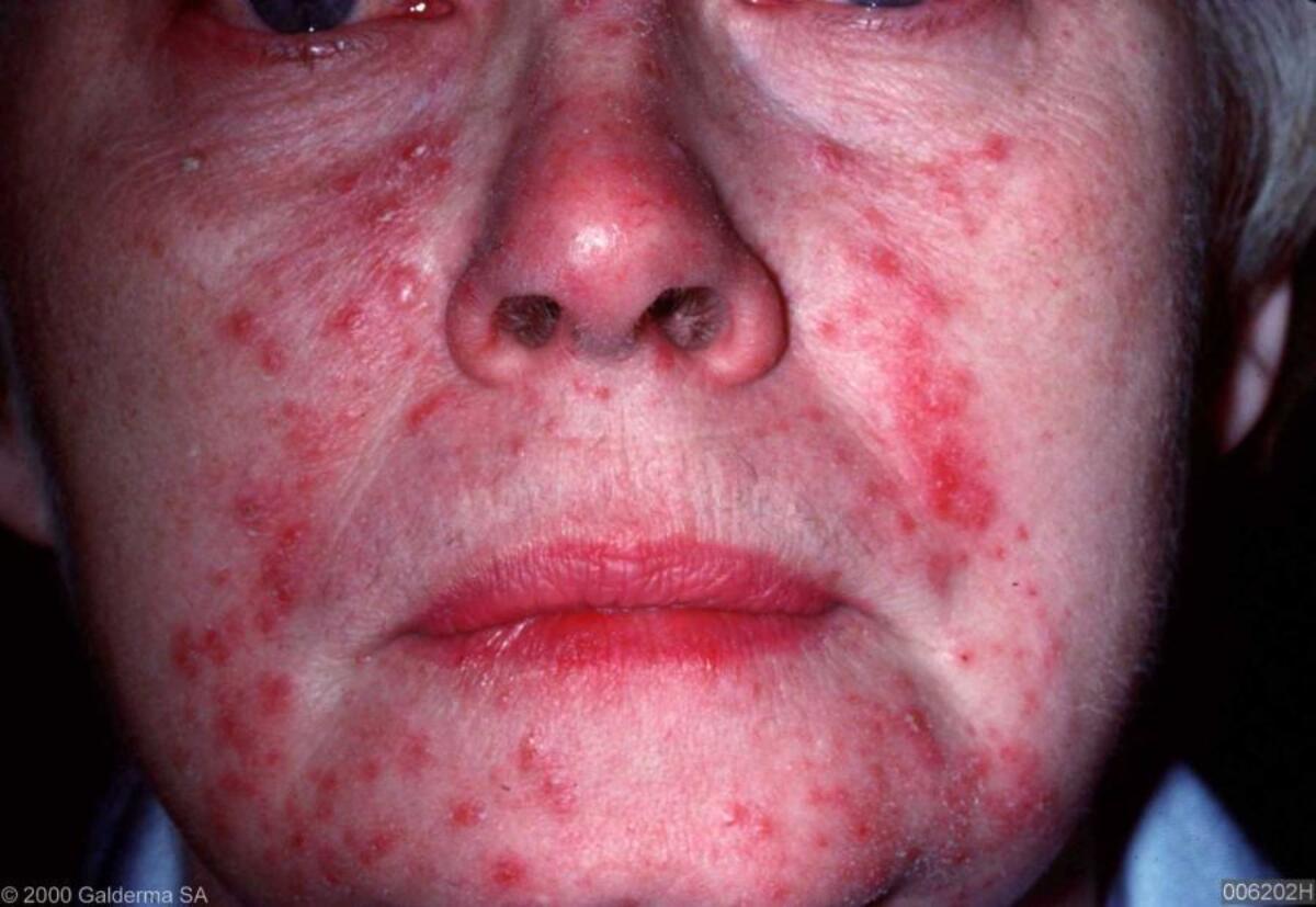
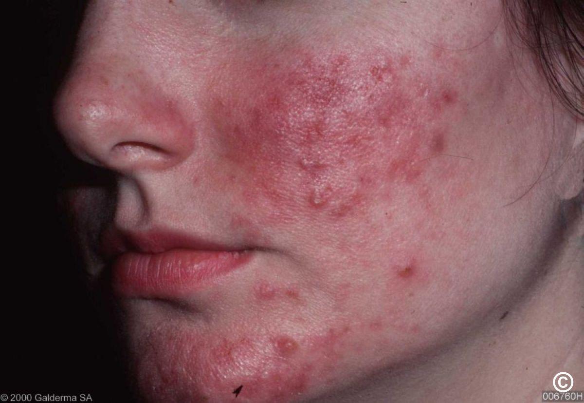
Inflammatory rosacea
Topical treatments - mild symptoms:
- First-line: Soolantra ® (ivermectin 10mg/g) cream OD for 3 months
- Second-line: options include azelaic acid BD (15% gel as Finacea ® or 20% cream; cream may sting less), or Rozex ® gel or cream BD
Systemic antibiotics - if topical agents fail or presenting symptoms more severe:
- First-line: the tetracyclines (contraindicated in pregnancy). Consider doxycycline 40mg OD, this dose reduces the risk of antibiotic resistance. Alternatively, lymecycline 408mg OD or doxycycline 100mg OD. Unlike oxytetracycline, these drugs can be taken with (or without) food
- Second-line: clarithromycin or erythromycin 250-500mg BD
- Duration: initially 3 months. For infrequent recurrences, repeat the course. For frequent recurrences, take standard dose until symptoms settle, then reduce to a maintenance dose, eg once-twice a week
Who to refer: moderate-severe symptoms responding poorly to treatment - consider for isotretinoin
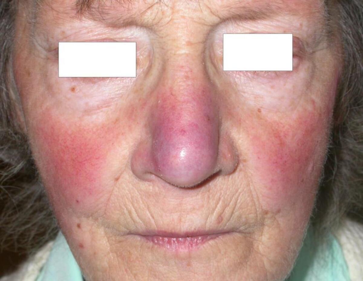
Vascular rosacea
- Does NOT respond to topical or systemic antibiotics
- Mirvaso ® (brimonidine) gel used once daily benefits some patients with persistent (fixed) erythema. Adverse reactions include erythema, flushing, burning sensation and contact dermatitis. NHS availability varies - check local prescribing arrangements
- Refer to the chapter Rosacea for systemic treatments
- Intense Pulse Light Therapy and Pulsed-dye Laser are often effective for fixed erythema, although symptoms will eventually recur. Limited NHS availability (check locally)
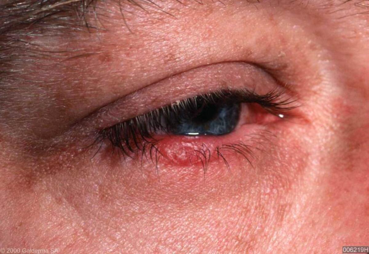
Ocular rosacea
- Clean eyelids using cotton wool soaked in cooled, boiled water
- Artificial tears - use regularly daytime. A lubricating ointment, sometimes containing an antibiotic, may be needed at night
- Systemic tetracyclines are the most effective treatment. Erythromycin can be used if unable to take tetracyclines
- Retinoids should be avoided in patients with significant ocular problems as they can lead to a severe keratitis
- Persistent, troublesome ocular symptoms should be referred to ophthalmology. Patients with more serious symptoms such as keratitis should be seen without delay
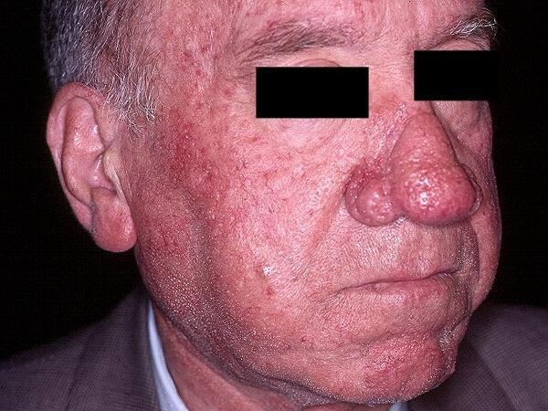
Rhinophyma
- CO2 laser ablation or shave excision of the affected tissue (performed by an individual experienced in the technique) are often effective, although there is limited NHS availability (check locally)
Additional resources
- Refer to the chapter Rosacea for more detail

Download the Patient Information Leaflet PDF: Rosacea
27. Scabies
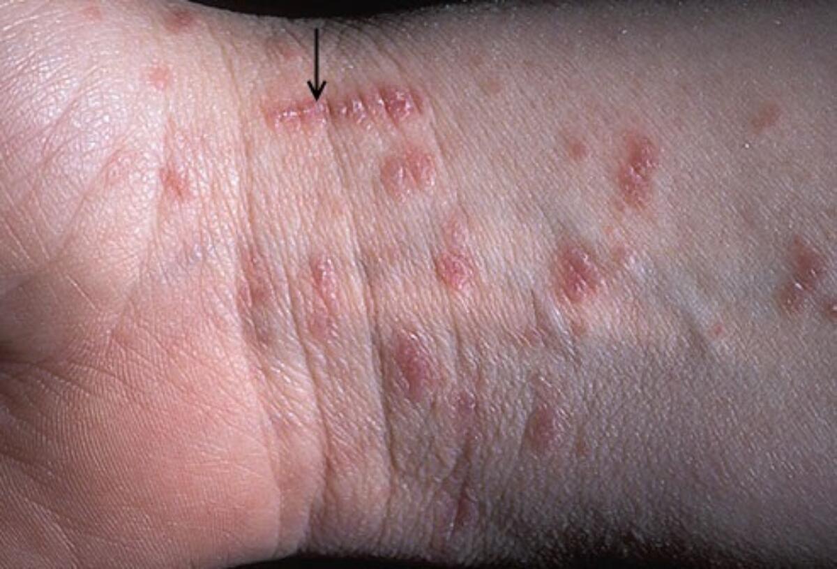
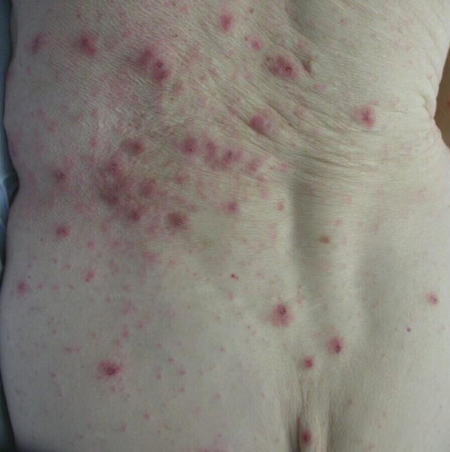
Key features
- Trunk and limbs the main sites affected. Face and scalp rarely involved other than in infants and bed-bound elderly patients
- A generalised rash with erythema, papular and urticated lesions
- Burrows (top figure) most common on sides/webs of fingers, borders of hands, wrists and feet
- Papules and nodules (bottom figure) - most common on penile shaft (pathognomonic), groins, axillae, and breasts. Can persist for several weeks after scabies eradicated
- Papules and pustules on palms and soles are characteristic of scabies in infancy
- Crusted scabies - much less itchy. A generalised scaly rash, especially finger webs, wrists, elbows, breasts, scrotum
- Differential diagnosis - see the General Dermatology Diagnostic Tool
Management
- 5% permethrin cream - two treatments one week apart for symptomatic individuals and one treatment for those in close contact
- All treatment information can be found in the patient information leaflet (below)
- Itchy papules and nodules may persist for several weeks requiring treatment with a potent topical steroid
- For detail refer to the chapter Scabies, including crusted scabies

Download the Patient Information Leaflet PDF: Scabies
28. Seborrhoeic eczema
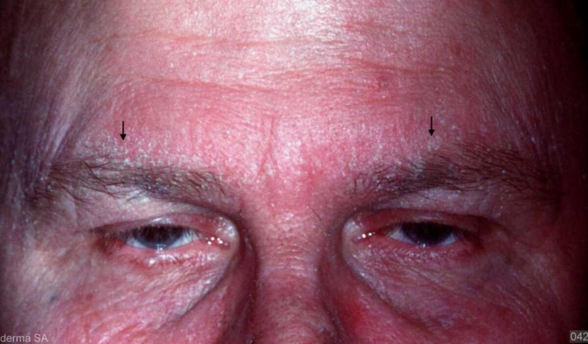
Key features
- Erythema and fine scale commonly affecting the central face (especially the glabella skin, eyebrows, sides of the nose and nasolabial folds), scalp, behind the ears, and occasionally the entire face
- Flexural involvement common, and the upper trunk may also be affected
- Differential diagnosis - refer to the General Dermatology Diagnostic Tool
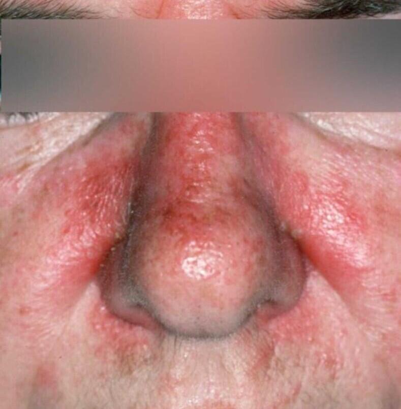
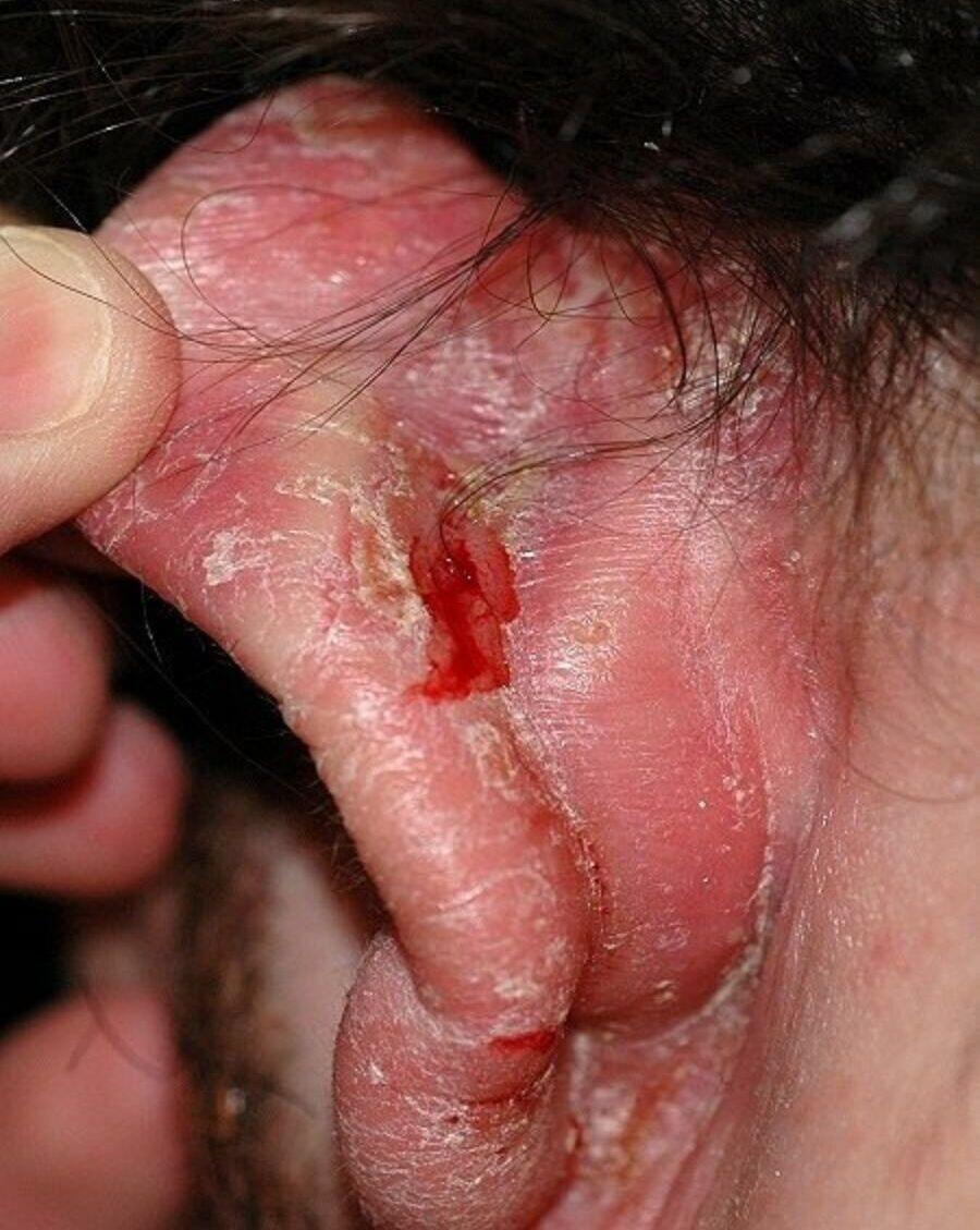
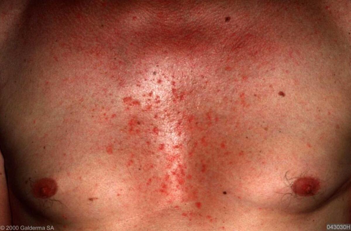
Management
- Refer to the bottom of the page for a patient information leaflet available through a QR code or printable PDF
- Test for HIV if more widespread, atypical features, no or minimal response to standard treatments
Scalp
- Ketoconazole shampoo twice weekly initially, then every 1-2 weeks for maintenance. Leave on scalp for 5-10 mins before rinsing off. Alternatives include over-the-counter only shampoos such as Dercos
- Persistent itch and erythema - steroid scalp application OD, eg betamethasone 0.1% scalp application. If stings use Synalar ® gel
Face and chest
- If symptoms very marked treat with itraconazole 200mg OD for 7 days
- Mainstay of treatment:
- Emollients several times a day
- Ketoconazole shampoo - use twice a week. Make into a lather to use as shampoo and also apply to facial skin for 5 minutes before washing off.
- Daktacort ® or Eumovate ® can be used BD for several days during a flare
- If steroid creams needed regularly on the face, change to calcineurin inhibitor, eg pimecrolimus cream OD-BD or tacrolimus ointment OD (both off licence), reverting back to topical steroids only for a flare
Intertriginous areas (body folds)
- Daktacort ® or Trimovate ® OD-BD
Other treatments
- Resistant cases - add in 8-week courses of lymecycline 408 mg OD (tetracyclines have an anti-inflammatory role in the skin)
- Refer to the chapter Seborrhoeic eczema for more detail
Patient Information Leaflet
Link: Seborrhoeic eczema

https://www.pcds.org.uk/patient-info-leaflets/seborrhoeic-eczema
Download the Patient Information Leaflet PDF: Seborrhoeic eczema
29. Urticaria and angioedema

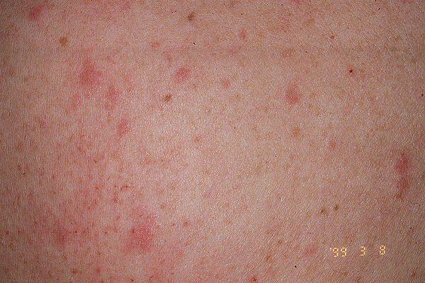
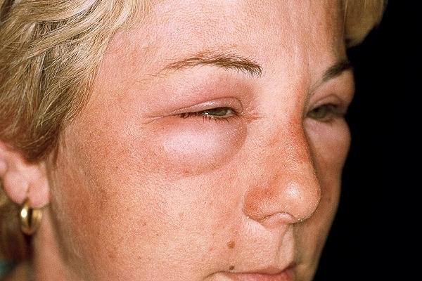
Key features of urticaria and angioedema
- Most lesions of urticaria have a wheal and flare - a smooth, elevated, blanched wheal surrounded by an erythematous flare (sometimes the wheal is erythematous), with the central aspects resolving to leave annular lesions. Each lesion generally lasts 30 minutes to 4 hours. Cases lasting more than 6 weeks with no obvious triggers are known as chronic spontaneous urticaria
- While some cases of chronic spontaneous urticaria have additional triggers (eg heat, pressure), other cases of chronic urticaria only occur as a result of specific stimuli and are called inducible/physical urticaria: dermographic urticaria (scratching) / delayed pressure urticaria (sites of sustained pressure eg over areas of tight clothing) / cholinergic urticaria (sweating) / cold urticaria (cold stimuli) / solar urticaria (sore, itchy lesions within minutes of UV exposure) / aquagenic urticaria (water, hot or cold) / contact urticaria (eg latex allergy)
- Angioedema - may or may not be associated with urticaria, causes transient swellings of deeper dermal, subcutaneous and submucosal tissues, often affecting the face (lips, tongue and eyelids)
Investigation of chronic urticaria (lasting more than 6 weeks)
- Blood tests are not routinely required nor usually helpful
- Cold, solar, or contact urticaria - need referring to dermatology for specialised tests
- Refer to the relevant chapter for patients with angioedema who do not have urticaria
- Patients should not be investigated for allergy unless there is a clear link between food and the onset of symptoms. Even then the gold standard is avoidance of the possible allergen
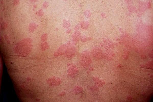
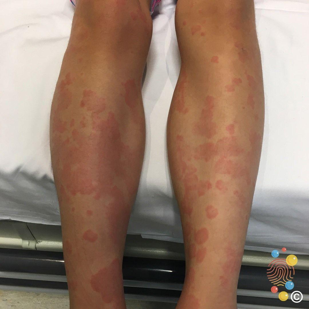
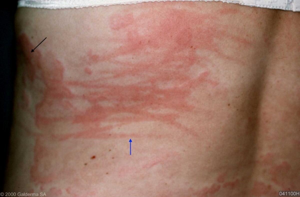
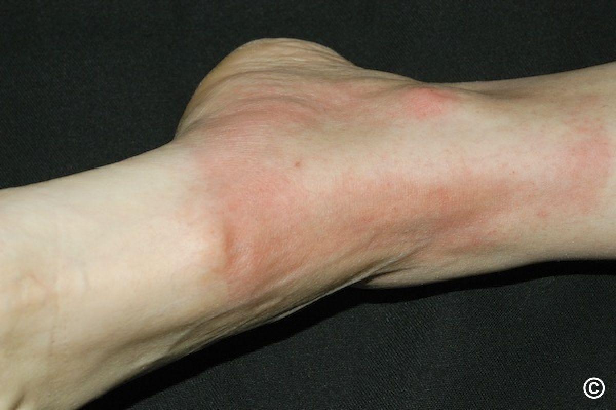
Management
- Refer to the bottom of the page for a patient information leaflet available through a QR code or printable PDF
- The mainstay of treatment are second-generation H1-antihistamines ('non-sedating' antihistamines):
- Start with a standard dose of one a day. The aim is to reduce frequency and intensity of flares, and improve symptoms
- Symptoms can be tracked via scoring systems, such as the UAS7 and DLQI
- If response inadequate within 2-4 weeks, increase the dose to one tablet BD. If response remains unsatisfactory, increase the dose as per NICE guidelines to up to one tablet QDS (some individuals will prefer, for concordance purposes to take two tablets twice a day)
- If response inadequate consider trying an alternative second-generation H1-antihistamine; there is no evidence to support combination therapy
- At higher doses consider co-existent morbidities such that fexofenadine (Telfast ®) 180 mg and cetirizine are excreted by the kidneys, and loratadine and desloratadine (Neoclarityn ®) are metabolised by the liver
- Treatment should be continued at the lowest effective dose until there is no further activity, but restarted if urticaria recurs. In general it is safe to take non-sedating antihistamines for as long as is needed, unless patients are on additional anticholinergic medications, which together may increase the risk of dementia - to help calculate the risk refer to the Anticholinergic Burden Scale
- Patients not responding to maximum dose antihistamines should be referred to Secondary Care
- Pregnancy and breast feeding - insufficient data. Consider loratadine or cetirizine
- Children - start with a standard dose of antihistamine. If inadequate response the dose can be increased gradually, and with caution. Licensed drugs in children:
- Under the age of 1 most specialists use weight-adjusted cetirizine (0.25mg/kg bd)
- Desloratadine from the age of 1 year
- Loratadine from the age of 2 years
- Fexofenadine from the age of 12 years
- For more information on the chronic inducible urticarias, some of which are more difficult to manage requiring referral, refer to the relevant clinical chapters - this is especially so for cold urticaria, which can be life-threatening
- For more information on urticaria with angioedema refer to the relevant clinical chapter, and for more information on angioedema without urticaria refer to the relevant clinical chapter
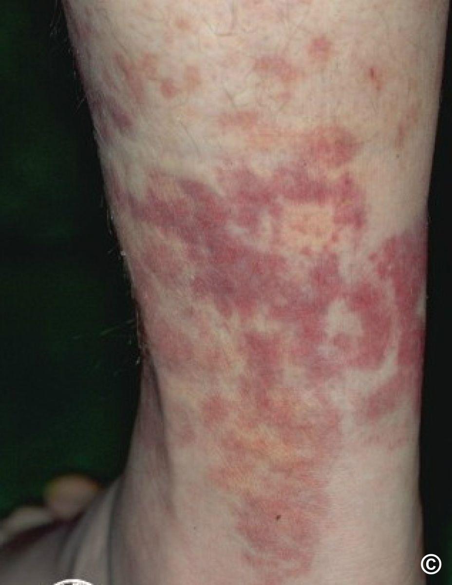
Vasculitic urticaria
- Presentation - urticaria-like lesions persist longer than 24 hours, and often demonstrate purpuric foci and post-inflammatory hyperpigmentation. Lesions are sometimes more angulated than those seen in classical urticaria
- Refer to the clinical chapter Urticarial vasculitis
Patient Information Leaflet
Link: Urticaria and angioedema

https://www.pcds.org.uk/patient-info-leaflets/urticaria-and-angioedema
Download the Patient Information Leaflet PDF: Urticaria and angioedema
30. Warts
Key features
- Hands / feet (verrucae) - often multiple. Sharply defined rounded lesions with a rough keratotic surface. On the feet often painful, on the hands can be associated with nailfold (periungual) involvement. Multiple pinpoint vessels, typically seen as black dots (naked eye / magnifying lens / dermatoscope) are a common feature, which help differentiate warts from other hyperkeratotic lesions, eg corns
- Mosaic - plaque/s of closely grouped warts on the plantar foot, tending not to be painful
- Filiform - often face, filiform appearance and may have a stalk
- Plane - mainly face or dorsal hands. Multiple small lesions (< 5mm), round, slightly elevated with a smooth surface
- Anogenital - often multiple, may appear pearly, filiform, fungating, cauliflower or plaque-like (refer to GUM; do not treat in Primary Care unless trained / extended scope of practice)
Management overview - for all
- Set patient expectations
- No treatment is a good first option - new warts often resolve spontaneously (30% within 10 weeks)
- Refer to the bottom of the page for a patient information leaflet available through a QR code or printable PDF, which provides advice on first steps and OTC options, before consideration of NHS treatment
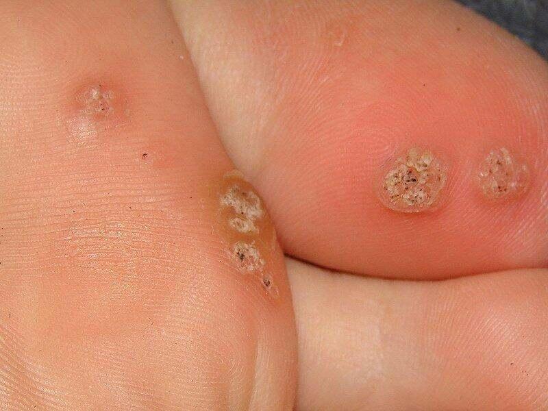
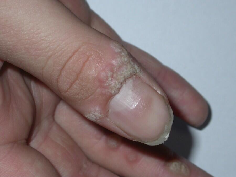
Common warts - feet, hands, trunk
Can be difficult to treat. Options include:
- Salicylic acid (SA) - topical
- Slow to work needing daily applications
- Examples - Bazuka/Salatac 12% gel with Lactic acid 4% or Verrugon/Wartex 50% ointment
- Use daily for upto 12 weeks - improved outcome with warm water soaks, drying the affected skin, then paring (filing down thick skin) pre-treatment
- If no response, consider occlusion with waterproof plaster/duct tape after application of SA gel/ointment and change every 4 days
- Cryosurgery
- Most trials comparing this with SA found no difference in effectiveness (cure rates 50-70% within 3 months)
- Suggested protocol - a single freeze-thaw cycle of 10-30 seconds, once a week. Stop if no significant improvement after 6 treatments
- Combination therapy of cryosurgery and then SA between cryotherapy sessions may be beneficial
- The following attachment provides detail on how to use cryosurgery and this link is a patient information leaflet
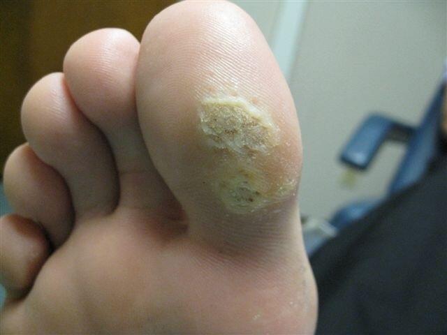
Mosaic warts
- Often very resistant to treatment
- Management as above
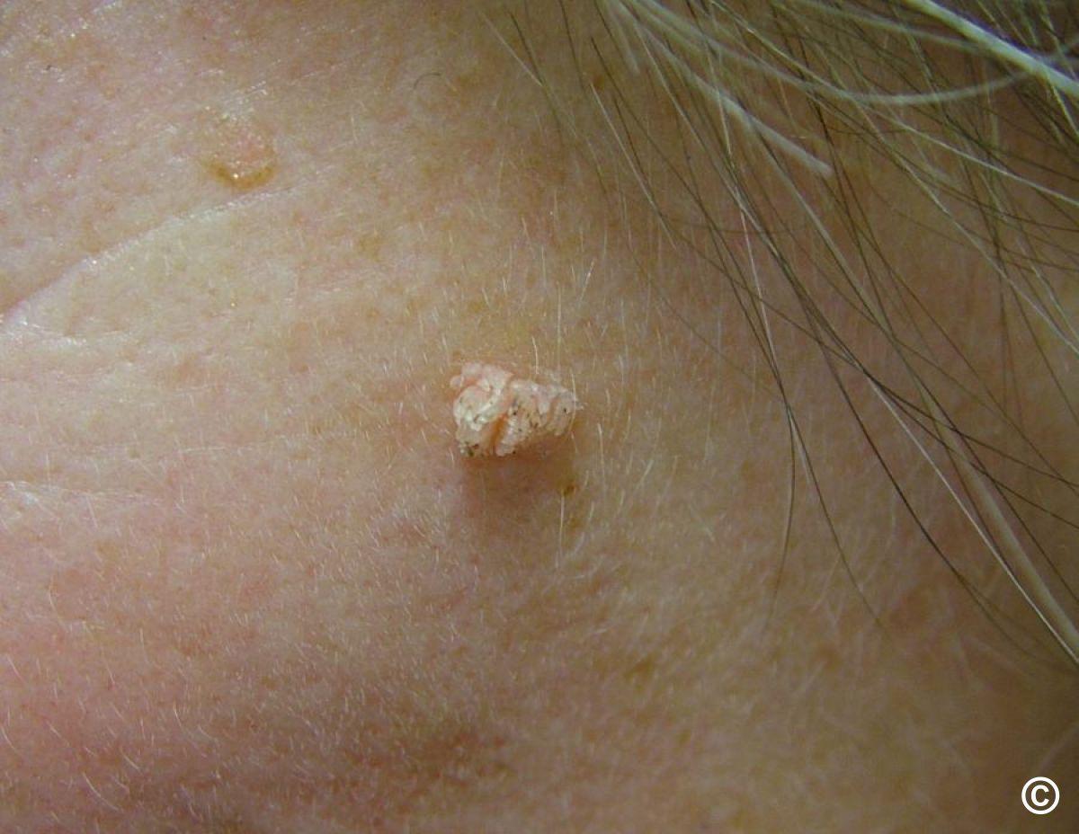
Filiform warts
- SA is not advised for this variant as too difficult to apply
- Cryotherapy or curettage & cautery seem to be most effective. NB NICE advise facial warts should not be treated in Primary Care
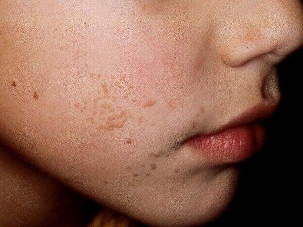
Plane warts
- Often multiple, thin and often in high-risk areas such as the face and hands
- Usually not suitable for SA or cryotherapy due to the risk of scarring, especially on the face
- Retinoid creams/gels can be tried, eg adapalene gel, once a day for six weeks (30% effective)
Management notes
- Referral of warts (as per NICE) may be considered in exceptional circumstances including - an uncertain diagnosis, a persistent facial wart, extensive recalcitrant warts (ie not responding to treatment). NB Most CCG’s will require an exceptional funding request
- There are several other treatment options available in Intermediate/Secondary Care, or through a GP experienced in treating warts, however, it is important to note that all the following are unlicensed and evidence for their effectiveness is limited - Imiquimod 5% cream x3/week (Mon/Wed/Fri) for up to 16 weeks, Efudix cream at night under occlusion with a review after 4 weeks, or Actikerall® gel applied daily for up to 12 weeks. Other treatments occasionally used include surgical removal with cautery or curettage (up to 65% effective but risks of scarring and recurrence rate > 30%), photodynamic therapy and pulsed-dye laser

Download the Patient Information Leaflet PDF: Warts
31. Over-the-counter treatments (for patients)
While many skin treatments can be purchased without a prescription (known as over-the-counter medications), if you have been diagnosed with a skin condition then you are entitled to getting relevant treatments prescribed. For example, if you have eczema you should be prescribed emollient moisturisers, whereas if you simply have skin that is a little dry, your health professional would expect you to buy the moisturiser.
There may be circumstances in which you decide to buy skin treatments over-the-counter (OTC) as opposed to getting a prescription, including:
- a simple skin complaint that you can manage yourself (or with the advice of a pharmacist) without needing to attend your GP surgery
- the treatment may be cheaper to buy than the price of a prescription; however, if you require several medications every month (including for other health conditions), and you pay for your prescriptions it will be cheaper for you to buy a prepayment certificate
A list of useful OTC products is listed below.
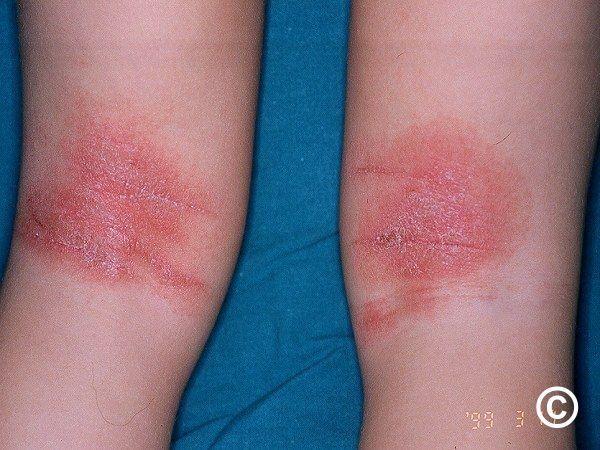
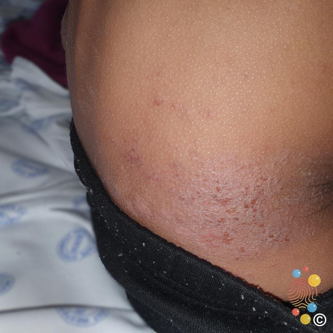
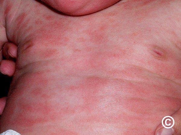
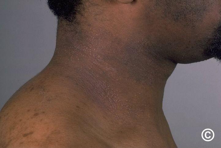
Dry, itchy skin conditions (eg atopic eczema and psoriasis)
- Emollient moisturising creams (and ointments) should be used at least twice a day - the best treatment is the one that suits you most
- This link provides a video on emollients and how to apply them
- If your skin feels very hot, creams containing menthol can be very cooling (eg Menthoderm® and Dermacool®)
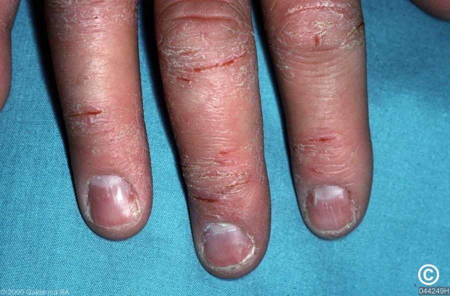
Inflammatory skin conditions
- Inflammatory skin conditions cause the skin to become red and often itchy. In skin of colour the affected skin may take on a darker colour
- In addition to using moisturising creams, mild steroid creams, eg 1% Hydrocortisone or Eumovate ®, can also be used once or twice a day
- If steroid creams are not helping and/or you are applying them to your face on a regular basis (more than twice a week) you must seek advice from a health professional
- Steroid creams must not be used close to the eyes
- This link provides a video on how to apply steroid creams
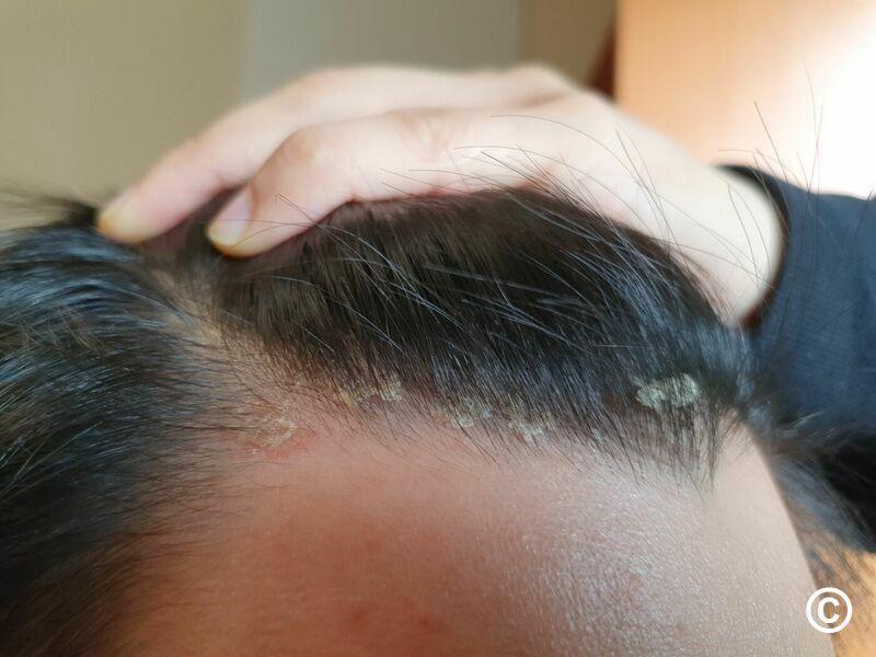
Scalp conditions - itchy, dry, scaly
- Fine scale (dandruff) can be treated with Nizoral ® Shampoo (ketoconazole)
- When the scale is thicker (eg scalp psoriasis) there are a number of options including Neutrogena® T/Gel® Therapeutic Shampoo, Polytar® Scalp Shampoo, and Capasal® Therapeutic Shampoo
- These shampoos should generally be used twice a week, if used too often they may start to irritate the scalp. On days in-between you can use a moisturising or other shampoo of your choice as long as it does not irritate the scalp
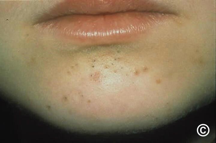
Acne
- Mild cases of acne vulgaris can be treated with one of several formulations of benzoyl peroxide gels / creams - seek advice from a pharmacist as to how to use these treatments
- You must see a health professional if your acne is not responding to treatment - moderate-severe acne can leave scars if not treated properly
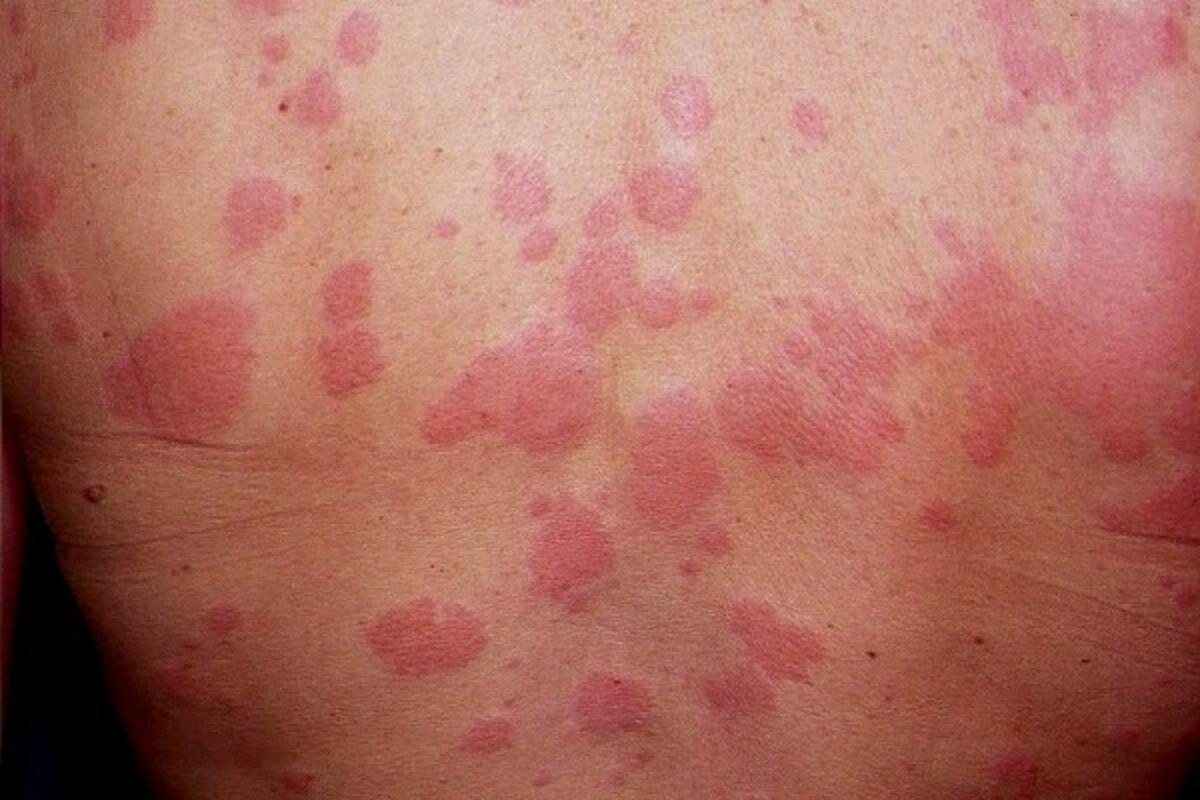
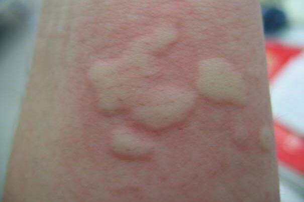
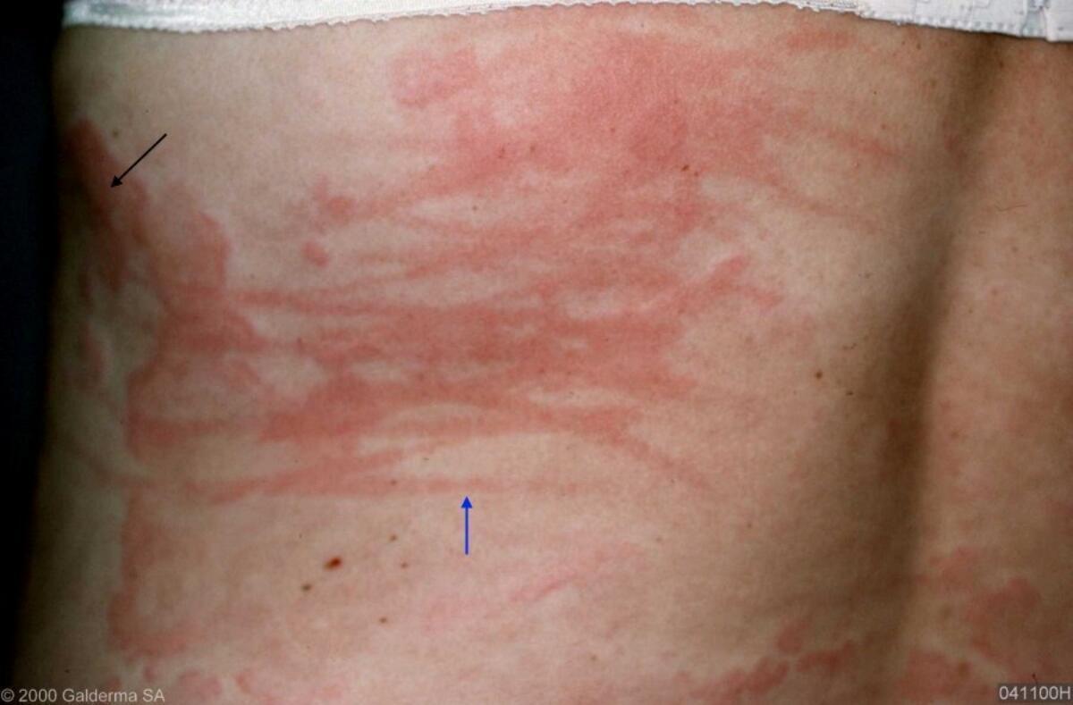
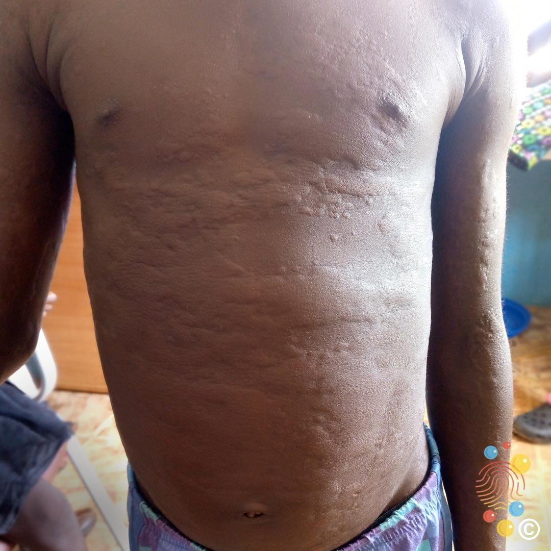
Urticaria
- Urticaria causes itchy lesions that generally last 30 minutes to 4 hours
- Lesions are swollen in the centre (a wheal) with surrounding redness (a flare) giving a nettle rash appearance (hives)
- Urticaria is treated with non-sedating (non-drowsy) antihistamines - ask your pharmacist for advice
- Occasionally, urticaria causes swelling of the lips or eyelids known as angioedema. In the unlikely event you develop a swelling of the tongue or throat and/or have problems breathing this could suggest a reaction known as anaphylaxis, in which case you must seek urgent medical attention
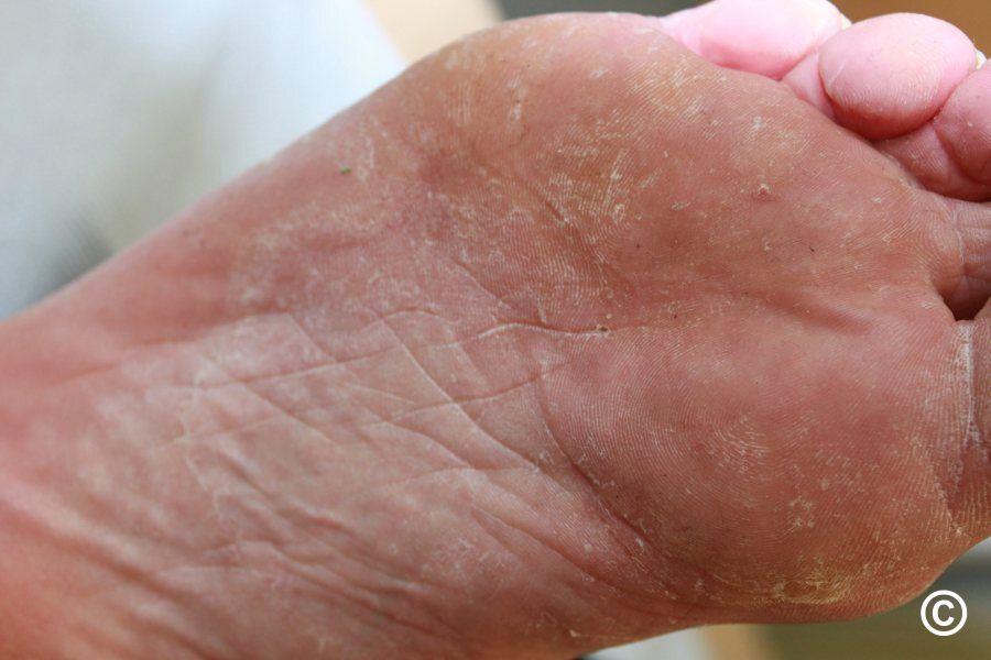
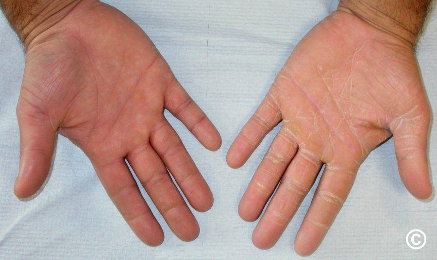
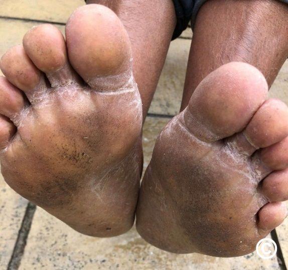
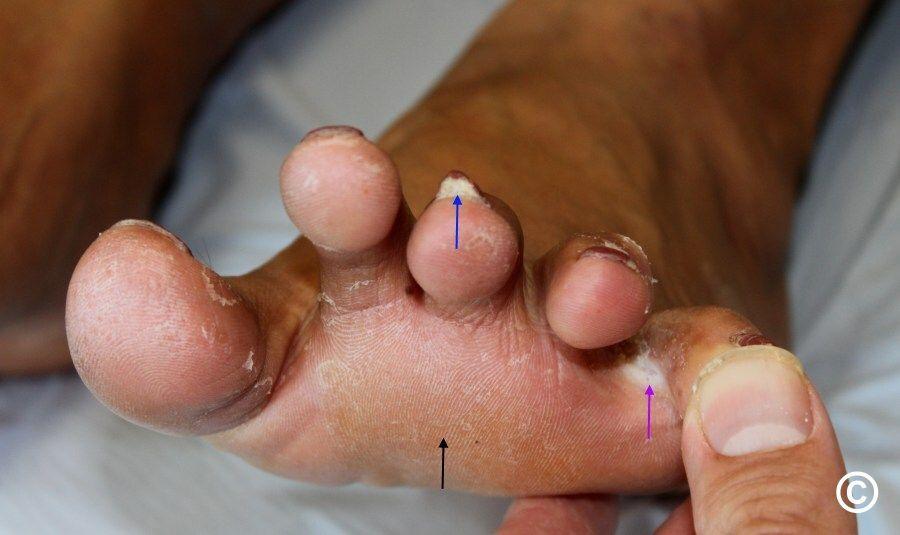
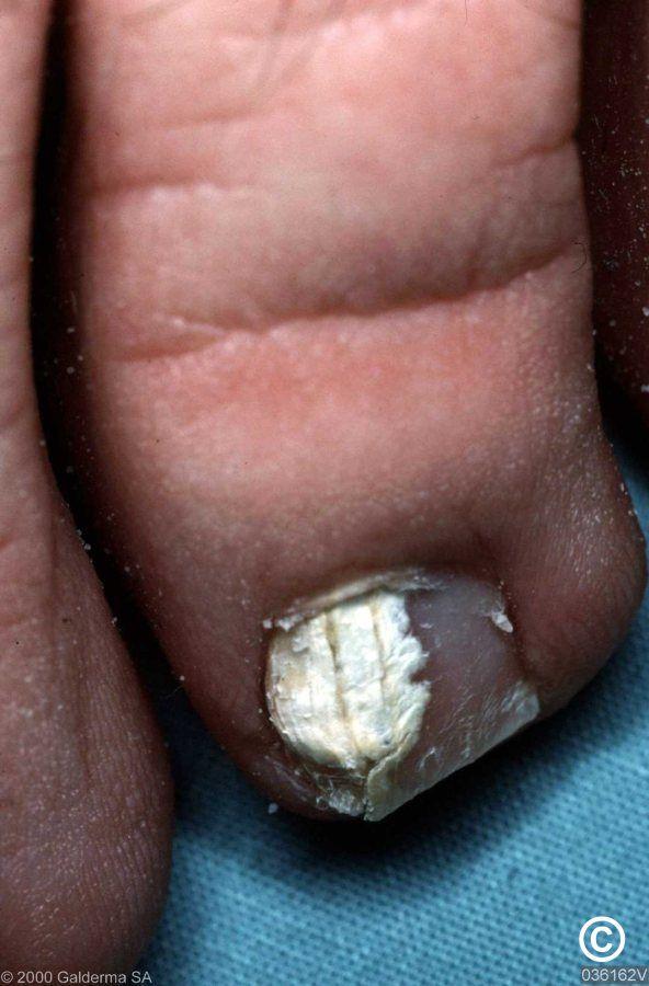
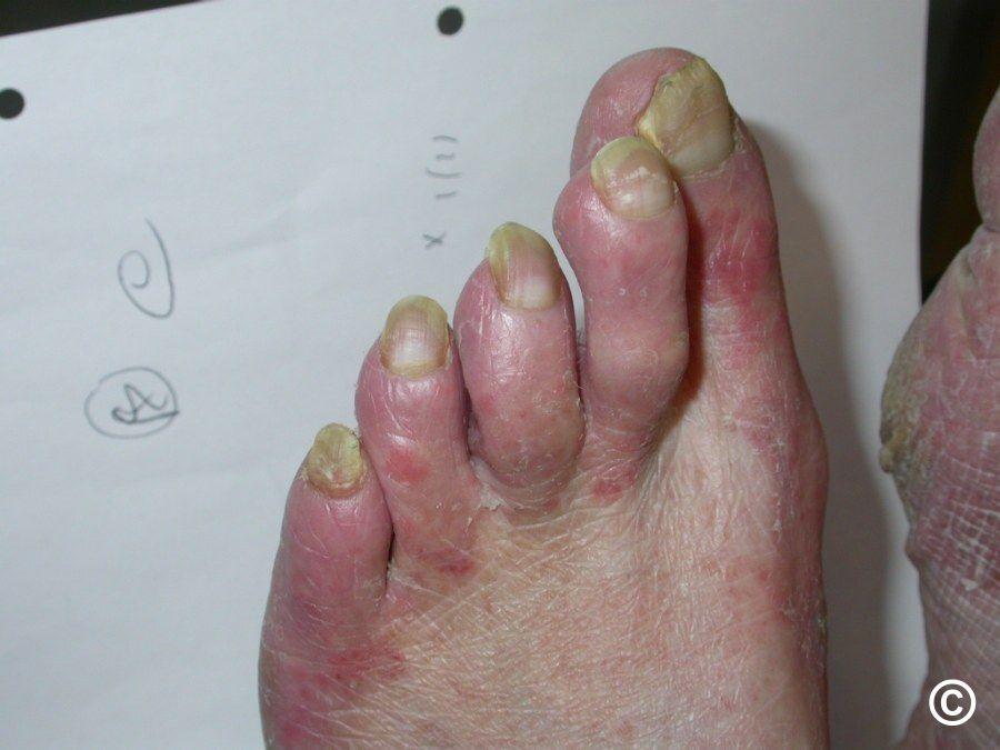
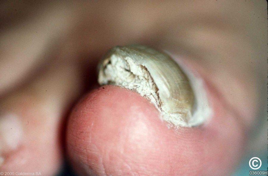
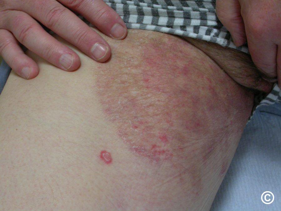
Athlete's foot and other fungal/yeast infections
Treatment of hands (tinea manuum), feet (tinea pedis ie Athlete's foot), or groin (tinea cruris)
- Treat twice a day until the scale disappears, and then carry on for one more week. Choice of treatment includes Lamisil® cream (terbinafine), Daktarin® cream (miconazole), or Canesten® cream (1% clotrimazole). If treating the feet it is important to also treat between the toes
- If athlete's foot is recurrent make sure you wear breathable footwear and leave shoes off around the home. Using the creams referred to above once-twice a week can also help
Treatment of nails (tinea unguium)
- Fungal nail infections only need treatment if bothersome and/or cause recurrent athlete's foot
- Treatment with a nail paint (lacquer) is only likely to help white nail infection (image 5) or distal nail infection (the end of the nail) - see your pharmacist for advice
- If the nails are yellow and thickened (images 6&7), you will need to see a health professional for a course of tablets
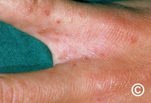
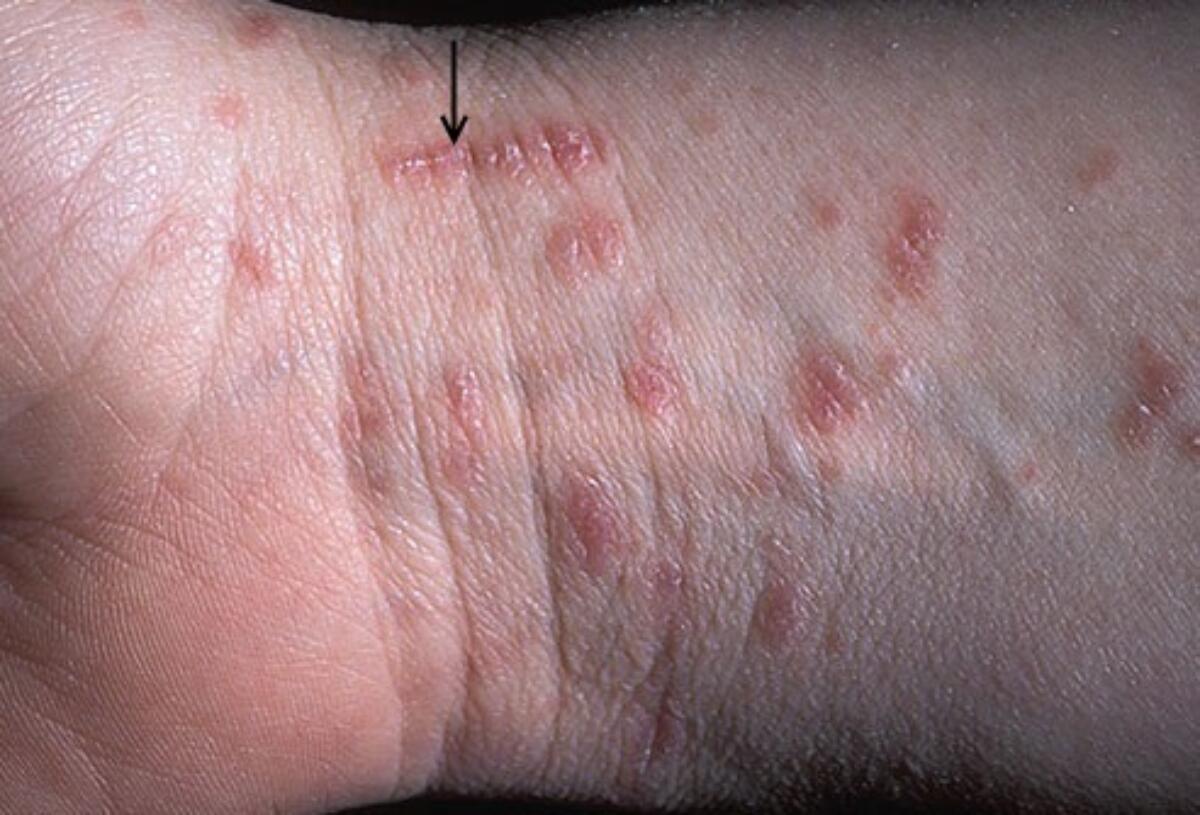
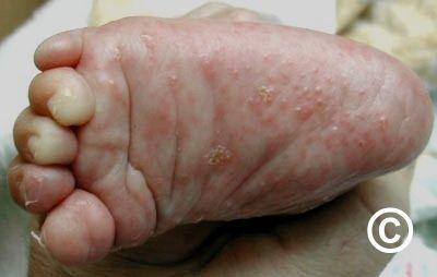
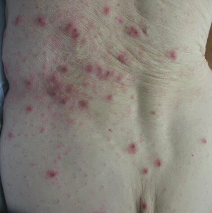
Scabies
- The main treatment is Lyclear® cream (5% permethrin cream) - two treatments one week apart are needed for those with symptoms of scabies, for other contacts who do not have symptoms only one treatment is needed
- Seek advice from a pharmacist and use this patient information leaflet
- Follow this link for more information


Molluscum contagiosum
- Most cases do not need treatment with lesions resolving over 6-9 months. Occasionally lesions can persist for up to 4 years
- If treatment is needed MolluTinc® can be used from the age of 2 years. It should be used twice a day until lesions become inflamed (appearing red in Caucasian skin), usually around day 5, at which stage treatment should be stopped. If there is no inflammation by day 14 the treatment will not work and should be stopped
- For more information follow this link on molluscum contagiosum


Pityriasis versicolor
- Pityriasis versicolor is a yeast infection of the skin
- It is treated with Nizoral® shampoo (ketoconazole), which should be made into a lather and left on affected areas of skin for 10 minutes before washing off. This should be done once a day for 5 days
- The treatment has been successful if the scale disappears. It will take several months for the skin colour to return to normal, on occasions the discoloration can be persistent
- If the scale persists you will need to see a health professional for a course of Sporanox® capsules
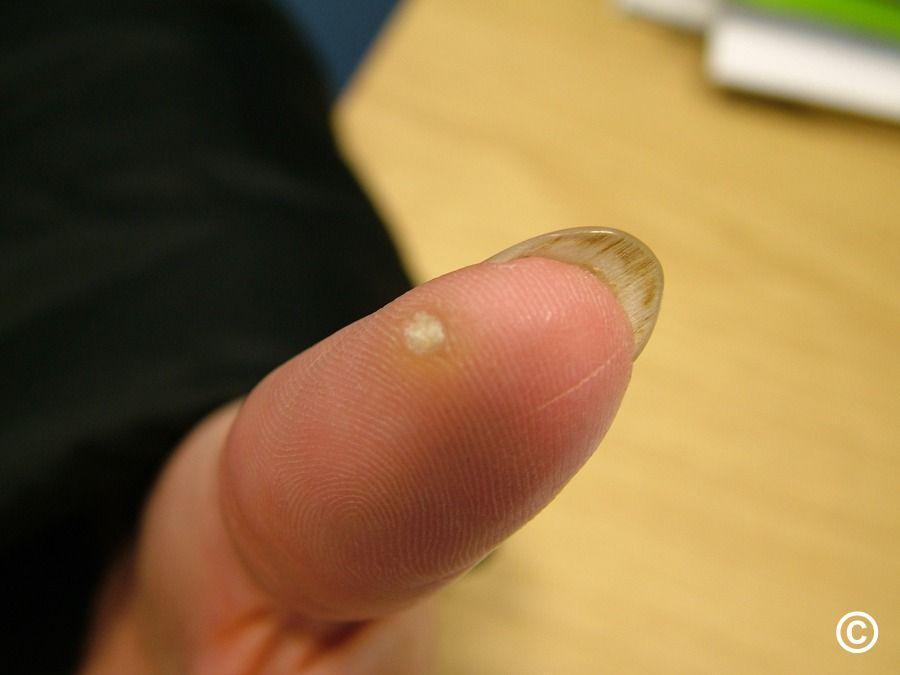
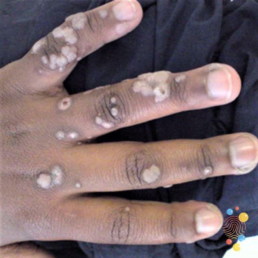
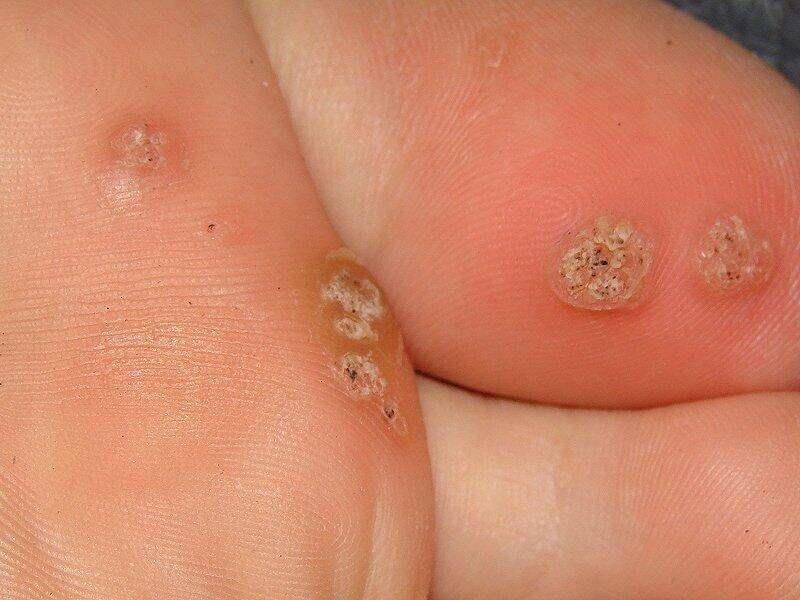
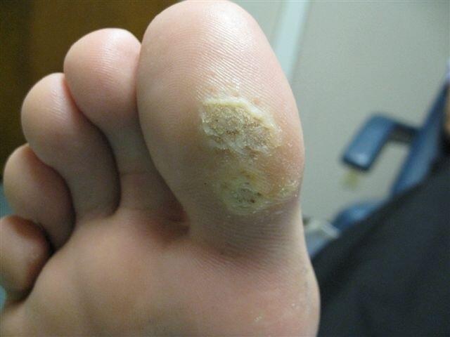
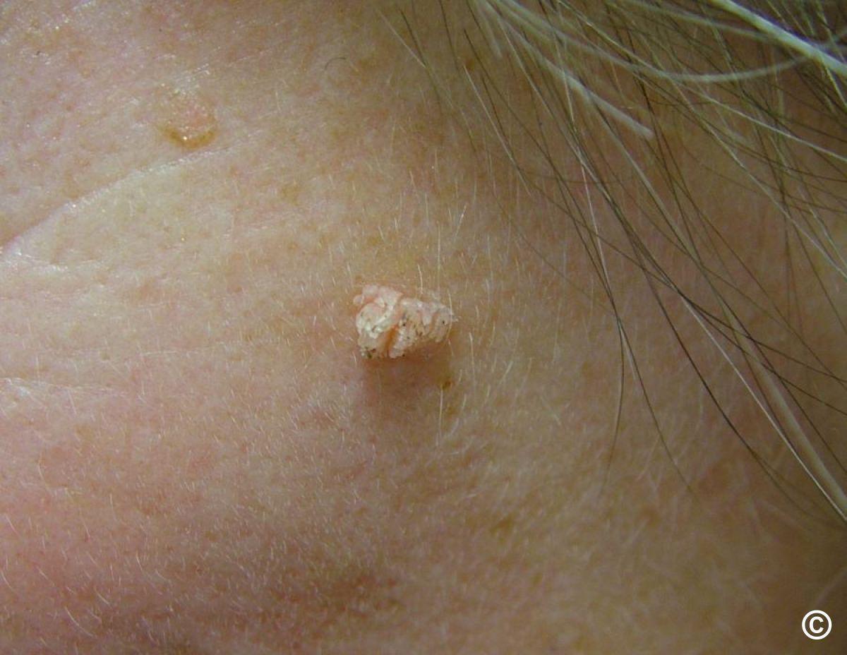
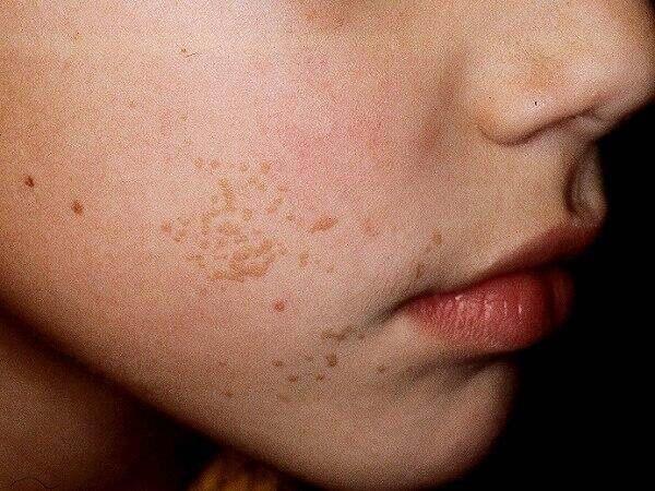
Wart preparations
- There are different types of warts
- Most warts do not require treatment and will eventually resolve. If warts on the feet (verrucae) are painful they can be pared - the affected skin should be soaked in warm water and the thickened skin filed away with a pumice stone or emery board
- If treatment is needed seek advice from a pharmacist about a wart paint or freeze spray (neither should be used on the face)
- In older children and adults, cryotherapy can be used but is painful, can blister, several treatments are likely to be needed, and not all cases respond. Cryotherapy is unlikely to be available on the NHS unless provided at your GP surgery (which is not often the case due to safety issues related to storage)
- For more information refer to this patient information leaflet
Disclaimer - the author PCDS cannot accept responsibility for any misleading or incorrect statements, and the management of individual patients remains the direct responsibility of the individual doctor. We do however hope that visitors to this site can contact us regarding comments that are considered misleading or incorrect so that we can continue to improve the site.
Image Rights - The PCDS would like to thank Dermatoweb, DermQuest (Galderma), and others who have contributed images. All named individuals and organisations maintain copyright for the relevant images.
The following pharmaceutical companies have had no involvement in the content of this website or in our conference programmes







