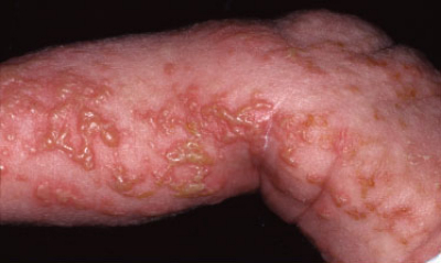
Blistering (bullous) disorders - an overview
LAST UPDATED: Apr 11, 2023
Introduction
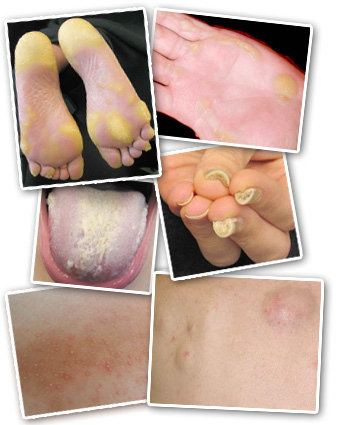
There are many dermatological conditions that can cause vesicles and bullae (blisters). In some of these conditions, eg the immunobullous disorders, the bullae are the primary feature, in others, eg leg oedema, the bullae are a secondary phenomenon.
This chapter, which is set out as below, provides an overview of vesicular and bullous disorders, with an aim to aid in diagnosis. More detailed information on the individual conditions can be found in the related chapters.
Aetiology
-
While there are many causes of vesicles and bullae, it is the immunobullous conditions, and epidermolysis bullosa, which commonly provoke most interest due to their dramatic impact on the patient and their relatives
-
Immunobullous conditions
-
These are characterised by pathogenic autoantibodies directed at target antigens whose function is either cell-to-cell adhesion within the epidermis or adhesion of stratified squamous epithelium to dermis or mesenchyme. These target antigens are components of desmosomes or the functional unit of the basement membrane zone known as the adhesion complex. Damage to this areas causes blisters
-
The main immunobullous conditions are the pemphigus and pemphigoid disorders
-
Epidermolysis bullosa (EB) comprises a group of genetically determined skin fragility disorders characterised by blistering of the skin and mucosae following mild mechanical trauma
History
Given the many causes of vesicles and bullae it is important to ascertain a thorough history, which includes:
-
Speed of onset
-
Was there any preceding itch / rash - this occurs in a number of conditions, especially bullous pemphigoid
-
Are the lesions itchy eg pompholyx, or painful eg cellulitis, or Stevens-Johnson syndrome / toxic epidermal necrolysis
-
Have any medications (prescribed or over the counter) been started recently
-
Pets, such as dogs and cats, if infested by fleas or mites can cause a papular urticaria with bullae
-
Recent travel - bed bugs can cause a papular urticaria
-
Is anyone else affected eg bullous impetigo, papular urticaria
-
Does the condition run in the family eg epidermolysis bullosa
-
Is there anything unusual about the history or clinical appearance eg burns / scalds resulting from child abuse, self-harm or domestic violence
Clinical findings
This section is divided in to three parts:
-
Red flags
-
Common causes of vesicles and bullae
-
Physical and chemical - friction blisters / UV-related: sunburn, polymorphic light eruption (prickly heat) / chilblains / bites & stings / burns & scalds / miliaria crystallina
-
Leg oedema
-
Infection - bacterial (bullous impetigo, cellulitis) / viral (herpes zoster, herpes simplex, hand foot and mouth disease, orf) / others - scabies and tinea
-
Eczema - pompholyx type eczema / contact allergic dermatitis
-
Papular urticaria
-
Uncommon / rare causes of vesicles and bullae - based on site
-
Trunk / wider distribution - epidermolysis bullosa / epidermolytic hyperkeratosis (syn. bullous congenital ichthyosiform erythroderma) / Staphylococcal Scalded Skin syndrome / linear IgA disease / bullous erythema multiforme / epidermolysis bullosa acquisita - generalised form / bullous systemic lupus erythematosus / pemphigoid gestationis / Stevens-Johnson syndrome and toxic epidermal necrolysis / subcorneal pustular dermatosis / pemphigus foliaceus / pemphigus vulgaris / paraneoplastic pemphigus / bullous pemphigoid / mucous membrane pemphigoid
-
Hands, feet and limbs - phytophotodermatosis / bullous erythema multiforme / bullosis diabeticorum / Gianotti-Crosti syndrome / localised bullous pemphigoid / porphyria cutanea tarda and pseudoporphyria / epidermolysis bullosa / epidermolysis bullosa acquisita / pachyonychia congenita
-
Scalp - pemphigus vulgaris, and some other forms of pemphigus and pemphigoid / epidermolysis bullosa / erosive pustular dermatosis
-
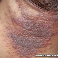
Flexures - Hailey-Hailey disease (syn. benign familial pemphigus) / pemphigus vulgaris / pemphigus vegetans
-
Mucosal surfaces - drug reactions / lichen planus / erythema multiforme / Stevens-Johnson syndrome and toxic epidermal necrolysis / mucous membrane pemphigoid / pemphigus vulgaris / paraneoplastic pemphigus / linear IgA disease / lupus erythematosus / epidermolysis bullosa / epidermolysis bullosa acquisita
-
Various sites - fixed drug eruptions / Grover's disease / dermatitis herpetiformis / lichen planus / neutrophilic dermatoses - some cases of pyoderma gangrenosum and Sweet's syndrome / lymphomatoid papulosis / incontinentia pigmenti
1. Red flags
There are a number of bullous conditions that are potentially serious, and it important to be aware of the red flags that could suggest such a condition:
-
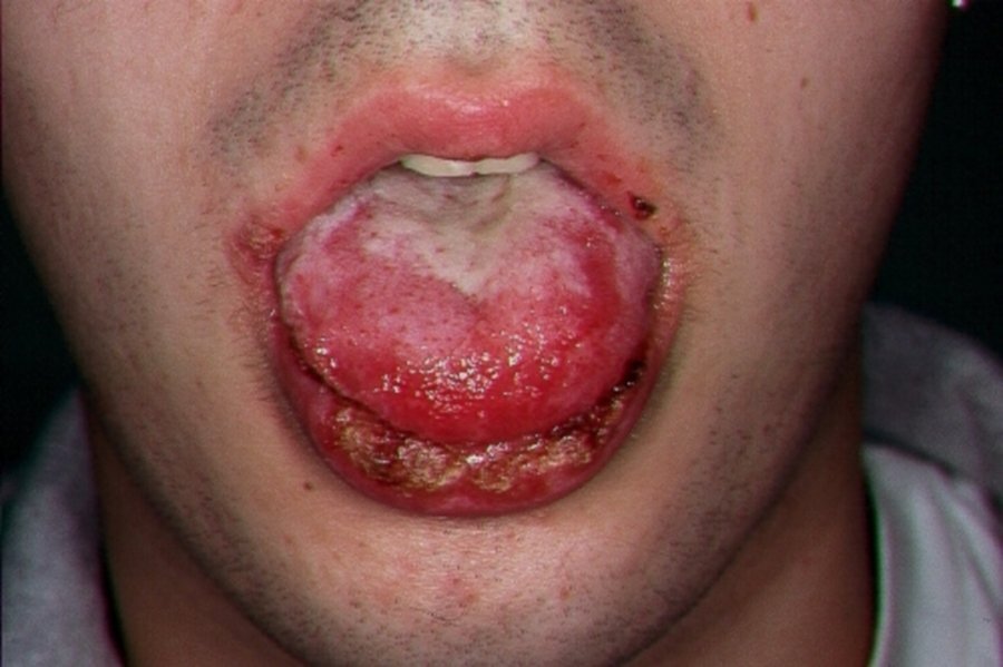
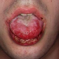
Involvement of the lips and mouth and/or the rapid progression of bullae could suggest a serious drug eruption such as Stevens-Johnson syndrome or toxic epidermal necrolysis
- Immunobullous conditions
- Increasing numbers of large tense bullae could be due to bullous pemphigoid - this is very uncommon under the age of 50 years
- The development of blisters in pregnancy could be due to pemphigoid gestationis
- Involvement of mucous membranes (eyes, mouth, genitalia) could be caused by a number of immunobullous conditions such as mucous membrane pemphigoid or pemphigus vulgaris
- The presence of erosions and crusting with or without flaccid blisters could represent pemphigus
- The PEM Friends website provides a good diagnostic tool for this group of conditions
-
Child abuse, self-harm and domestic violence - look out for an inconsistent history, as well an unusual appearance / distribution of burns or scalds
It is important to note:
-
The majority of patients that present to a health care professional with bullae will have a relatively harmless, self-limiting condition
-
In order to make a diagnosis it is important to take a logical approach to the patient with vesicles / bullae, as described below in sections 2 & 3
2. Common causes of vesicles and bullae
Physical / chemical
-
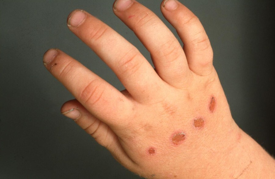
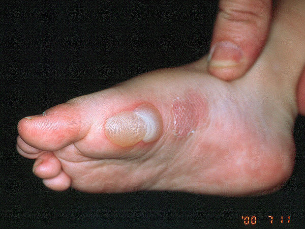
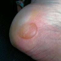
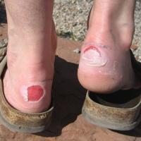
Friction blisters (figures 1-2)
-
Are most commonly seen on the heels and soles of feet, and on the palms of the hands
-
Beware that epidermolysis bullosa causes friction blisters, and in some cases may be quite subtle. Refer to section 3 for more information
-
UV-related
-
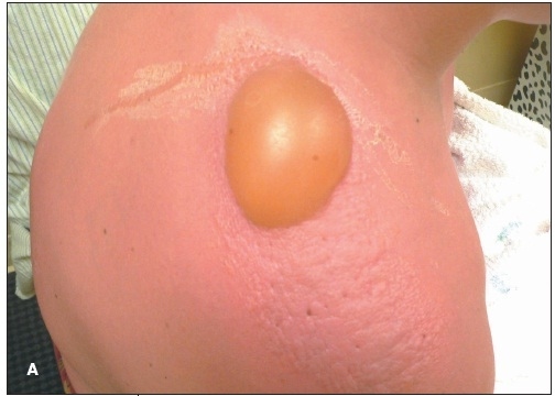
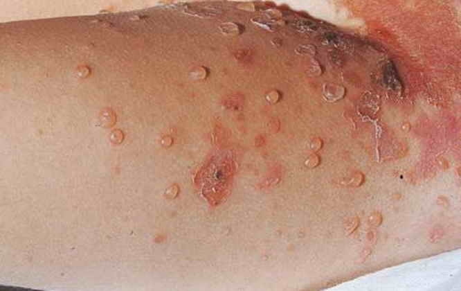
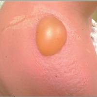
Sunburn (figure 3)
-
Is unfortunately a common cause of blistering. All episodes of sunburn increase the risk of skin cancer
-
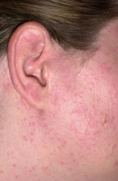
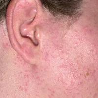
Polymorphic light eruption - syn. prickly heat (figure 4)
-
Symptoms commonly develop 24 hours after UV exposure but can occur anywhere between two hours and five days after
-
Affects exposed areas of skin, although less exposed sites can also be affected
-
Lesions are iIl-defined papules, occasionally vesicular. The background skin is usually normal
-
In severe cases symptoms may be much more easily provoked and cause a burning sensation. Affected areas can be erythematous, oedematous and blister. Such patients may even be affected by light coming through a car window
-
Refer to the chapter Polymorphic light eruption
-
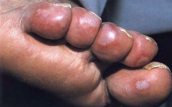
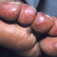
Chilblains syn. perniosis (figure 5)
-
Arise several hours after being exposed to the cold and affect the extremities ie toes, fingers, nose and earlobes
-
The skin is usually red at first but may become purple
-
Lesions are itchy and often have a burning sensation. The skin may become oedematous, blister and ulcerate
-
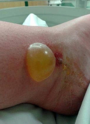
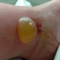
Bites and stings (figure 6)
-
Can produce local skin reactions, including small and large bullae
-
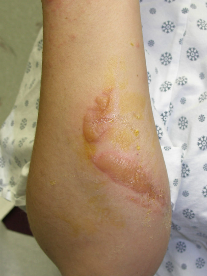
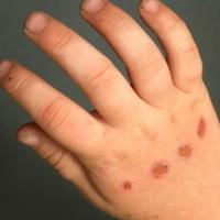
Burns and scalds (figure 7)
-
Can result from either accidental or non-accidental injury
-
Look out for an inconsistent history, as well an unusual appearance and/or distribution
-
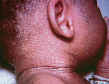
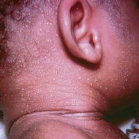
Miliaria crystallina (figure 8)
-
Is the mildest form of heat rash, affecting the sweat ducts in the topmost layer of skin. Although it is common in newborns, adults can also develop it
-
Distribution - in infants the rash is mainly found on the neck, shoulders and chest, but it can also occur in the armpits, elbow creases and groin. Adults usually develop symptoms in folds of skin and wherever clothing causes friction
-
Morphology - small, clear, vesicles that break easily
-
This type of heat rash usually clears on its own but can come back if hot, humid weather persists
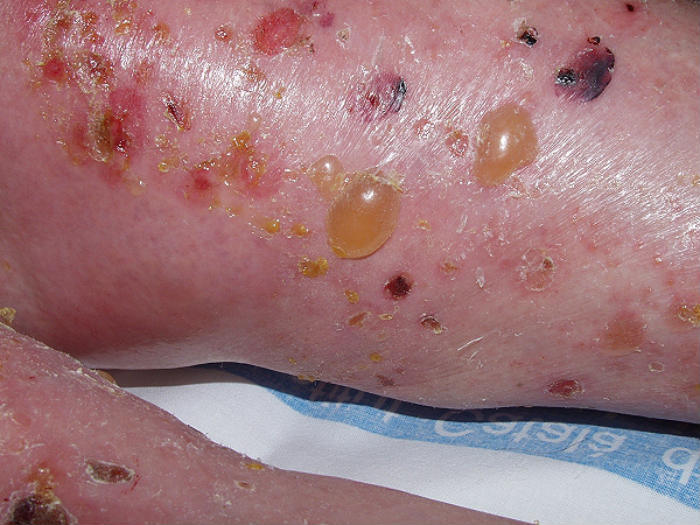
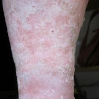
Leg oedema (figures 9-10)
- Oedema / lymphoedema, with or without cellulitis, is a common cause of bullae
-
It is important to manage the underlying cause
-
Dressing may be required - refer to the section on Leg dressings and other bandaging techniques
Infection
-
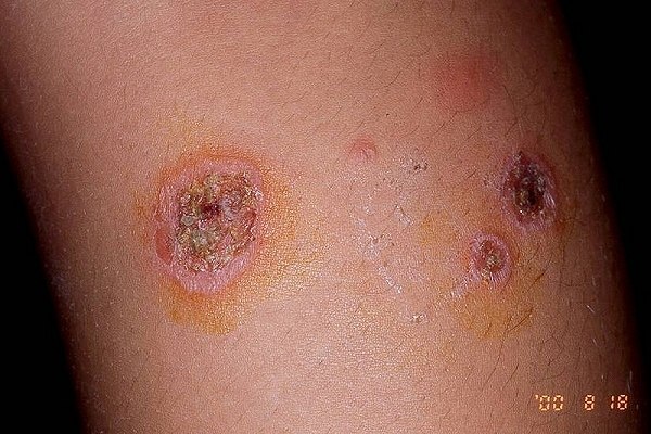
Bacterial infection
-
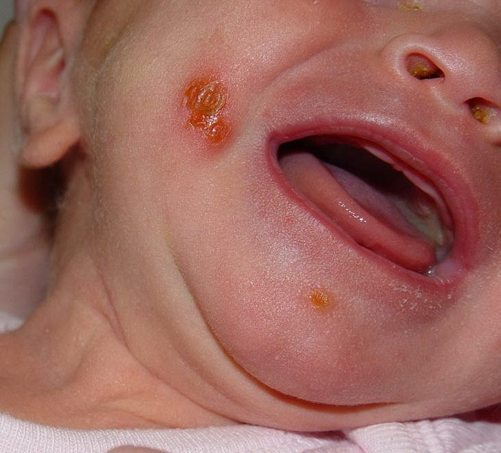
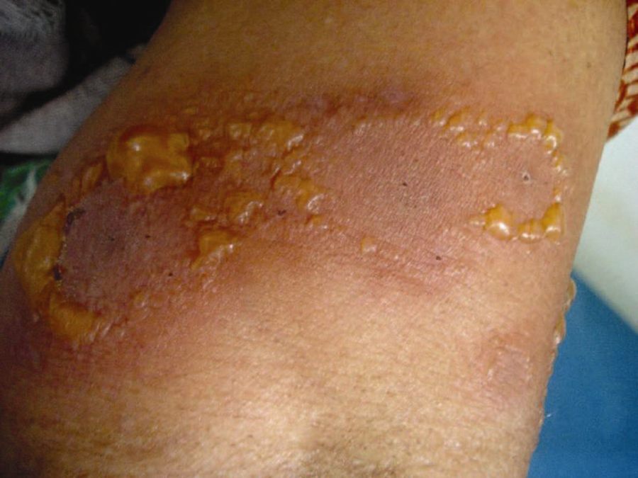
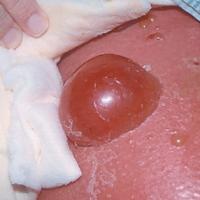
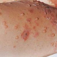
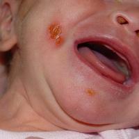
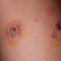
Bullous impetigo (figures 11-13)
-
Usually caused by streptococcus pyogenes and / or staphylococcus aureus
-
Small or large bullae arise over a short period of time, usually spreading locally on the face, trunk, extremities, buttocks, or perineal regions and may reach distal areas
-
Bullae are fragile and burst spontaneously to leave a yellow crust that usually heals without scarring
-
Refer to the chapter Bullous impetigo
-
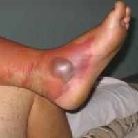
Cellulitis (figure 14)
-
Usually caused by streptococcus pyogenes and / or staphylococcus aureus
-
Commonly affects a limb but can occur anywhere on the body
-
The underlying skin is erythematous and feels hot. In more severe cases patients are systemically unwell
-
Refer to the chapter Cellulitis
-
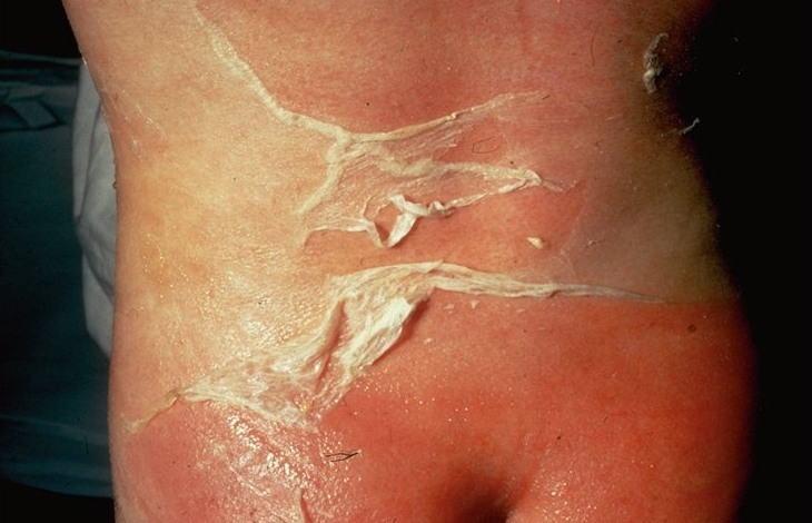
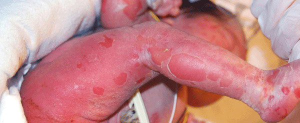
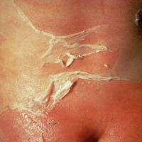
Staphylococcal Scalded Skin syndrome (figure 15)
-
An uncommon condition
-
It is caused by exotoxins produced by Staphylococcus aureus
-
Children presents with fever, irritability and widespread erythema. Within 24-48 hours fluid-filled bullae form, which rupture easily, leading to a diffuse exfoliation (peeling) of the epidermis
-
Refer to the chapter Staphylococcal scalded skin syndrome
-
Viral infection
-
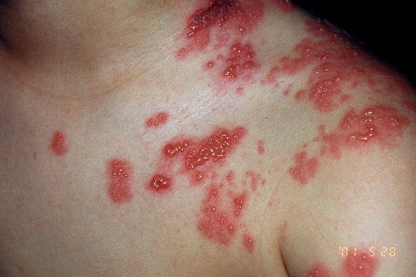
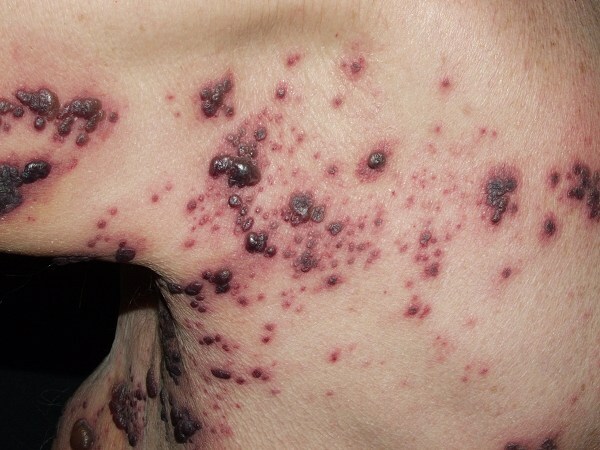
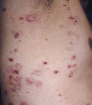
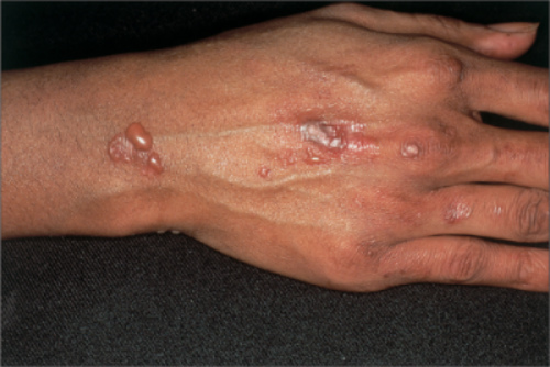
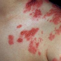
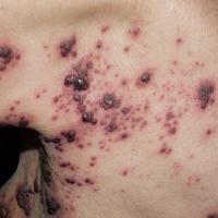
Herpes zoster (figures 16-17)
-
Patients normally have preceding pain
-
Closely grouped red papules, rapidly becoming vesicular and then pustular, develop in a continuous or interrupted band in the area of one or occasionally two and, rarely, more contiguous dermatomes
-
Lesion can become bullous and necrotic
-
Immunosuppressed patients can develop widespread zoster
-
Refer to the chapter Herpes zoster
-
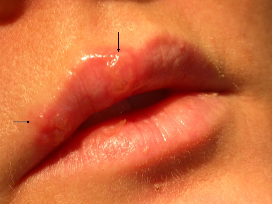
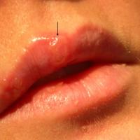
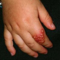
Herpes simplex (figures 18-19)
-
Patients normally have preceding tingling, itching or burning
-
Most commonly affects the mouth and genitalia, although any site can be affected
-
Morphologically the rash consists of small, closely grouped vesicles on a red base
-
Eczema herpeticum - causes clustered vesicles / punched-out monomorphic erosions, which may coalesce. Any site may be affected, most commonly the face and neck. Lesions can occur in normal skin or in sites actively or previously affected by atopic eczema or other skin conditions. New patches form and spread over a period of 7 to 10 days, and can become widespread
-
Refer to the chapter Herpes simplex
-
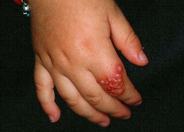
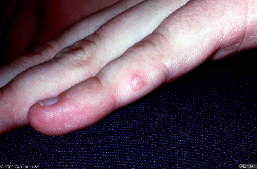
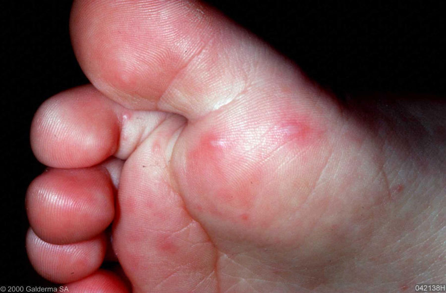
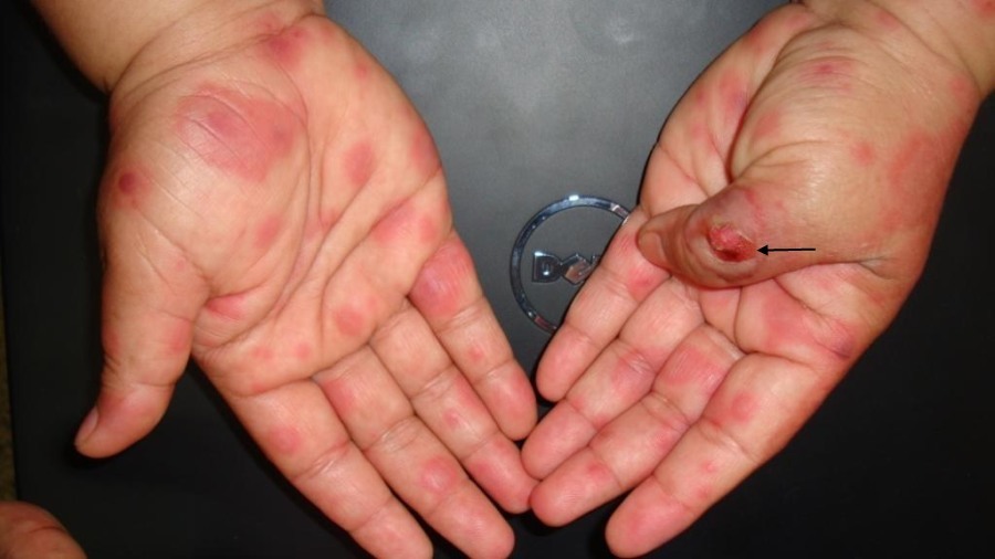
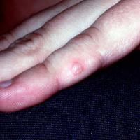
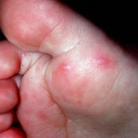
Hand, foot and mouth disease (figures 20-21)
-
Is most commonly caused by the Coxsackie A16 virus
-
It predominantly affects children under 10 years of age. Older children and adults are sometimes affected
-
There is a prodrome with high fever and a sore throat
-
Initially macular lesions appear on the buccal mucosa, tongue, and / or hard palate, which rapidly progress to vesicles that erode and become surrounded by an erythematous halo
-
Cutaneous lesions, arising as tender macules or vesicles on an erythematous base, develop in 75% of cases. They mainly affect the hands and feet, but the buttocks and genitalia can also be involved
-
It takes around five to seven days for the infection to resolve completely
-
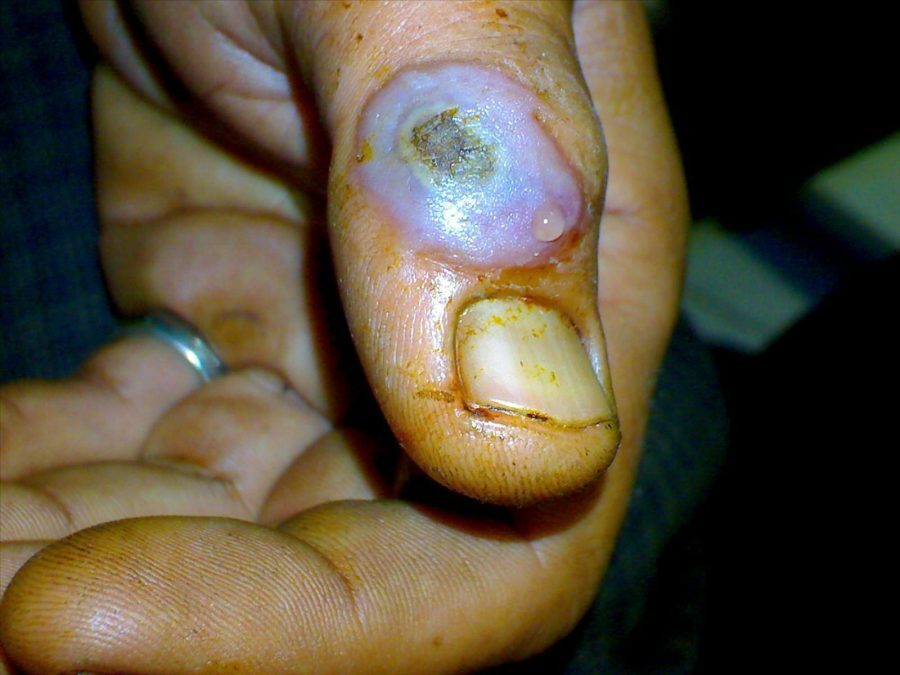
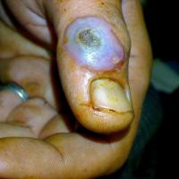
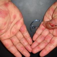
Orf (figures 22-23)
-
Orf is caused by the parapox virus, which mainly infects young lambs and goats. Human infection is caused by direct contact
-
Lesions occur most commonly on the fingers, hands or forearms but can appear on the face
-
Lesions are generally solitary, but can be a few in number. Orf presents as a small, firm, red or red-blue papule, which enlarges to form a flat-topped, blood-tinged pustule or blister. Lesions are often 2-3 cm in diameter but may be as large as 5 cm
-
The condition is self-limiting and usually clears within six weeks. The lesion may be covered to prevent contaminating the environment or other people, although person-to-person spread is very uncommon
-
Other infections
-
Scabies can occasionally cause bullae on the feet in infants
-
Tinea can occasionally cause localised bullae
Others common causes of vesicles and bullae
-
Eczema
-
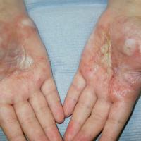
Pompholyx (figures 24-25) - a type of eczema that usually presents periodically with intensely itchy vesicles, and occasionally large bullae, that predominate on the palms and sides of fingers - refer to the chapter Eczema: hand (and foot) eczema
-
Contact allergic dermatitis can occasionally cause blistering
-
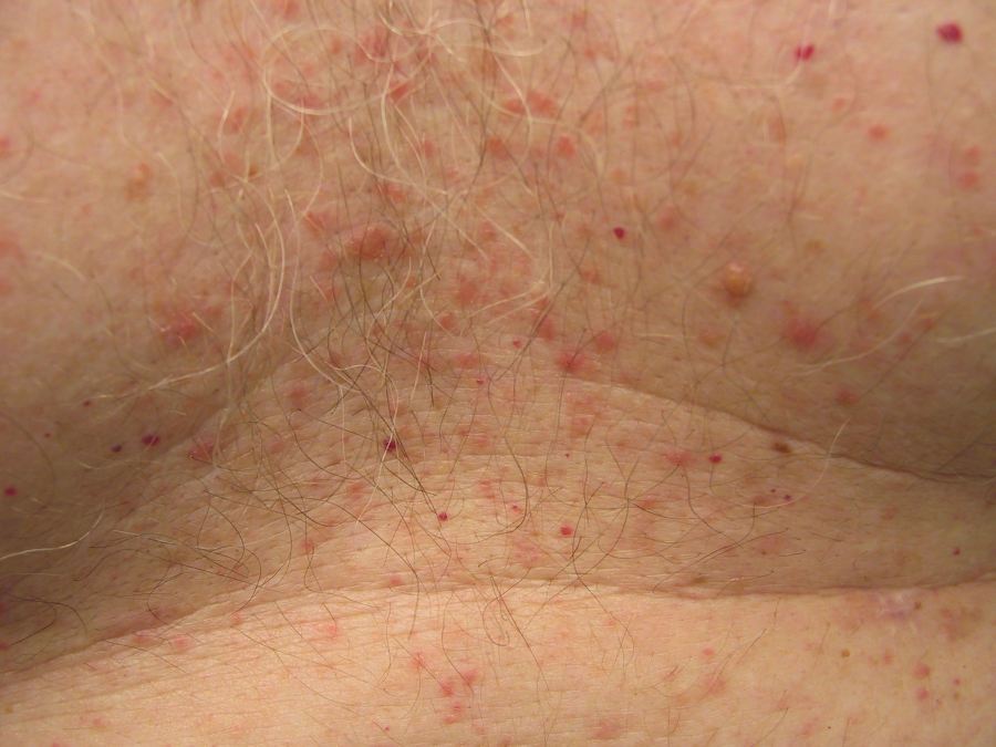
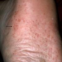
Papular urticaria (figure 26)
-
Is a hypersensitive reaction to various insect bites
-
It is more common on the lower legs, and other exposed sites
-
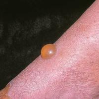
Bullae can be small or large and are often grouped together, sometimes in a circular or linear arrangement
-
Refer to the chapter Papular urticaria
3. Uncommon / rare causes of vesicles and bullae, based on site
Trunk / wider distribution
The following list is set out chronologically, such that those conditions tending to present at a younger age are listed higher up. The main exceptions are Stevens-Johnson syndrome / toxic epidermal necrolysis, and paraneoplastic pemphigus, which can present at any age.
-
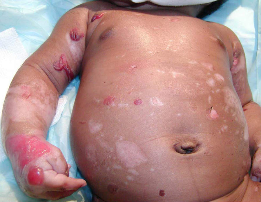
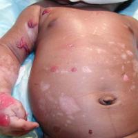
Epidermolysis bullosa (figure 27)
-
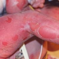
Epidermolytic hyperkeratosis syn. bullous congenital ichthyosiform erythroderma (figure 28)
-
A form of congenital ichthyosis inherited as an autosomal-dominant trait and characterised by generalised blistering, erythroderma and severe hyperkeratosis. It is manifested at birth by widespread bullae followed by the appearance of thickened, horny, verruciform scales over the entire body, but accentuated in flexural areas
-
Staphylococcal Scalded Skin syndrome
-
Refer to the notes in section 1 above, under infection
-
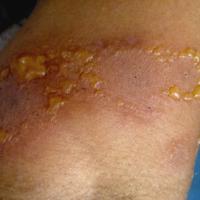
Linear IgA disease (figure 29)
-
Lesions can affect many body sites, although in children they favour the lower abdomen and anogenital areas
-
Bullae arise in urticated plaques and papules. In children, and sometimes adults, lesions are annular / polycyclic, often with blistering around the edge known as the 'string of pearls' sign
-
Refer to the related chapter Linear IgA disease
-
Erythema multiforme
-
Can become generalised - refer to the notes in the section below on hands / forearms
-
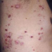
Epidermolysis bullosa acquisita - generalised inflammatory type (figure 30)
-
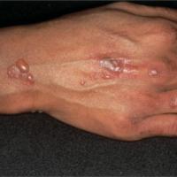
Bullous systemic lupus erythematosus (figure 31)
-
Is an immunobullous condition, usually arising in patients with well-established SLE
-
The blistering is widespread, although in some patients the eruption is photosensitive
-
Lesions vary in size from vesicles to large bullae
-
Mucosal lesions are uncommon
-
Histologically the blisters are subepidermal. Direct immunofluorescence shows IgG, IgA, IgM and C3 in the basement-membrane zone
-
Bullous SLE is often transient
-
Refer to the chapter Systemic lupus erythematosus
-
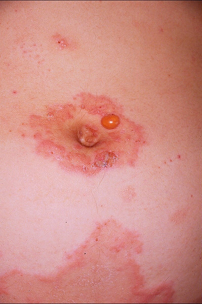
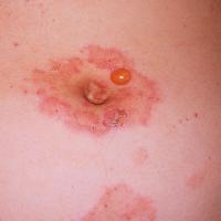
Pemphigoid gestationis, syn. pemphigoid of pregnancy (figure 32)
-
Is a rare pregnancy-associated, immunobullous condition, characterised by an itchy rash that develops in to blisters
-
It may arise at any time between four weeks gestation and five weeks postpartum, with the majority presenting in the second and third trimesters
-
Initially there are itchy urticated lesions around the umbilicus. Within days to weeks, the rash spreads to other parts of the body including the trunk, back, buttock, and arms. The face, scalp, palms, soles and mucous membranes are usually unaffected. The rash then evolves in to vesicles and large, tense bullae
-
Refer to the related chapter Pemphigoid gestationis
-
Stevens-Johnson syndrome / toxic epidermal necrolysis
-
Symptoms often start within a few days of a drug being commenced although it can take a little longer with anticonvulsants. Patients have fever, malaise, myalgia and arthralgia
-
Painful / tender erythema with local erosions and blisters quickly progress to areas of confluent erythema with sheet-like skin and mucosal loss
-
Mucosal involvement includes the eyes, lips / mouth, oesophagus, upper respiratory tract, and genitalia and gastrointestinal tract resulting in diarrhoea
-
Refer to the notes in the section below on mucosal surfaces
-
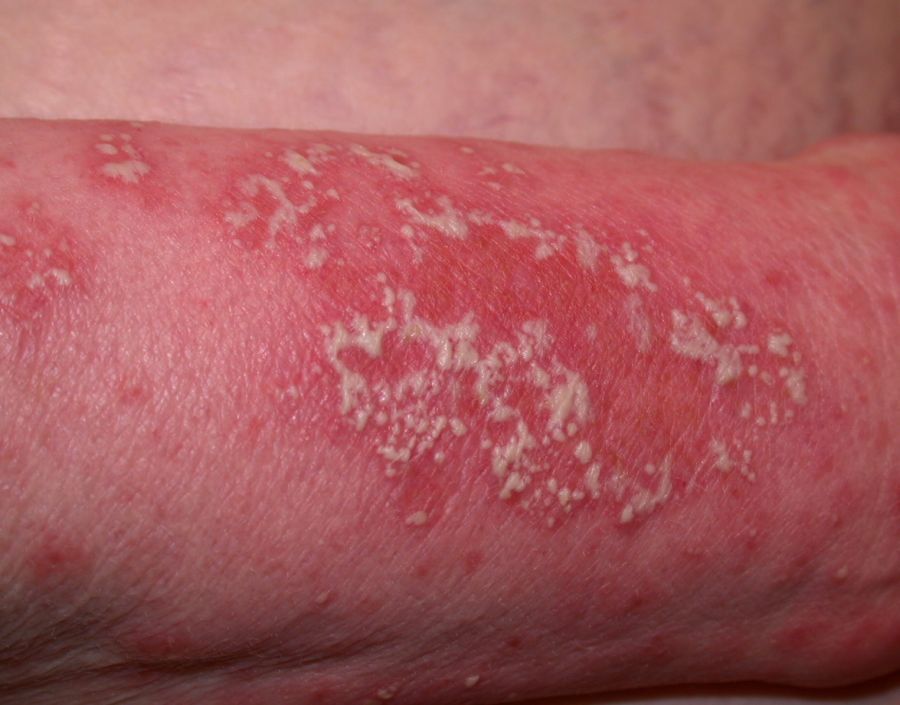
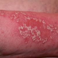
Subcorneal pustular dermatosis (figure 33)
-
The usual distribution is the flexural areas of the trunk and proximal extremities
-
Lesions arise as numerous small, sterile pustules. Characteristically the pus accumulates in the lower half of the pustule. Pustules easily rupture and tend to coalesce, forming annular or serpiginous patterns with a scaly edge
-
Refer to the related chapter Subcorneal pustular dermatosis
-
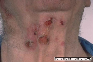
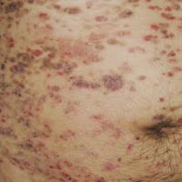
Pemphigus foliaceus (figure 34)
-
The onset is usually insidious with scattered, scaly lesions with a 'seborrhoeic' distribution, ie the scalp, face and upper trunk
-
Erosions, crusting and erythema are common. Bullae may not be obvious as they easily rupture. The Nikolsky sign is positive
-
Refer to the related chapter Pemphigus foliaceus
-
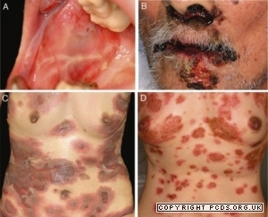
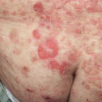
Pemphigus vulgaris (figure 35)
-
The most commonly affected sites include the scalp, face, axillae, groins and pressure points. The trunk can also be involved
-
The primary lesion of pemphigus vulgaris is a flaccid blister filled with clear fluid that arises on healthy skin or on an erythematous base. Bullae are fragile and may rupture, producing painful erosions (the most common skin presentation). Lesions heal without scarring
-
Mucosal involvement is common and may precede cutaneous signs
-
Refer to the notes below on mucosal surfaces
-
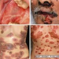
Paraneoplastic pemphigus (figure 36)
-
Skin lesions occur anywhere on the body, especially the upper body, and are highly variable in appearance
-
Lesions can be inflamed papules, scaly plaques, fluid-filled bullae or ulcerated. Palmoplantar target lesions are another feature
-
Refer to the related chapter Paraneoplastic pemphigus
-
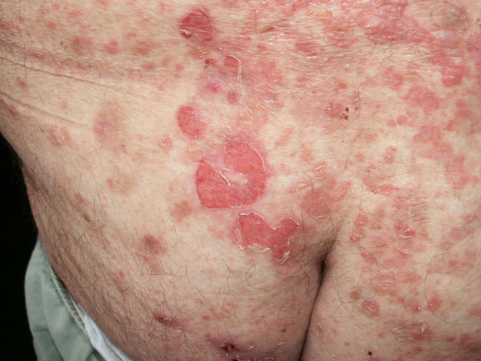
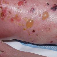
Bullous pemphigoid (figure 37)
-
Mainly affects older patients
-
There may be a prodromal phase with itch and fixed urticarial-like lesions
-
Tense bullae can develop anywhere on the skin and are especially widespread on the trunk, proximal limbs and flexures
-
Mucosal symptoms tend to be mild
-
Refer to the related chapter Bullous pemphigoid
-
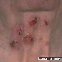
Mucous membrane pemphigoid (figure 38)
-
Mainly affects mucosal surfaces, however, the skin is involved in approximately one-quarter of cases
-
In the skin it can cause either a generalised bullous eruption similar to bullous pemphigoid, or localised areas of scarring blisters, predominantly of the scalp and of the skin close to affected mucosal surfaces
-
Refer to the notes below on mucosal surfaces
Hands, feet and limbs
-
Hands only
-
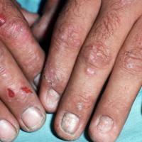
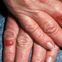
Porphyria cutanea tarda and pseudoporphyria (figures 39-40)
-
Causes skin fragility and bullae on the backs of hands and bald areas of the scalp
-
Lesions heal slowly and often scar
-
Milia and areas of hyperpigmentation may develop
-
Pseudoporphyria, in which porphyria-like blistering of exposed skin on the extremities occurs in the absence of abnormal porphyrin metabolism, may be caused by a number of drugs, especially furosemide, nalidix acid and naproxen
-
Refer to the chapter Porphyria
-
Hands and feet
-
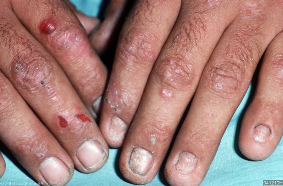
Epidermolysis bullosa
-
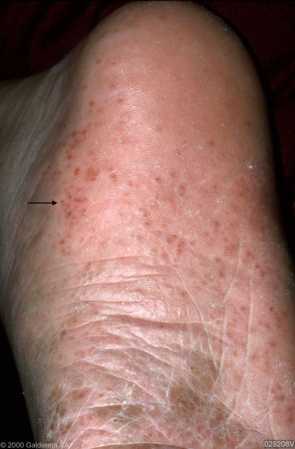
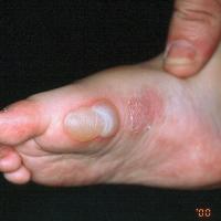
Epidermolysis bullosa simplex (figure 41)
-
Is characterised by painful blisters on the palms of the hands and soles of the feet that develop after mild or moderate physical activity. Many patients have blisters only on the feet
-
Symptoms usually become apparent during early childhood, although mild cases may go undiagnosed until early adult life
-
Hyperhidrosis of the feet is common
-
The blisters usually heal without significant scarring or milia
-
Refer to the related chapter Epidermolysis bullosa
-
Dominant dystrophic epidermolysis bullosa
-
A mild form of this condition can cause dystrophy of both fingernails and toenails, with very few other cutaneous features
-
Refer to the related chapter Epidermolysis bullosa
-
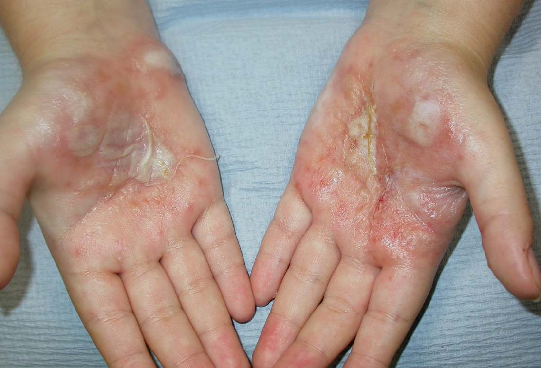
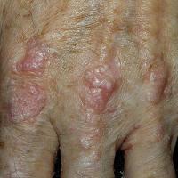
Epidermolysis bullosa acquisita - mechanobullous type (figure 42)
-
Tense vesicles and bullae (serous or haemorrhagic) primarily on areas of trauma such as the extensor surfaces of hands, elbows, knees, ankles and feet
-
Lesions heal with significant scarring, milia and hyperpigmentation
-
Refer to the related chapter Epidermolysis bullosa acquisita
-
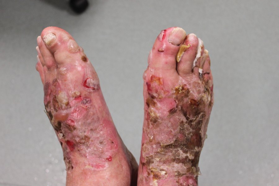
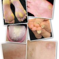
Pachyonychia congenita (figure 43)
-
Is a rare, autosomal dominant condition that primarily affects the nails and skin
-
The signs and symptoms of this condition usually become apparent within the first few months of life, although a rare form of the condition known as pachyonychia congenita tarda appears in adolescence or early adulthood
-
Almost everyone with pachyonychia congenita has hypertrophic nail dystrophy, which causes the fingernails and toenails to become thickened and abnormally shaped. Many affected children also develop very painful bullae and calluses on the soles of the feet and, less commonly, on the palms of the hands
-
Limbs, and hands / feet
-
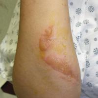
Phytophotodermatosis (figure 44)
-
Is caused by a reaction between contact with naturally occuring plant psoralens, eg common hogweed, giant hogweed, cow parsnip, and UV exposure
-
The blistering rash, which is often streaky, arises some 24 hours later
-
Refer to the chapter Photodermatoses: drug/chemical-induced
-
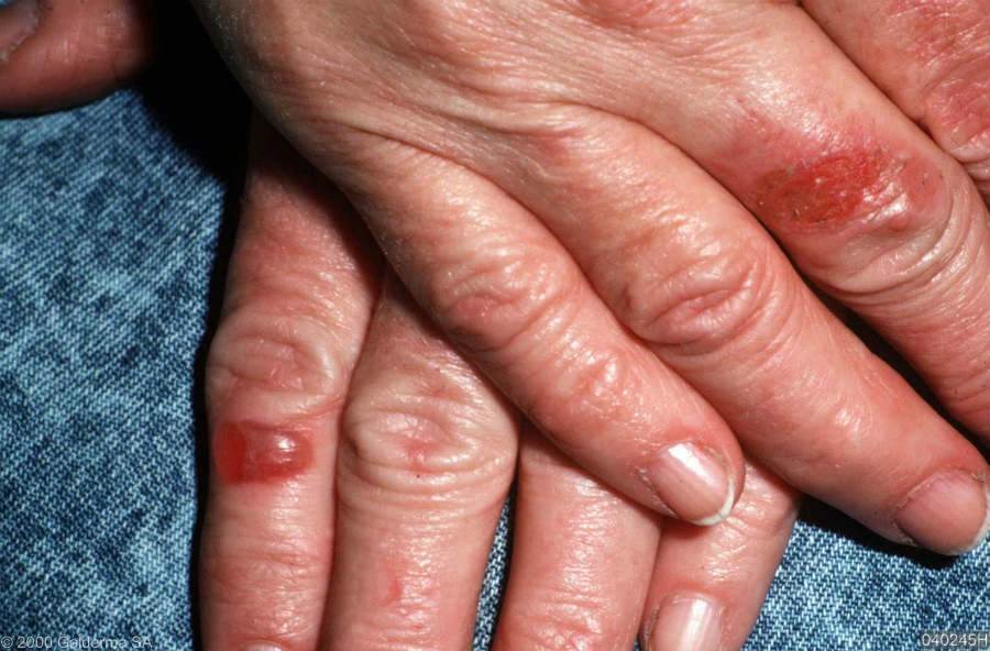
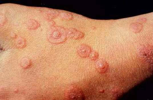
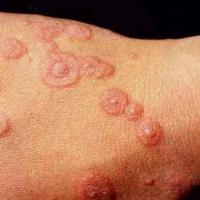
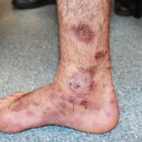
Erythema mulitforme (figures 45-46)
-
The lesions are symmetrical and first seen on the backs of hands and / or dorsal aspects of the feet, and then spread along the limbs towards the trunk. The upper limbs are more commonly affected than the lower limbs. The palms and soles may be involved, and the rash can become more generalised
-
The initial lesion is a dull-red, purpuric macule or urticarial plaque that expands slightly to a maximum of 2 cm over 24-48 hours. In the centre, a small papule, vesicle, or blister develops, flattens, and then may clear. An intermediate ring develops and becomes raised, pale, and oedematous. The periphery gradually changes to become cyanotic or violaceous and forms a typical concentric, “target” lesion
-
Mucosal involvement may occur
-
Refer to the chapter Erythema multiforme
-
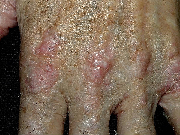
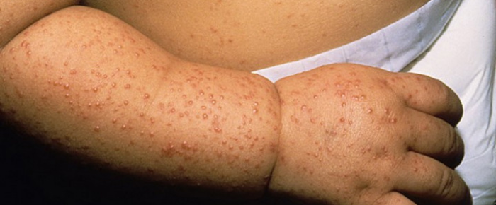
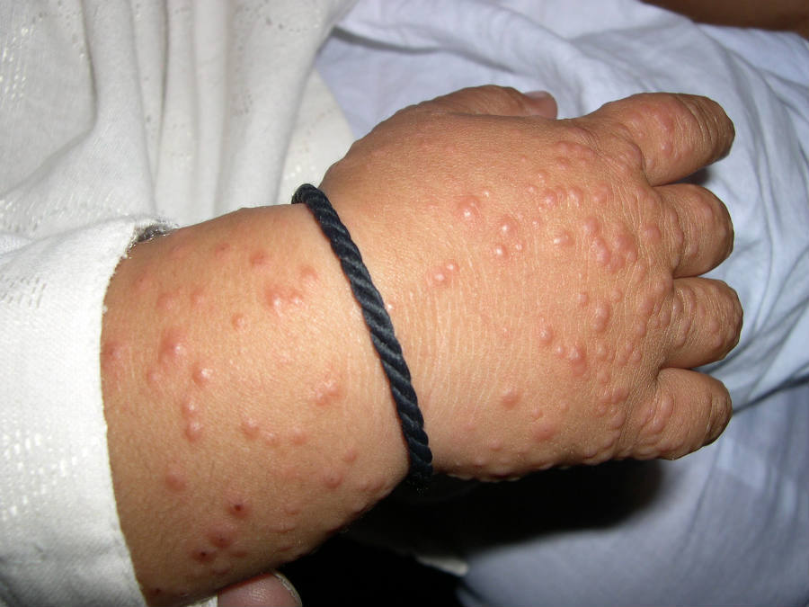
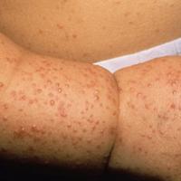
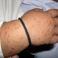
Gianotti-Crosti syndrome (figures 47-48)
-
Is a self-limited childhood exanthem that manifests in a characteristic acral distribution, and is rarely associated with systemic findings
-
The rash is distributed symmetrically and acrally over the extensor surfaces of the extremities, the buttocks, and the face
-
Lesions arise as monomorphous pale, pink-red or skin-coloured papules or papulovesicles, up to 1 cm in size
-
Refer to the chapter on Gianotti-Crosti syndrome
-
Lower legs (predominantly)
-
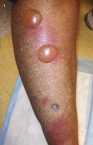
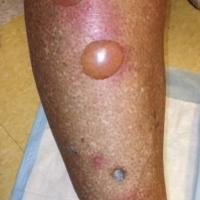
Bullosis diabeticorum (figure 49)
-
A distinct, spontaneous, non-inflammatory, bullous condition of acral skin that is unique to patients with diabetes mellitus
-
Although bullae typically occur on the feet or lower legs, they also may occur on fingers, toes, hands, and arms
-
Non-tender bullae, up to 2 cm in diameter, arise abruptly on non-erythematous skin
-
Lesions heal spontaneously within 2-6 weeks
-
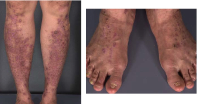
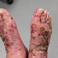
Bullous pemphigoid - localised form (figure 50)
-
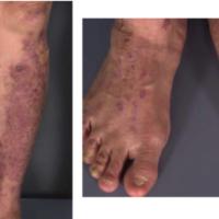
Dominant dystrophic epidermolysis bullosa - pretibial variant (figure 51)
-
This condition, as the name implies, almost exclusively involves the anterior lower legs
-
Individual lesions, which tend to be papular or plaque-like, are often somewhat violaceous, suggesting the clinical diagnosis of lichen planus. Bullae and scarring are also present
-
Refer to the related chapter Epidermolysis bullosa
Scalp
-
Pemphigus vulgaris and many of the other forms of pemphigus / pemphigoid cause scalp lesions
-
Epidermolysis bullosa can affect the scalp
-
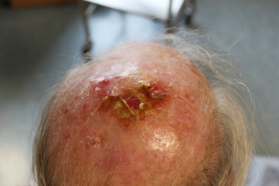
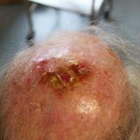
Erosive pustular dermatosis (figure 52)
-
An uncommon condition affecting UV-damaged areas of the scalp in older patients. The risk appears to be increased with the subsequent treatment of actinic keratoses, especially with cryotherapy
-
Clinically there is varying degrees of scarring associated with yellow-brown crusts, pustules, lakes of pus, erosions and ulceration
-
Refer to the chapter Actinic keratosis (management section)
Flexures
Partly as a result of friction, blisters are seldom seen on flexural sites.
-
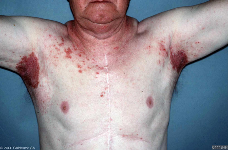
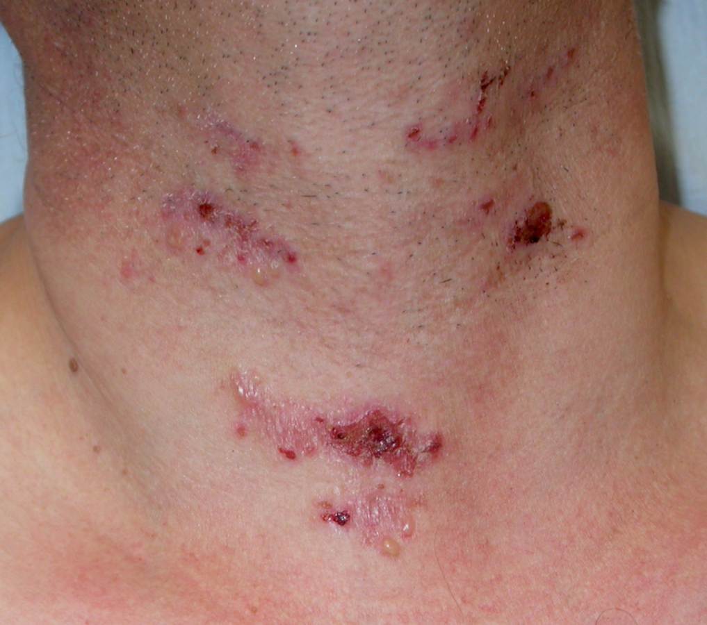
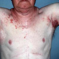
Hailey-Hailey disease - syn. benign familial pemphigus (figure 53)
-
Typically begins as a painful erosive skin rash on areas exposed to friction such as the neck, axilla, under the breasts, the groins and in between the buttocks
-
Lesions extend peripherally, healing in the centre without scars
-
If the lesions are present for some time they may become thickened and malodorous with soft vegetations, and fissures
- Refer to the chapter Hailey-Hailey disease
-
Pemphigus vulgaris
-
Can have flexural involvement
-
Pemphigus vegetans (figure 54)
Oral lesions
Chronic erosions and ulcers, which can be the endpoint of bullae, are seen more commonly than the bullae themselves.
-
Lichen planus - erosive type
-
Erythema multiforme
-
Can cause intermittent mucosal eruptions, often with sparing of the gingiva
-
Lots of small lesions arise, which then coalesce
-
Stevens-Johnson syndrome / toxic epidermal necrolysis (figure 55)
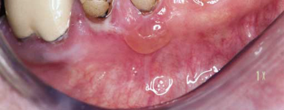
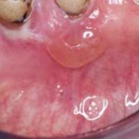
- Mucous membrane pemphigoid (figure 56)
- Gum involvement is common, and as with lichen planus the gums are bright red
- Compared to other conditions affecting the mouth, vesicles or small bullae may remain intact for some time. When erosions form they are slow to heal
- Lesions can be persistent and extensive in the buccal mucosa, especially the hard palate
- Refer to the related chapter Mucous membrane pemphigoid
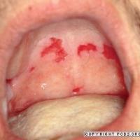
-
Pemphigus vulgaris (figure 57)
-
Mucosal lesions may precede cutaneous lesions by months, or may be the only manifestation of the condition
-
Bullae are rarely seen, instead, ill-defined, painful erosions mainly involving the lips, buccal mucosa and palate, which are slow to heal. Lesions on the border of the soft and hard palate are almost pathognomonic. There is significant whitening of the mucosa. Erosions can be seen on the gingival tips
-
The mucosa returns to normal when the condition is in remission
-
Refer to the related chapter Pemphigus vulgaris
-
Paraneoplastic pemphigus
-
Linear IgA disease
-
Mucosal involvement, which can precede cutaneous signs, is common with bullae and ulceration on the lips and inside the mouth in approximately 50% of cases
-
Lesions may spread to the pharynx
-
Refer to the notes in the section above on trunk / wider distribution
-
Epidermolysis bullosa and epidermolysis bullosa acquisita
-
Others - drugs reactions and lupus erythematosus
Conjunctival erosions / bullae / scarring
- Erythema multiforme
-
Stevens-Johnson syndrome / toxic epidermal necrolysis
-
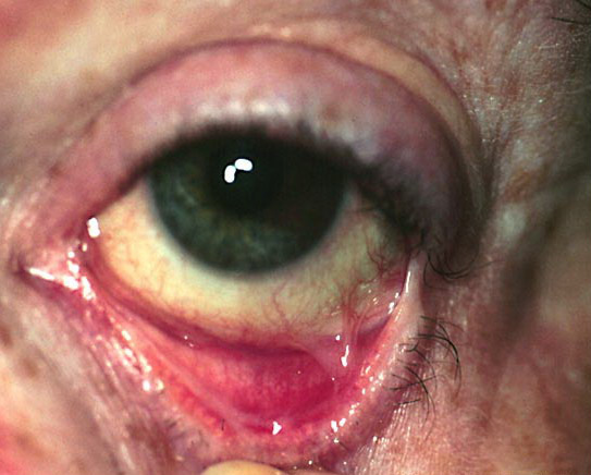
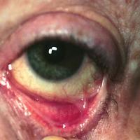
Mucous membrane pemphigoid (figure 58)
-
In some cases patients present with conjunctivitis and complain of grittiness or pain. This may come and go over a few years, before then progressing
-
On occasions painful erosions and bullae are the presenting feature, although the bullae may be difficult to visualise
-
Once established, lesions erode and heal to leave scar tissue, which impairs vision, or even causes blindness. Sometimes scarring can occur without any preceding symptoms
-
Refer to the related chapter Mucous membrane pemphigoid
Anogenital erosions / bullae / scarring
- Drug eruptions
- Refer to the notes below on Various sites
-
Lichen sclerosus
-
Causes white patches, scarring and architectural changes, bullae are rare
-
Refer to the chapter Lichen sclerosus
-
Erosive lichen planus
-
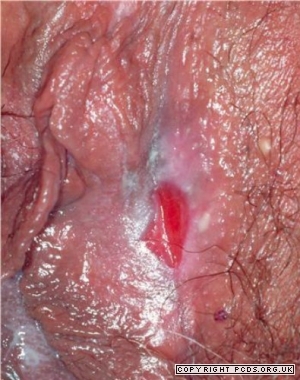
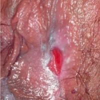
Mucous membrane pemphigoid (figure 59)
-
Painful blisters and erosions on the clitoris and labia can lead to architectural changes similar to those seen in lichen sclerosis
-
Penile involvement can lead to adhesions between the prepuce and glans penis
-
Linear IgA disease
-
In children, blisters have a predilection for the lower abdomen and anogenital regions
-
Lesions are often and annular / polycyclic lesions with blistering around the edge known as the 'string of pearls' sign
-
Refer to the notes in the section above on trunk / wider distribution
-
Pemphigus vulgaris and paraneoplastic pemphigus can affect the genitalia
-
Epidermolysis bullosa can also affect the genitalia
Various sites
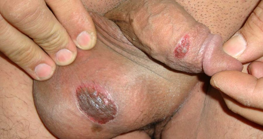
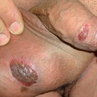
- Drug eruptions - fixed drug eruptions (figure 60)
-
Symptoms occur within 48 hours of commencing the medication
-
Clinical features
-
Distribution - lips, face, distal limbs, hands & feet, glans penis and perianal skin. Occurs at same site on re-exposure
-
Morphology - well-demarcated erythematous plaque that may blister
-
Tend to heal with post-inflammatory hyperpigmentation
-
Large numbers of drugs implicated. The most commonly associated drugs are tetracyclines, sulphonamides, NSAID and quinine
- Bullae can occasionally occur in other Adverse Cutaneous Drug Eruptions
-
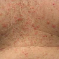
Grover's disease (figure 61)
-
An acute eruption of itchy, erythematous to red-brown papules or papulovesicles on the trunk, predominantly in middle-aged or elderly white patients
-
Refer to the chapter Grover's disease
-
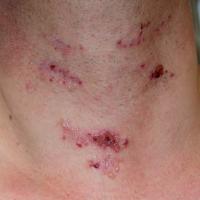
Dermatitis herpetiformis (figure 62)
-
Distribution - symmetrical involvement of the extensor surfaces of limbs (classically the knees and just below the elbows), scalp, scapula and natal cleft are characteristic
-
Morphology - small papules or vesicles, which are often grouped together on erythematous patches of skin. Bullae 1-2 cm in diameter are less common. The intense itch of dermatitis herpetiformis normally provokes vigorous scratching, which frequently destroys areas of vesicles / bullae and leaves behind erosions
-
Refer to the related chapter Dermatitis herpetiformis
-
Lichen planus - is associated with two bullous eruptions, both of which are rare
-
Bullous lichen planus is mainly seen on the legs. Bullae are tense and may be multilocular. Bullae formation may be due to the extensive liquefaction and vacuolation of the basal layer. Histopathologically, biopsy from the bullous lesion is characterised by a subepidermal bulla accompanied by classical changes of lichen planus. Direct and indirect immunofluorescence testing is negative
-
Lichen planus pemphigoides is characterised by bullae developing over lesions of lichen planus and also on normal appearing and erythematous skin, especially during an acute episode of lichen planus. The condition occurs in a much younger age group than bullous pemphigoid. It is commoner in men and usually occurs on the extremities, though dissemination can occur. As in bullous lichen planus, the blister is subepidermal but features of lichen planus are not evident and inflammatory cells are mainly neutrophils and lymphocytes with a few eosinophils. Direct immunofluorescence testing of perilesional skin shows linear deposits of C3 and IgG along the basement membrane zone. Circulating antibodies have been demonstrated against 130, 200 and 180 kda antigens. Healing may leave behind post-inflammatory hyperpigmentation. Lichen planus pemphigoides can also be associated with various drugs such as angiotensin-converting enzyme inhibitors, cinnarizine, simvastatin, or following psoralen combined with ultraviolet A therapy (PUVA)
-
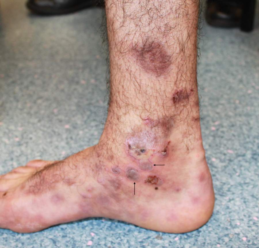
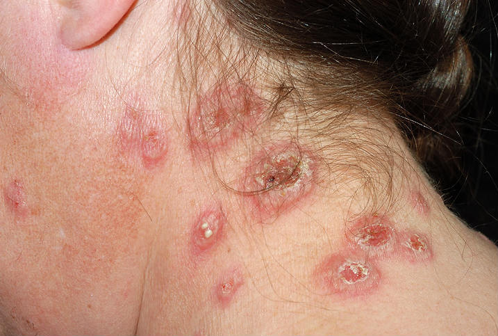
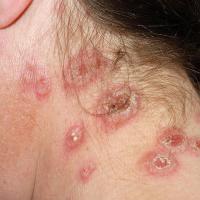
Sweet's syndrome (figure 63) and other neutophilic dermatoses
-
Sweet's syndrome
-
Is characterised by a rash, most commonly arising in middle-aged women, and systemic features of fever, malaise, arthralgia and sore eyes
-
The lesions of Sweet's syndrome may be few in number or numerous. They are typically characteristically tender and may be extremely painful, persisting from days to weeks. The limbs and neck are the most commonly affected sites, but other areas of skin and mucosa may be involved
-
Morphologically the lesions can be vesicles, bullae, plaques, nodules, and erosions / ulcers
-
Other neutophilic dermatoses, eg pyoderma gangrenosum, can also occasionally cause blistering
- Refer to the chapter Neutrophilic dermatoses
-
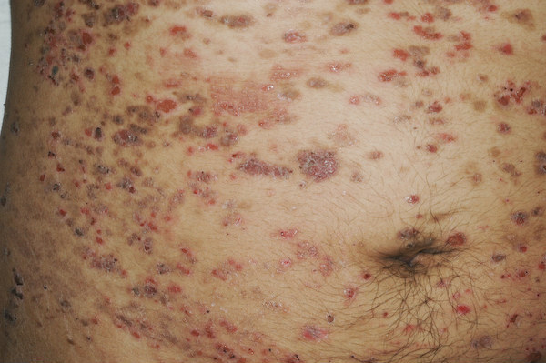
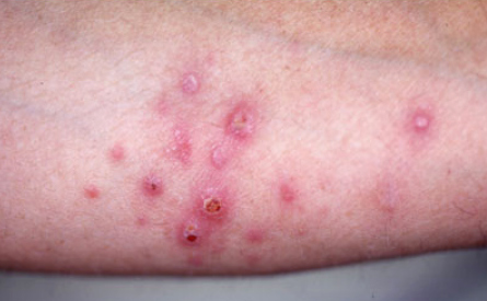
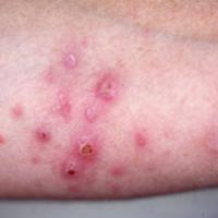
Lymphomatoid papulosis (figure 64)
-
Is characterised by recurrent crops of pruritic papules at different stages of development that predominantly arise on the trunk and limbs
-
Each erythematous papule evolves into a red-brown, often haemorrhagic, papulovesicular or papulopustular lesion over days to weeks. Some lesions develop a necrotic eschar before healing spontaneously over 1-2 months. Lesions usually leave slightly depressed oval scars
-
The skin distribution of lesions, characteristically, is on the trunk and extremities, although the palms and / or soles, face, scalp, oral mucosa, and anogenital area also may be involved
-
Refer to the chapter on Lymphomatoid papulosis
-
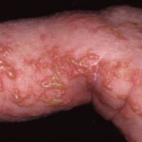
Incontinentia pigmenti (figure 65)
-
Incontinentia pigmenti is characterised by skin abnormalities that evolve throughout childhood and young adulthood. Many affected infants have a blistering rash at birth and in early infancy. Abnormalities of the teeth, nails and hair are very common.
-
There are four main stages
-
Stage 1: vesicular - characterised by the development of red papules and vesicles on an erythematous base that follow Blaschko's lines. Lesions are seen predominantly on the extremities but may also occur on the trunk or on the head and neck. The vesicular stage has been reported to occur in 90-95% of patients. In most patients lesions are present at birth or develop within the first 2 weeks of life
-
Stage 2: verrucous - the blisters heal and develop in to wart-like skin growths
-
Stage 3: hyperpigmented - in early childhood the skin develops grey or brown patches with a swirled pattern
-
Stage 4: atrophic / hypopigmented - the hyperpigmented patches fade with time, and adults with incontinentia pigmenti usually have lines of unusually light-coloured skin on their arms and legs
Clinical Images
Please refer to notes on image rights at bottom of the page with regards to individual image
ownership.
1
2
3
4
5
6
7
8
9
10
11
12
13
14
15
16
17
18
19
20
21
22
23
24
25
26
27
28
29
30
31
32
33
34
35
36
37
38
39
40
41
42
43
44
45
46
47
48
49
50
51
52
53
54
55
56
57
58
59
60
61
62
63
64
65
Disclaimer - the author PCDS cannot accept responsibility for any misleading or incorrect statements, and the management of individual patients remains the direct responsibility of the individual doctor. We do however hope that visitors to this site can contact us regarding comments that are considered misleading or incorrect so that we can continue to improve the site.
Image Rights - The PCDS would like to thank Dermatoweb, DermQuest (Galderma), and others who have contributed images. All named individuals and organisations maintain copyright for the relevant images.