Lichen sclerosus - female
LAST UPDATED: Mar 16, 2025
Introduction
Lichen sclerosus (LS) is an uncommon condition characterised by white sclerotic macules and patches, commonly affecting the genital and perianal skin, although it can affect non-genital skin separately or together. LS can cause permanent scarring of the genitalia, and in approximately 5% of cases is associated with squamous cell carcinoma, mainly of the female genitalia.
This chapter is set out as follows:
Aetiology
-
The suspected prevalence varies between 0.1% and 3% for children and post-menopausal women respectively, and 0.07% for males
-
The exact aetiology is unknown
-
An autoimmune cause has been suggested as LS is commonly found alongside other autoimmune conditions
- LS can develop after an injury to the affected area (the Koebner phenomenon)
History
-
LS is six times more common in women than in men
-
Although LS is most often seen in women around and after the time of the menopause, it can affect any age group - up to 15% of cases are in children with the majority affecting the vulva (a small percentage of boys present with phimosis)
-
Non-genital lesions are often asymptomatic, whereas genital lesions are often itchy or sore, sometimes severely so
Clinical findings
Extra-genital lichen sclerosus
-
LS can affect non-genital skin with or without genital involvement
-
Lesions are generally asymptomatic
-
Distribution
-
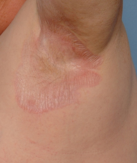
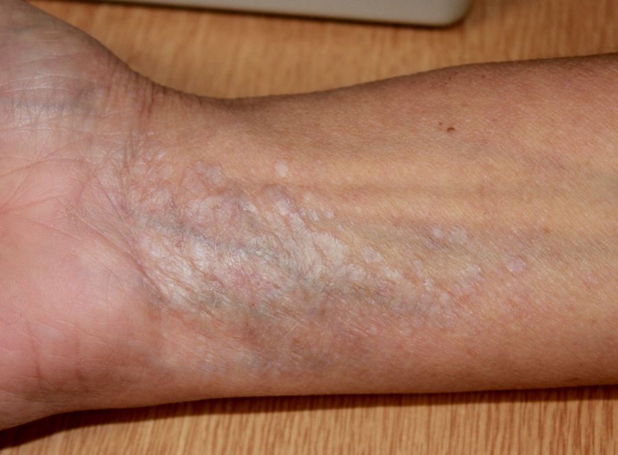
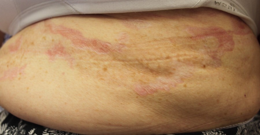
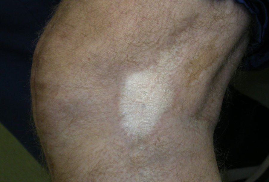
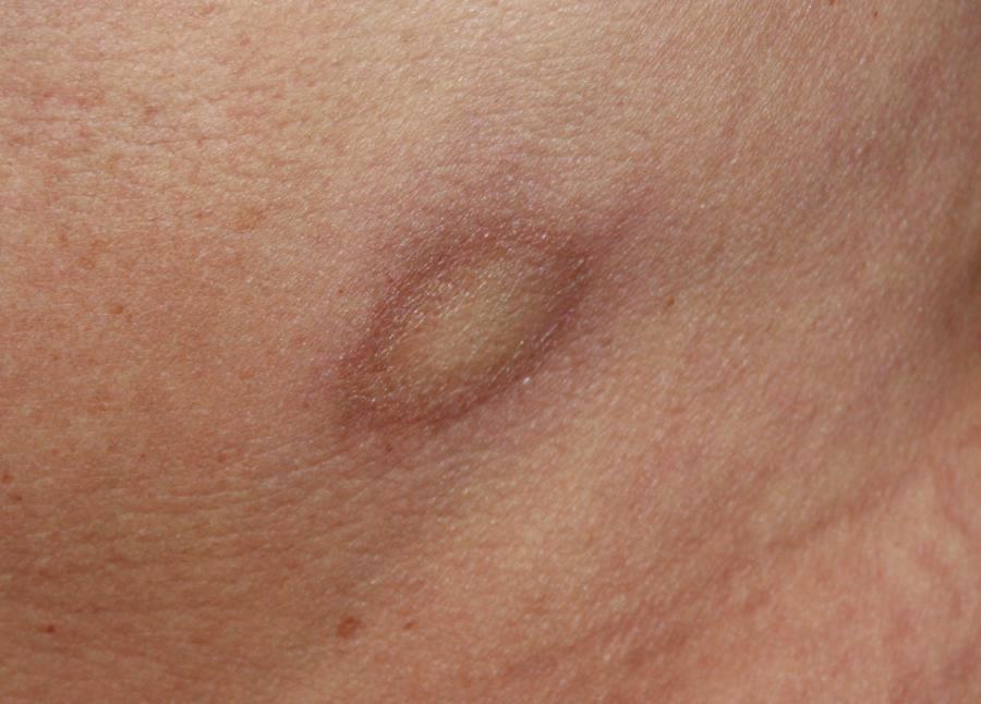
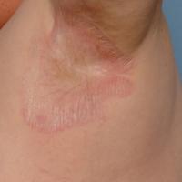
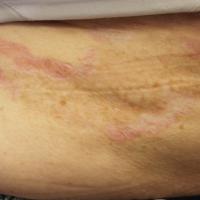
A few to many lesions most commonly affecting the upper trunk and neck, the axillae, around the umbilicus, the wrists and other flexural sites
-
Morphology
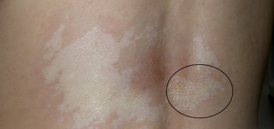
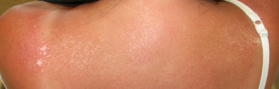
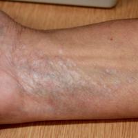
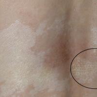
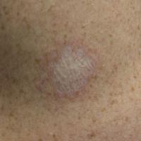
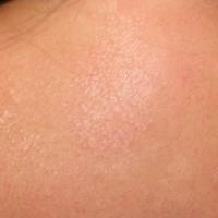
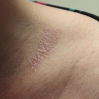
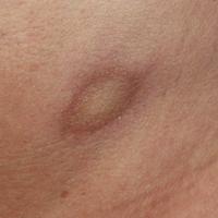
- Typical lesions are ivory or porcelain-white, shiny, polygonal macules or flat-topped papules
-
Lesions may be pitted
-
Many of the lesions coalesce to form larger patches-plaques
-
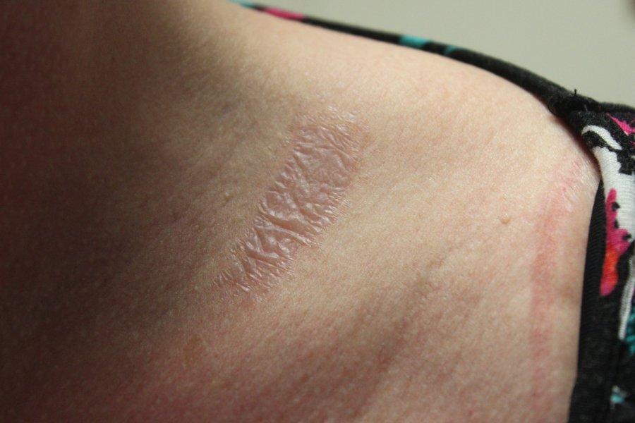
Later on the lesions develop a characteristic wrinkled appearance
-
Other features occasionally seen - thickened with a warty appearance, lichenification, bullae
-
Non-genital LS is not associated with SCC
-
The main differential diagnosis is Morphoea
Anogenital lesions in women (and children)
-
Patients usually complain or soreness and dyspareunia, or itch, all of which can be very distressing
-
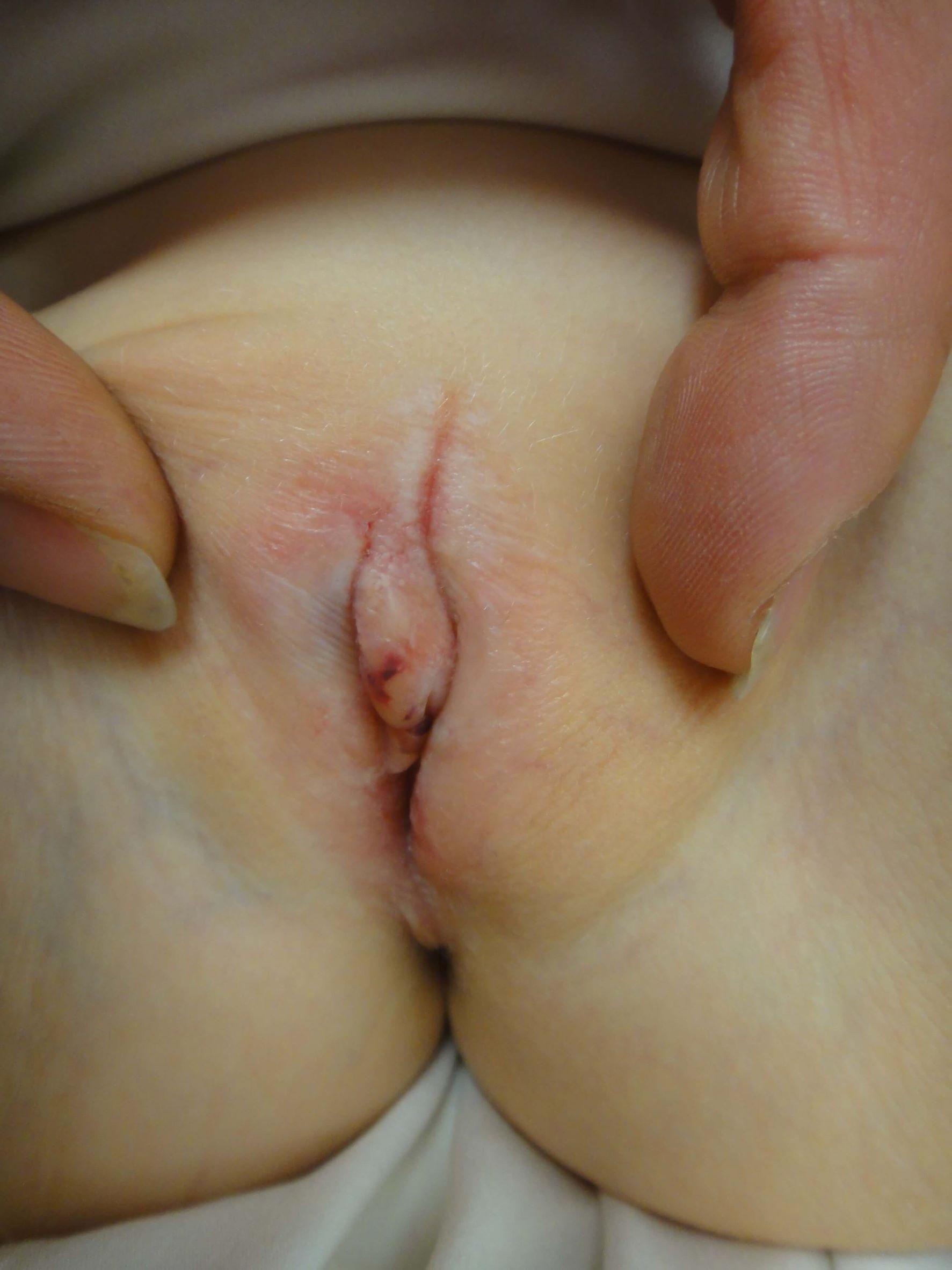
In children the majority of cases are pre-pubertal and patients are often asymptomatic, although some patients will complain of itch, discharge, dysuria, or constipation
- Early signs can include pallor and oedema
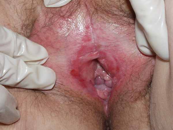
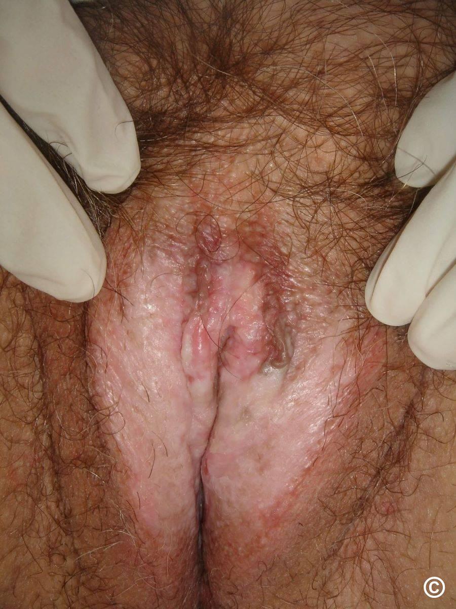
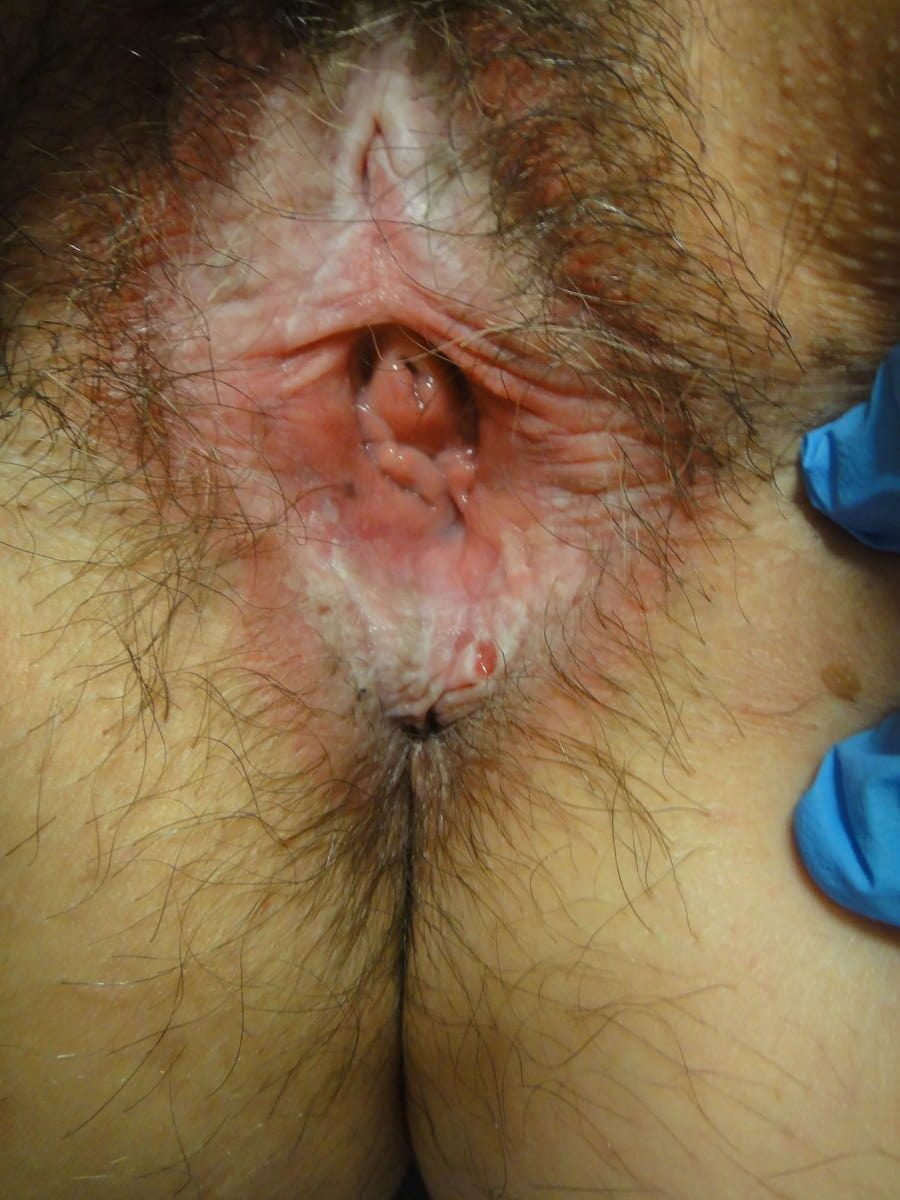
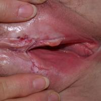
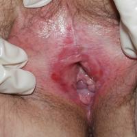
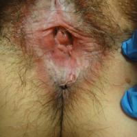
- Lesions may be localised or much more extensive affecting the vulva and surrounding skin, the perineum, and around the anus. If the affected areas extend around the vulval and perianal skin a figure-of-eight pattern can be seen
-
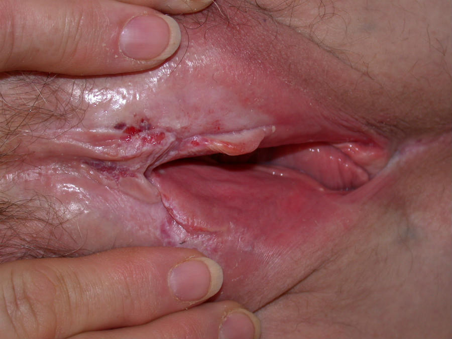
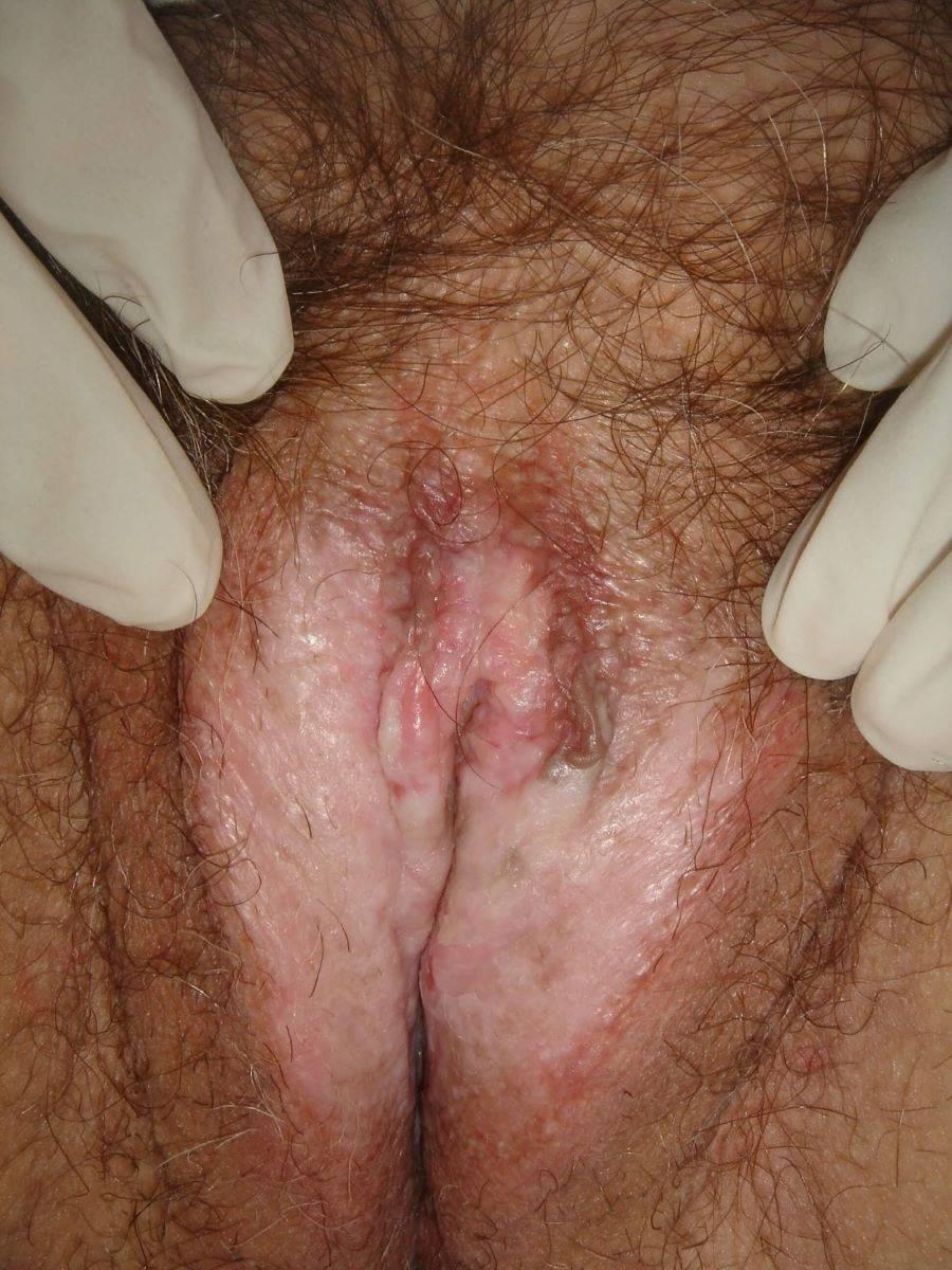
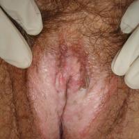
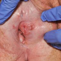
The affected skin is ivory-white, and may contain areas of telangiectases and purpura
-
As a result of friction the skin often breaks down to form a red raw surface
-
Other features occasionally seen include hyperkeratosis, vesicles, bullae, and ulcers
-
Atrophy is a common feature and sometimes there may be marked shrinkage of the vulva, especially the clitoris and labia minora, and the vaginal introitus. Lichen sclerosus never affects inside the vagina
-
Significant perianal involvement may cause discomfort, bleeding, and constipation, particularly in children
-
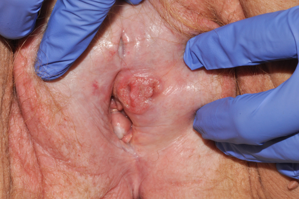
LS is associated with pre-malignant change and squamous cell carcinoma (SCC), which arises in 5% of patients. SCC presents mainly as a nodule, tending to arise on sclerotic tissue, especially the anterior vulva. It can also present as a thickening or an ulcer. Any patient with lichen sclerosis is at risk, regardless of whether their symptoms are well-controlled or are more recalcitrant to treatment. The risk of SCC is increased if the patient smokes
-
The main differential diagnoses of LS in women are:
Clinical Images
Please refer to notes on image rights at bottom of the page with regards to individual image
ownership.
1
2
3
4
5
6
7

8
9
10
11

12
13
14
15
Investigations
-
In many cases the diagnosis is clinical, however, if a biopsy is needed the characteristic feature is a band of hyalinization of the dermal collagen below the epidermis
Management
Extra-genital lichen sclerosus
-
Topical treatments are generally much less effective than when used to treat genital LS. A number of treatments have been used including Dermovate ® cream/ointment (Clobetasol proprionate), calcipotriol cream (Dovonex ®), and tacrolimus (Protopic ®) 0.1% ointment
-
Various forms of phototherapy including narrowband UVB, psoralen-UVA (PUVA) and UVA1 have been reported as successful in some case studies
-
It is important to see if there is co-existing genital involvement
Genital lichen sclerosus
General measures
- Most adults with uncomplicated lichen sclerosus can be managed in Primary Care, but if the diagnosis is in doubt the patient should be referred to Secondary Care. Children should be referred to Secondary Care from the onset
-
Patients should wash gently in a shower / bath using an emollient, and try to avoid rubbing and scratching. Some patients find it helpful to apply an emollient cream several times a day to relieve dryness or itching
Medical treatment
- The mainstay of treatment is that of Dermovate ® cream / ointment (Clobetasol proprionate)
- Adults - initially prescribe 30 g to use once a day for month 1, alternate days for month 2, twice weekly for month 3, and then review. Children - OD for three months
- Maintenance treatment is then required once to twice a week, and more often for a flare - if a patient is using more than 30 g over a 6 month period then a review is needed
- Such a regime not only improves symptoms in the majority of patients, but also stops scarring
-
Other treatments sometimes used include the topical calcineurin inhibitors - tacrolimus 0.1% ointment (Protopic ®) and pimecrolimus cream (Elidel ®)
- Long-term follow-up in Secondary Care is appropriate for patients with genital LS associated with troublesome symptoms, localised skin thickening (associated with a higher risk of malignant change), or VIN
-
Stable disease
-
Should be reviewed annually except in well-counselled patients who control their symptoms well
-
If review is by the General Practitioner this should be communicated to the patient and GP by the clinic
-
Written instruction should be given to the patient at the time of their discharge from the clinic warning them that any persistent area of well-defined erythema, bleeding/ulceration, or skin thickening/lump must be reported to their family practitioner straight away, who will then make an urgent referral back to an appropriate specialist, as such an occurrence could represent malignant transformation
Other resources
Patients
Healthcare professionals
Disclaimer - the author PCDS cannot accept responsibility for any misleading or incorrect statements, and the management of individual patients remains the direct responsibility of the individual doctor. We do however hope that visitors to this site can contact us regarding comments that are considered misleading or incorrect so that we can continue to improve the site.
Image Rights - The PCDS would like to thank Dermatoweb, DermQuest (Galderma), and others who have contributed images. All named individuals and organisations maintain copyright for the relevant images.