Actinic keratosis (syn. solar keratosis)

LAST UPDATED: Jun 05, 2026

Patient Information Leaflet
Link: Actinic (solar) keratosis

https://www.pcds.org.uk/patient-info-leaflets/actinic-solar-keratosis
Introduction

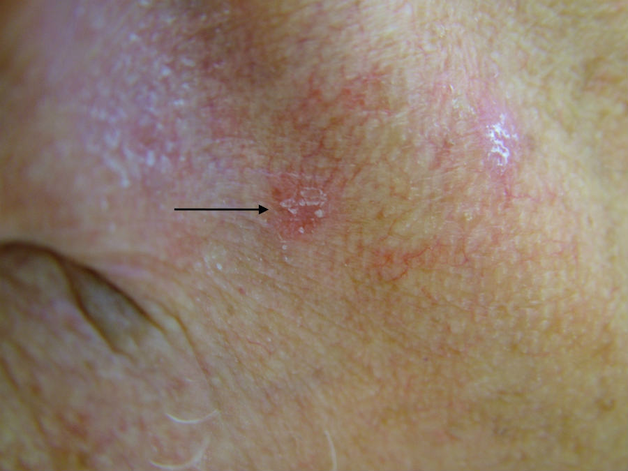
An actinic keratosis (AK) is a common sun-induced scaly or hyperkeratotic lesion, which has the potential to become malignant. NICE estimates that over 23% of the UK population aged 60 and above have AK. Although the risk of an AK transforming into a squamous cell carcinoma (SCC) is very low, this risk increases over time and with larger numbers of lesions. The presence of ten AK is associated with a 14% risk of developing an SCC within five years.

This chapter is set out as follows:


Aetiology

  • AK are a consequence of cumulative long-term sun-exposure:
    • Lesions are very uncommon under the age of 45 years
    • The incidence increases with age
    • The exceptions are patients with xeroderma pigmentosum and albinism who can develop AK at a very young age
  • Genetic factors play a role and individuals with fair skin, blue eyes and blonde hair are at higher risk, whereas lesions are exceedingly rare in patients of skin types IV-VI
  • Artificial UV radiation such as UVB and PUVA, used to treat psoriasis and a number of other skin conditions, as well as the use of sun beds, increase the risk
  • Men are more affected than women

History

  • Lesions are normally asymptomatic
  • Recent growth, pain / tenderness, bleeding or ulceration are suggestive of transformation into an SCC

Clinical findings

Distribution  

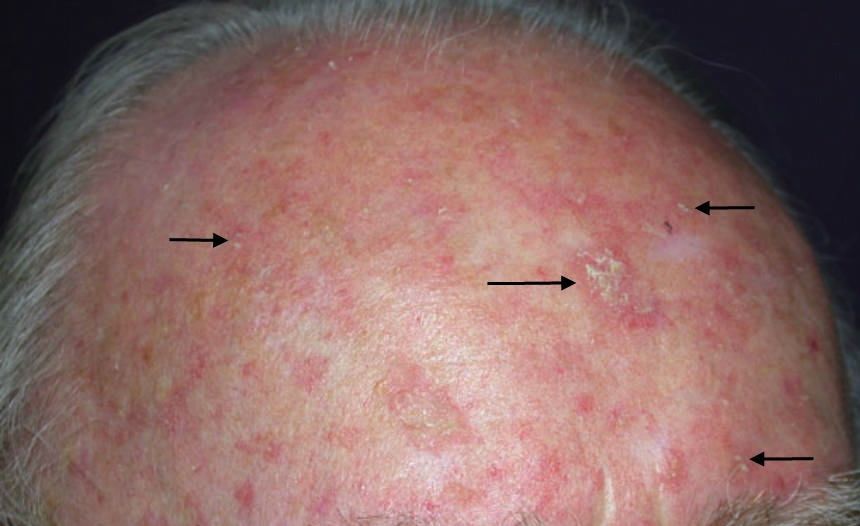
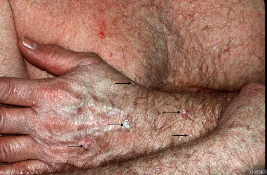
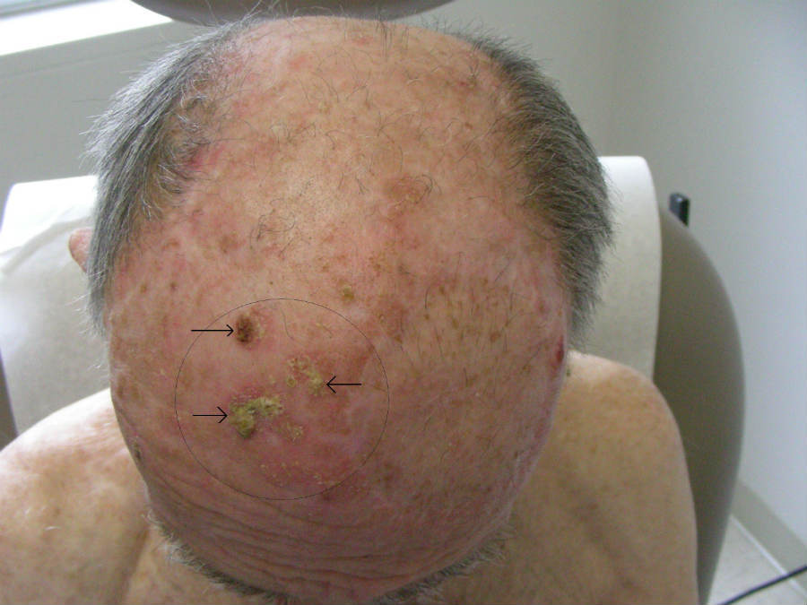
  • Reflects the intensity of sun-exposure with the greatest number of lesions occurring on the head, neck, forearms and hands
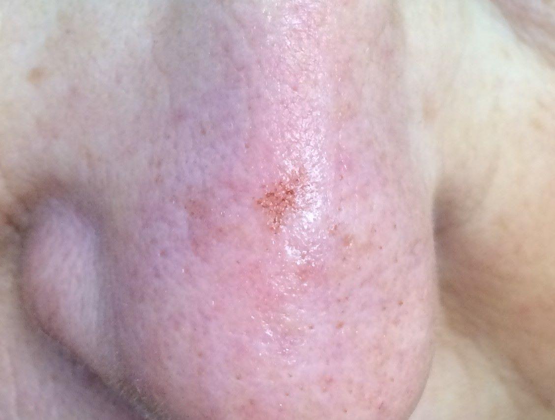
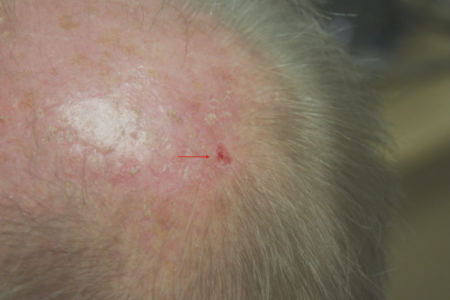
  • There is often a background of significant sun-damaged skin with pigment irregularity, telangiectasia, erythema and collagenosis (a yellow papularity of the skin)

Morphology

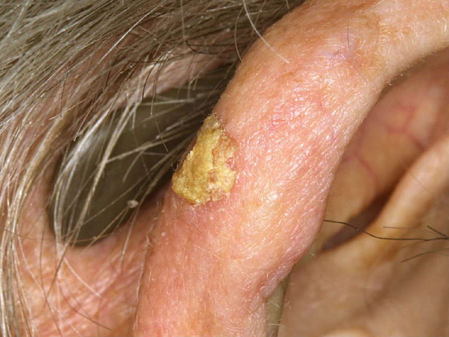
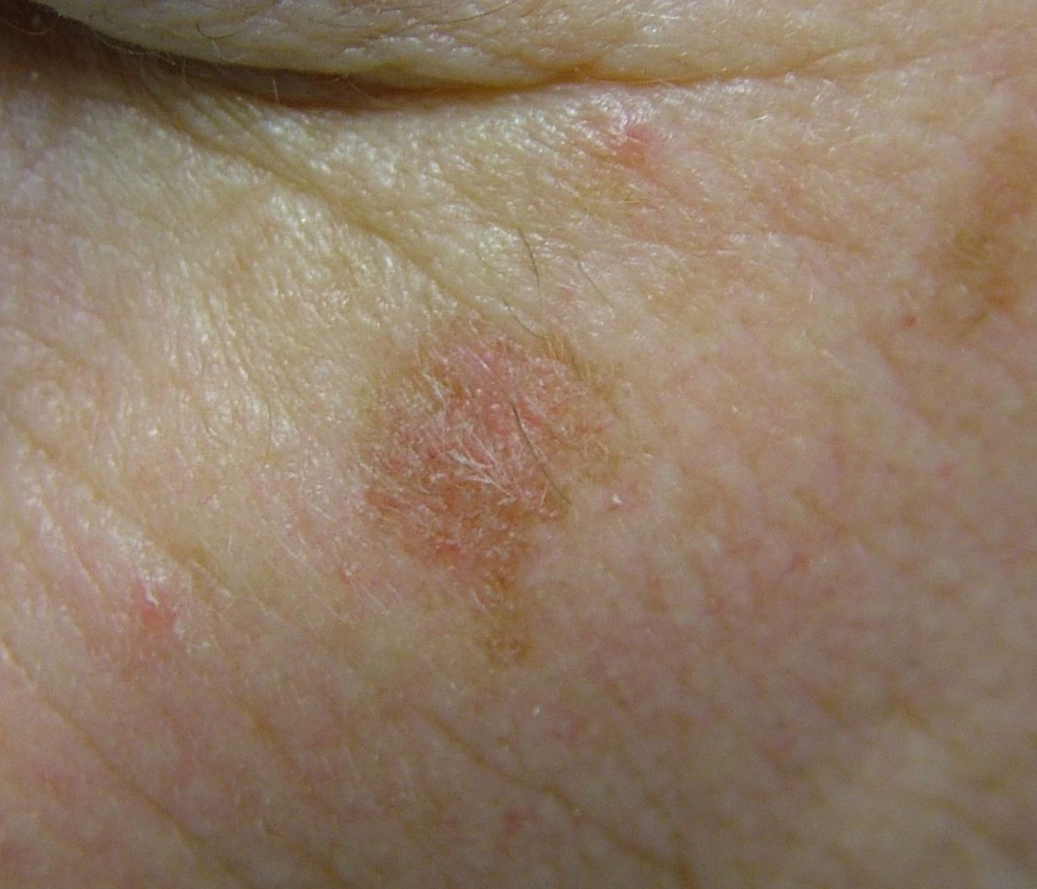
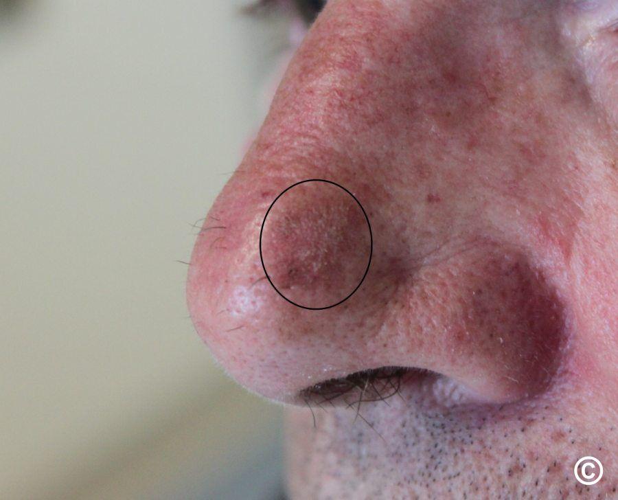
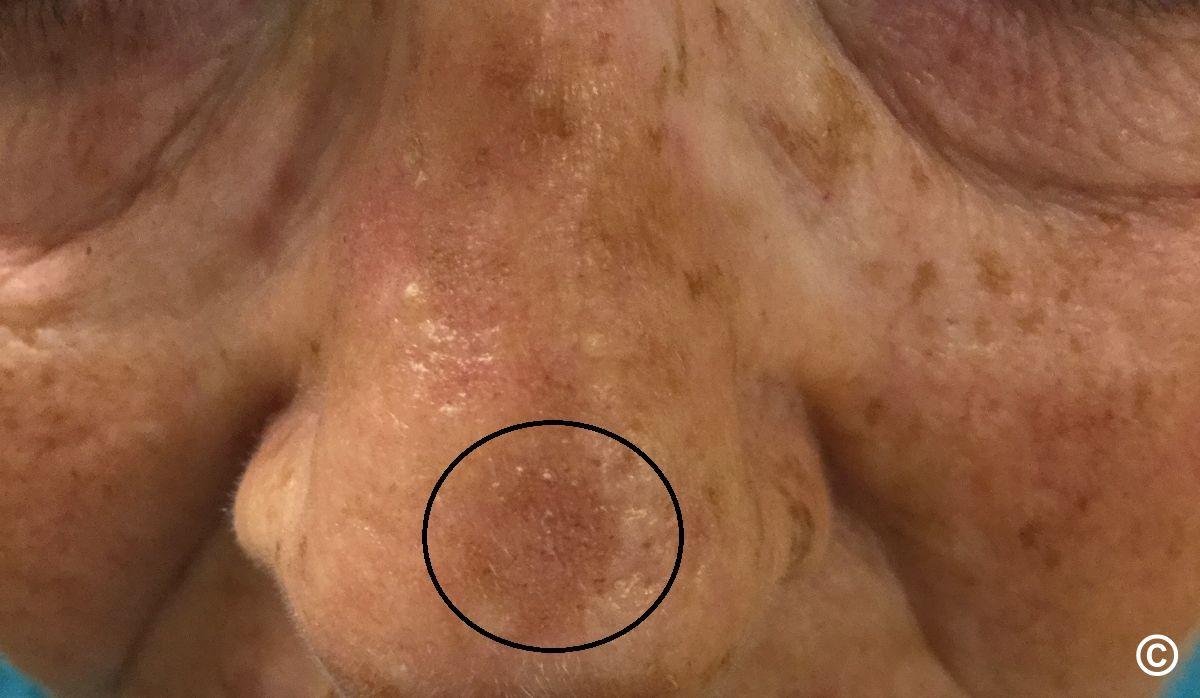
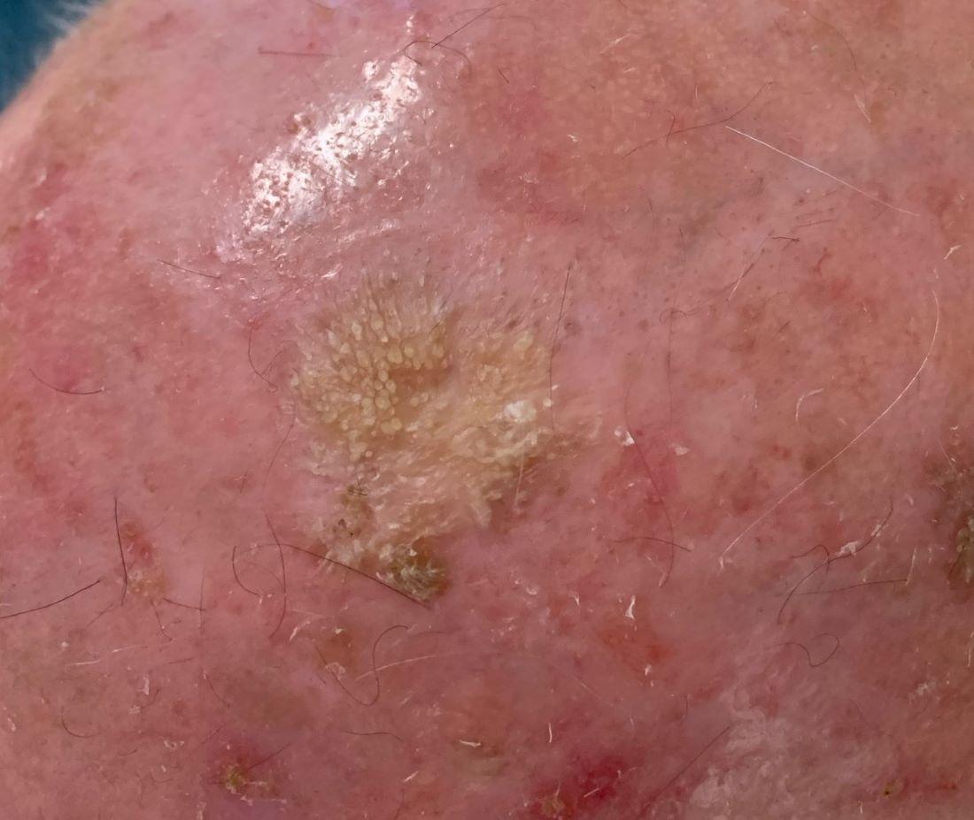
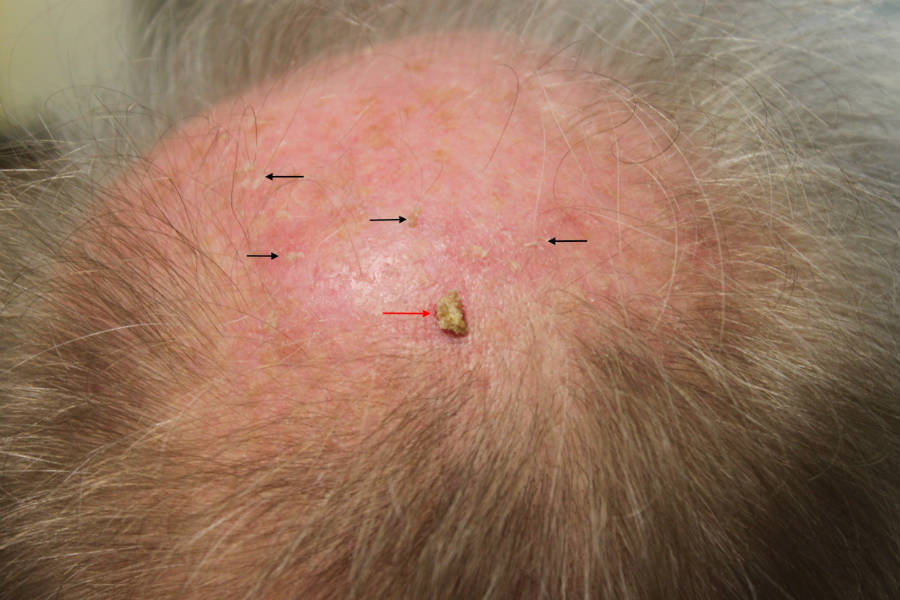
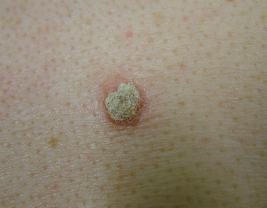
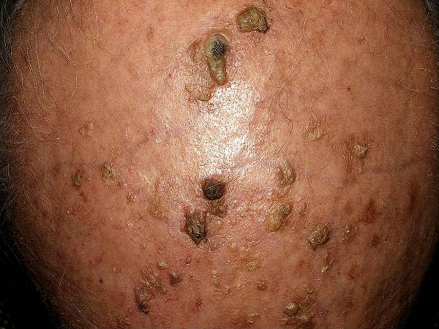
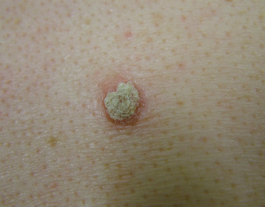
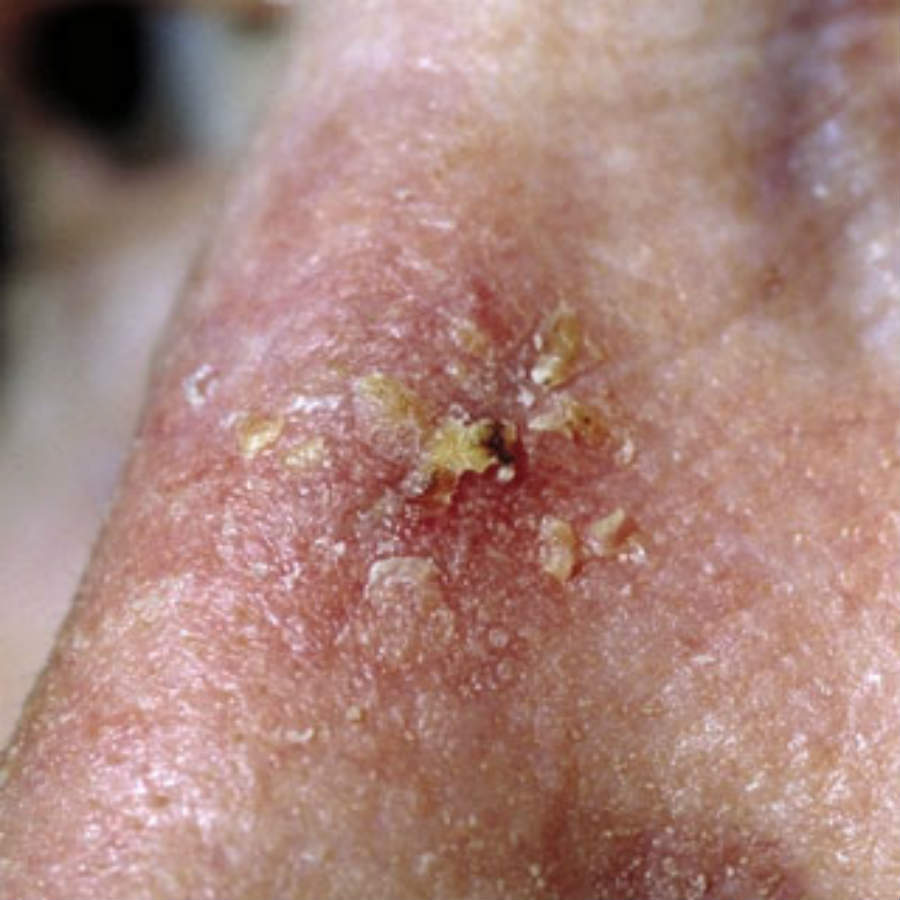
  • Lesions usually take on a similar appearance and seldom exceed more than 1 cm in diameter
  • Rough surface scale - usually white, although in patients with skin type I AK are often more easily felt than seen
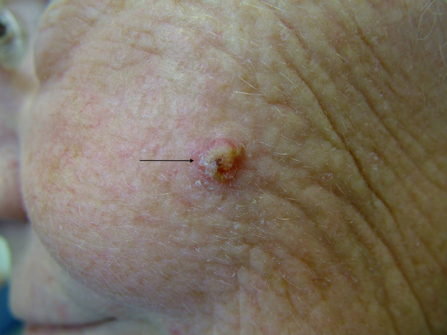
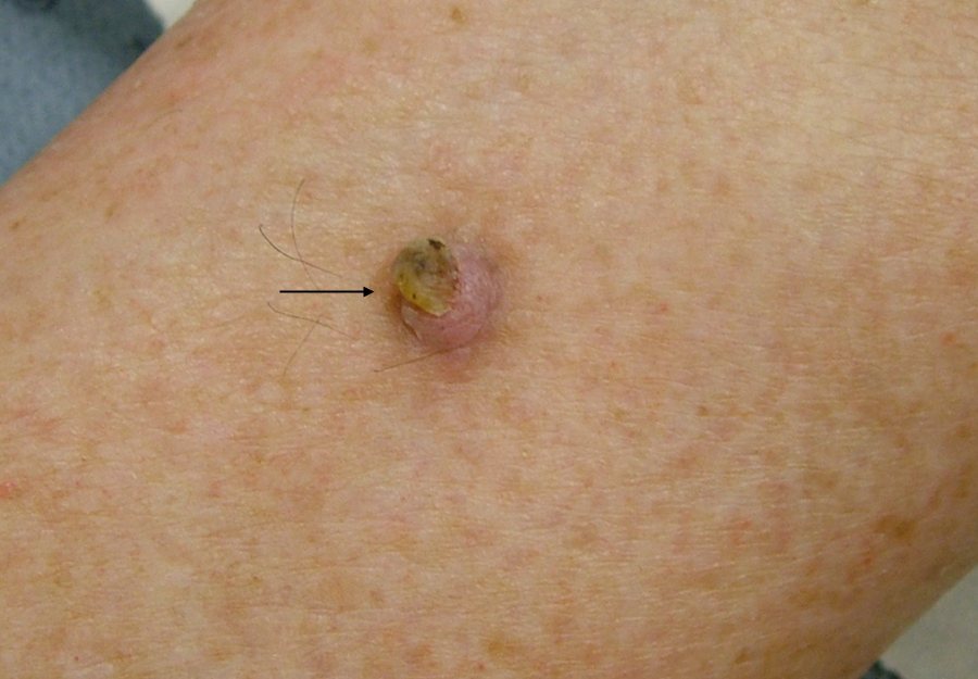
  • Often referred to as flat, although some lesions can have significant amounts of scale (hypertrophic or Bowenoid AK), which may be elevated

Dermoscopic features 

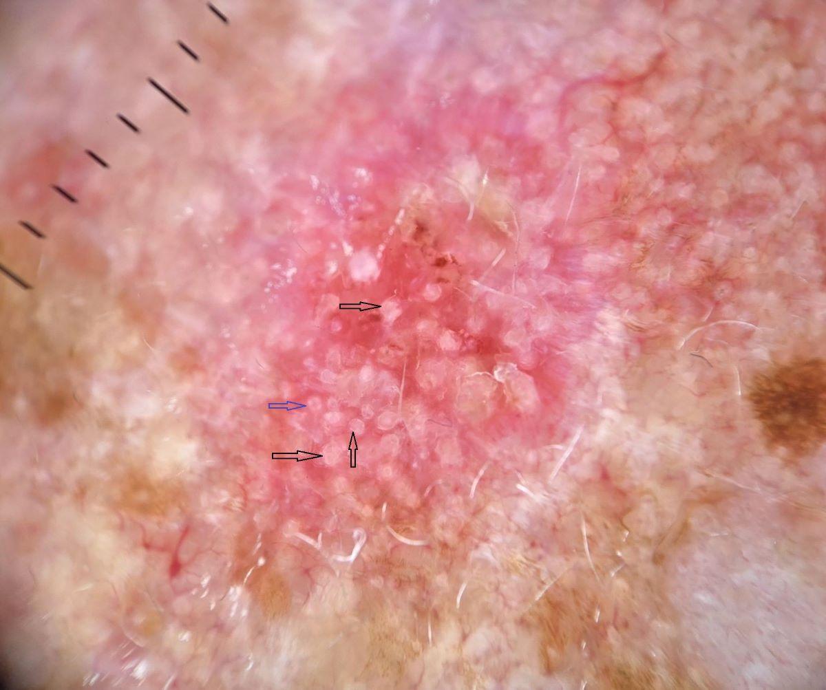
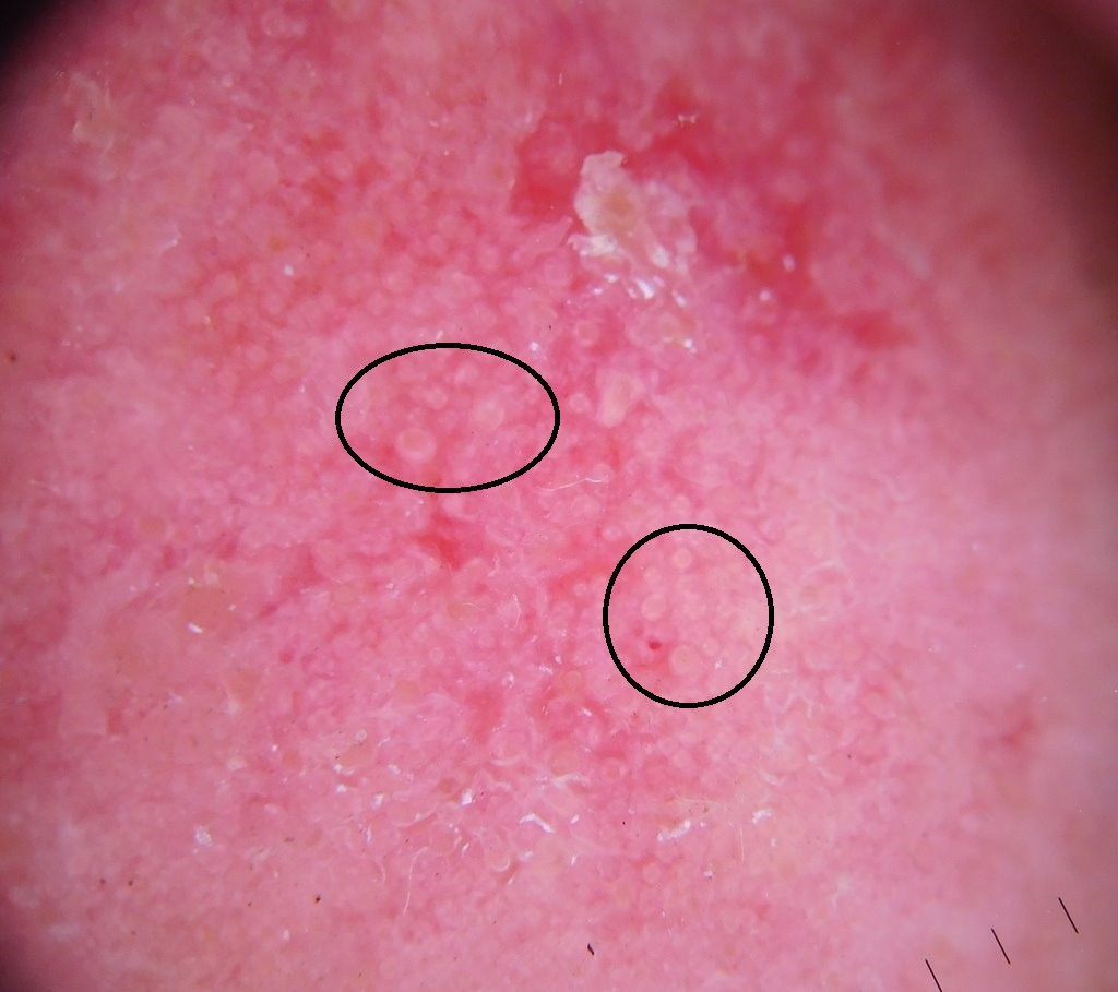
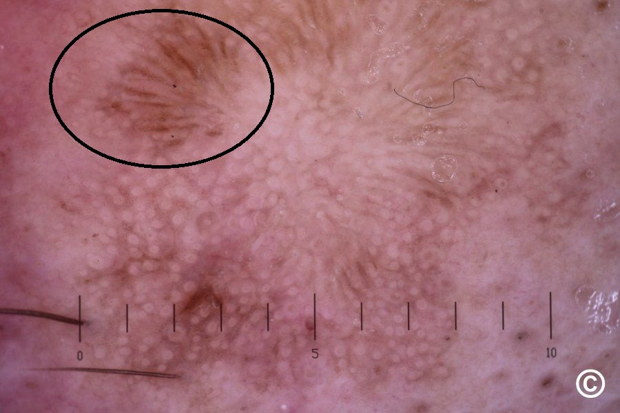
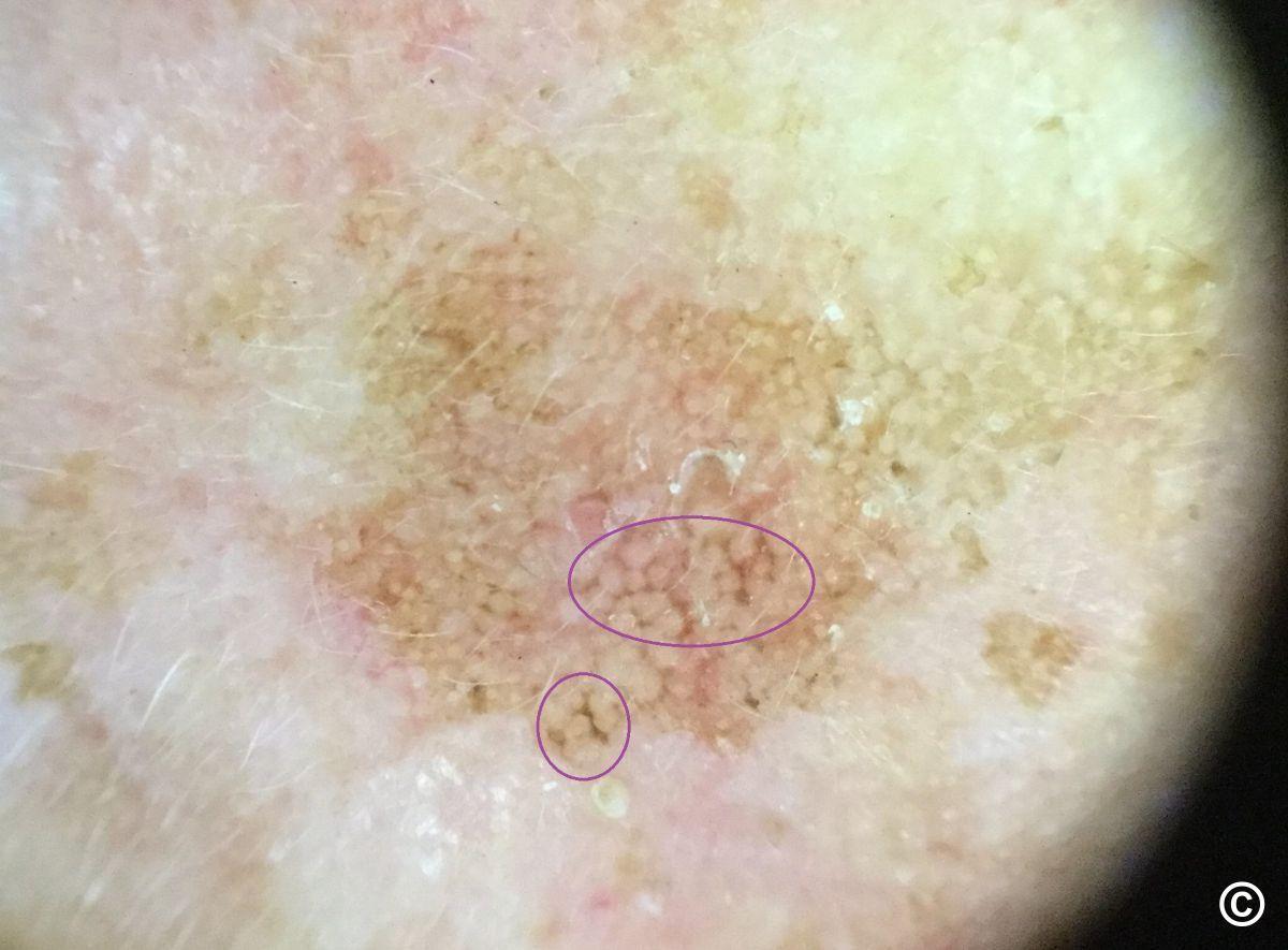
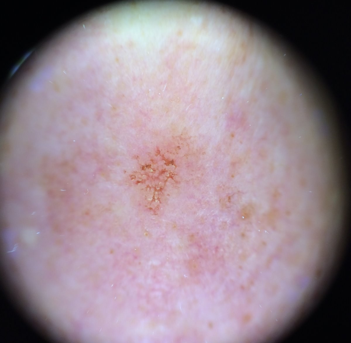
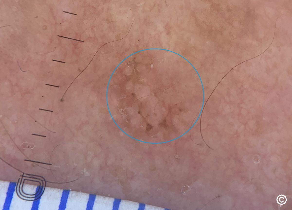
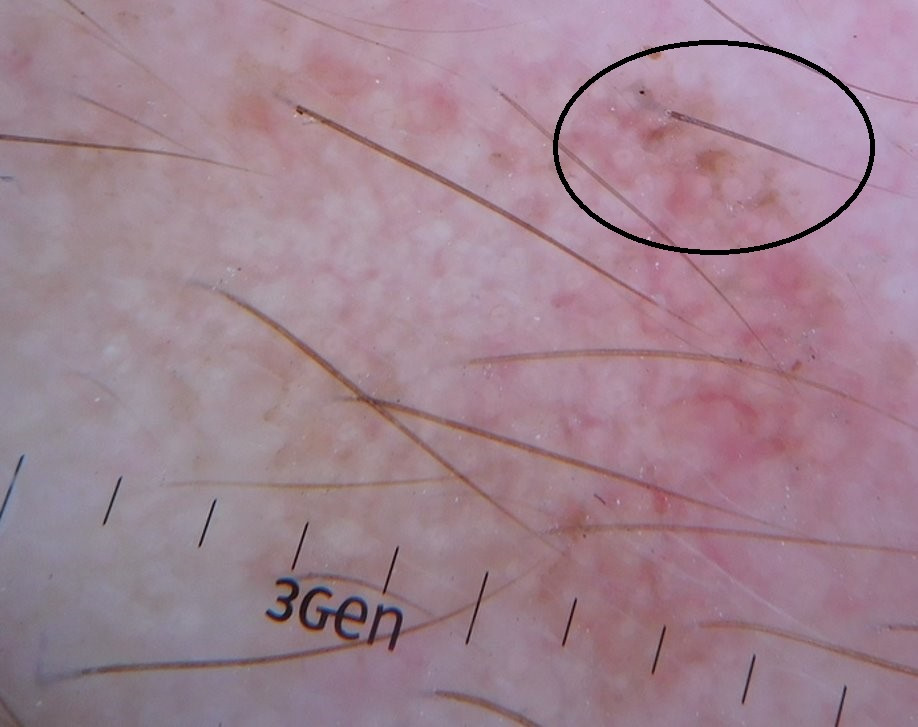
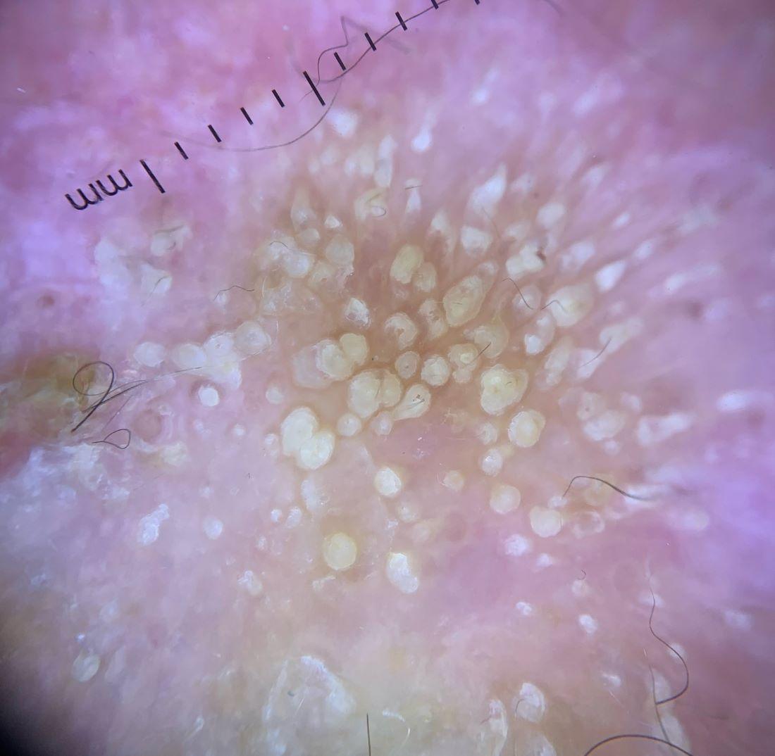
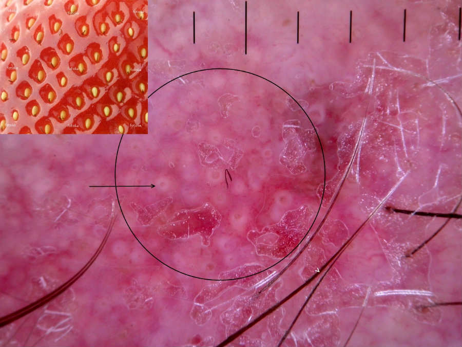
  • White-yellow scale
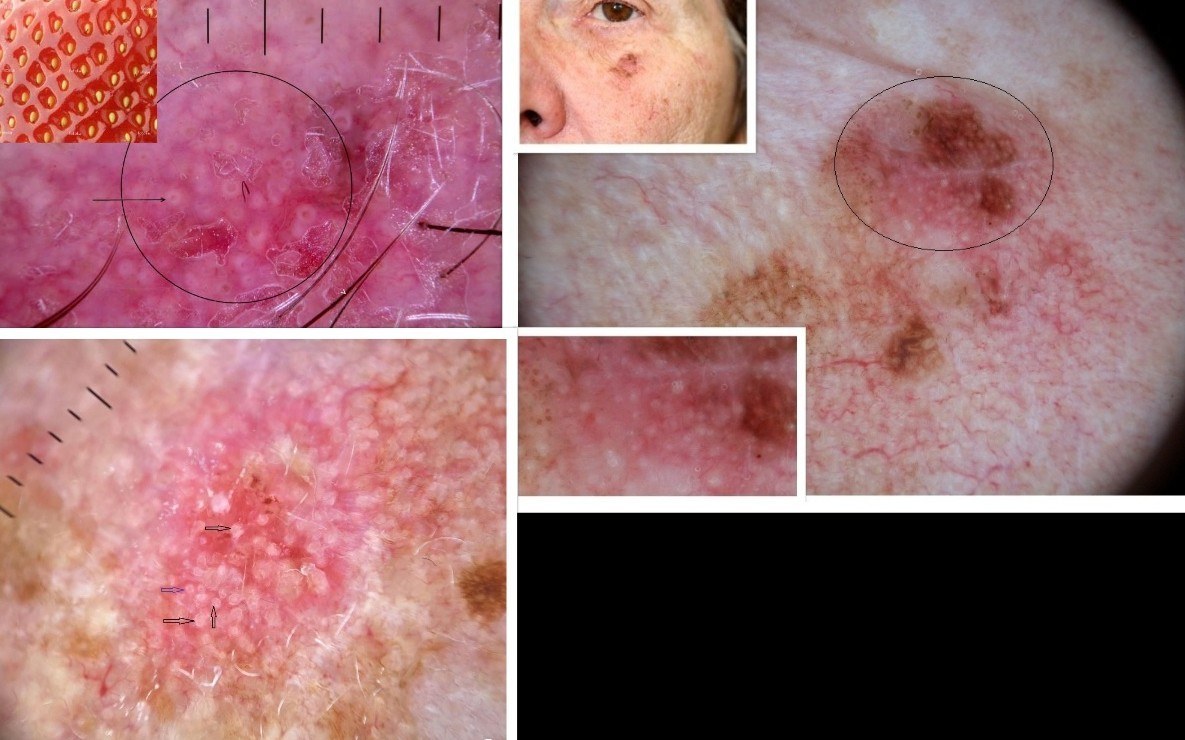
  • A red pseudonetwork giving a strawberry-like appearance
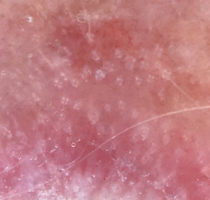
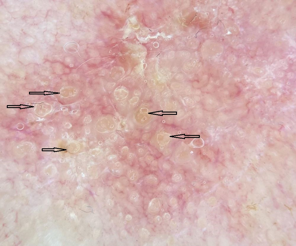
  • Enlarged hair follicles sometimes showing up as white circles with a ring of keratin at the periphery
  • The use of polarised light can make white circles look like rosettes comprised of four grouped dots/globules (clods)
  • The term 'follicular keratosis' is sometimes used to describe lesions with extensive involvement of the hair follicles, such lesions are often resistant to topical therapy
  • Features of pigmented actinic keratoses can also include grey confluent dots arranged in lines or grey to brown linear structures located between follicles, sometimes with small brown triangular structures. One of the differentials of a pigment AK is lentigo maligna
  • Structures may not be visible if there is a lot of scale 

Dermoscopic erythema in facial lesions 

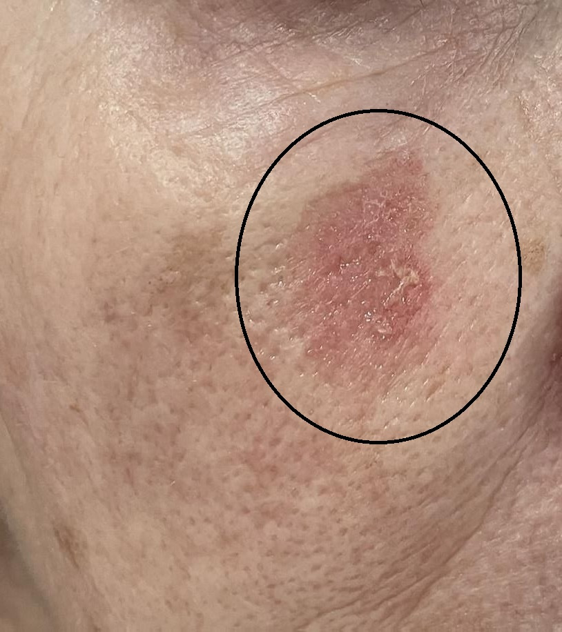
  • Erythema is a common feature of actinic keratoses (strawberry-like pattern)
  • A pink-to-red hue or erythematous pseudo-network can also be found in some cases of lentigo maligna and melanoma. In facial lesions with erythema, melanoma should be considered a differential when:
    • The lesion lacks features of an actinic keratosis such as surface roughness and/or enlarged hair follicles
    • Additional features of lentigo maligna / melanoma such as an annular granular pattern, angulated structures, or atypical vessels are present

AK variants

  • Actinic cheilitis - these are actinic keratoses affecting the lips
  • Lichenoid AK - smooth and shiny, mainly occurring in areas of friction 

Clinical Images

Please refer to notes on image rights at bottom of the page with regards to individual image ownership.
Investigations

  • Diagnosis is usually clinical. If a diagnostic biopsy is required the primary histologic feature is partial thickness atypia / dysplasia of the keratinocytes in the basal layers of the epidermis. This is often accompanied by parakeratosis, thinning of the granular layer, buds of atypical epidermis extending toward the papillary dermis, dermal solar elastosis, and inflammation. If squamous cell carcinoma is a differential the patient must be referred urgently to Secondary Care without a biopsy

Management

Who should manage AK?

The majority can be managed in general practice. The following groups should be referred: 

  • To a GPwER / accredited GPwSI, or a dermatologist
    • Diagnostic uncertainty
    • Patients with more widespread / severe actinic damage
       
  • If an SCC is suspected refer to Secondary Care as urgent (2-week wait). The following could suggest transformation from an AK into an SCC:
    • History - recent growth / pain / bleeding
    • Examination - an elevated lesion (if significant surface scale, remove to see if there is a palpable lump underneath), ulceration, induration, tenderness, surrounding inflammation
    • Beware lesions on lips - SCC can be very subtle at this site
    • Have a low threshold for referring immunosuppressed patients (in particular post-transplant) who are at high risk of developing SCC that tend to metastasise quicker
    • Very young patients presenting with AK - consider xeroderma pigmentosum 

Treatment of AK

For a simplified management plan please refer to the PCDS AK Treatment Pathway. The Karger group have also produced a Fast Facts learning course on actinic keratoses.

Step 1: general measures - appropriate for all patients
  • AK are a marker of sun damage and so a thorough skin examination is needed to look for more serious sun-related skin tumours such as melanoma
  • Moisturisers - it can sometimes be difficult to differentiate between early AK and dry scaly areas of normal skin. The use of a moisturiser two to three times a day can be helpful in differentiating between areas of normal and abnormal skin
  • Refer to the top right of the page for a patient information leaflet available through a QR code or printable PDF  - this provides the patient with all necessary information including a place for the health professional to provide treatment notes, advice on UV-protection and vitamin D, self-management of actinic keratoses, and links regarding self-examination 
Step 2: observation
  • Not all patients need treating eg patients with smaller numbers of lesions, especially if they have a reduced life expectancy - such patients should be given a choice or whether or not they wish to have their lesions treated 
Step 3: lesion specific treatment - a few lesions or larger numbers that are widely distributed (eg dotted around the scalp, face, hands); please view images at bottom of page 

Treat the individual lesions and not the normal surrounding skin. Options are:

  • 5-fluorouracil cream (5-FU) - 5% 5-FU cream or 4% 5-FU cream (Tolak®) applied once a day for four weeks. Wash hands thoroughly after application. Leave treated areas uncovered and wash off after 8 hours. Patients should be advised to expect redness, crusting, and some discomfort during treatment. It can take 6-8 weeks for the skin to fully settle. Please provide a patient information leaflet
     
  • Klisyri ® cream (tirbanibulin) - apply once a day for 5 days. Patients should be advised to expect redness, crusting, and some discomfort during treatment, which usually settles within 10 days of completing the treatment 
     
  • Actikerall ® solution combines 5% 5-FU with salicylic acid and as such it is also suitable for treating moderately thick (hyperkeratotic) AK - it should be used once a day for 6-12 weeks. Actikerall tends to leave a film on the skin, which should be washed/peeled off before the next application
     
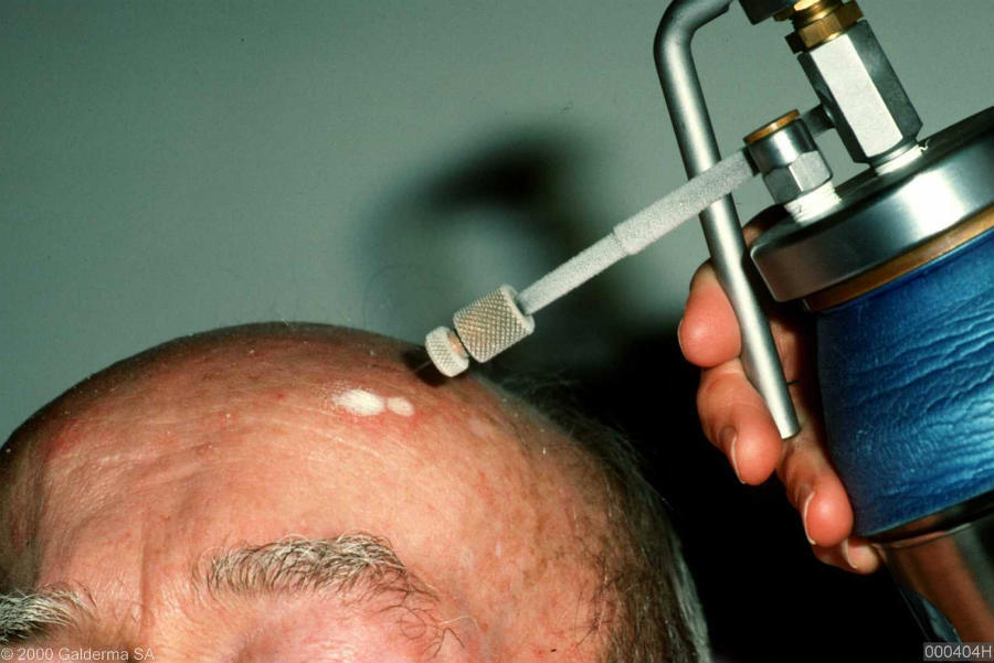
  • Cryotherapy - a single freeze-thaw cycle of approximately ten seconds (avoid the gaiter are of the legs due to risk of leg ulceration, and warn patients if used on the face it can cause hypopigmentation). For more information refer to the chapter Cryosurgery 
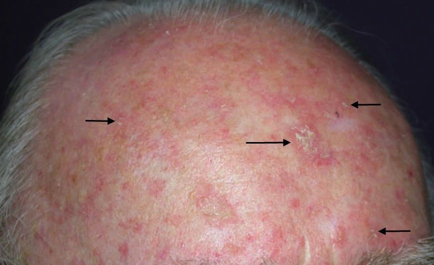
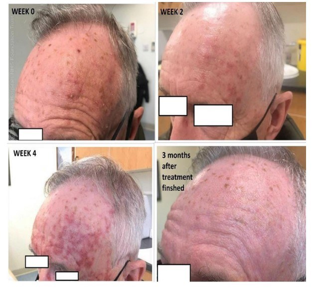
Step 4: field change - please view images at the bottom of the page

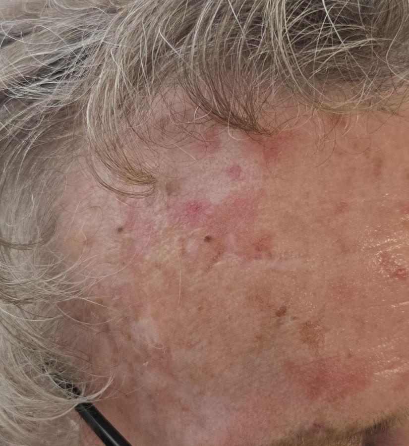
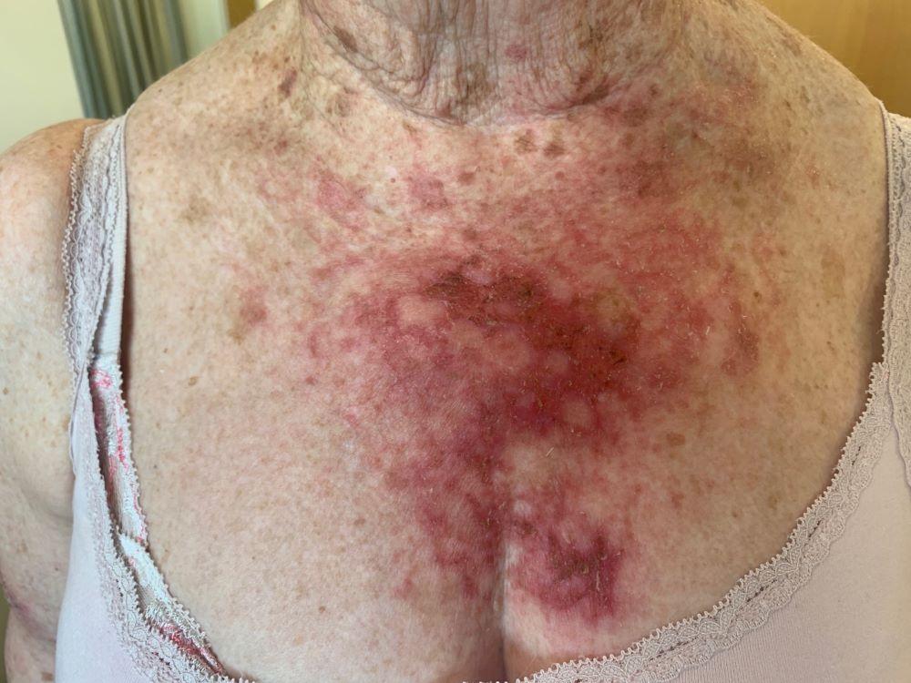
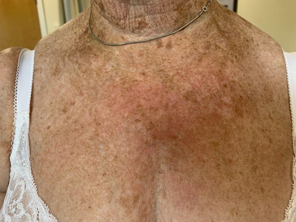
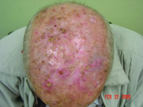
Field change refers to areas of skin that have multiple AK associated with a background of erythema, telangiectasia and other changes seen in sun-damaged skin. These areas are probably more at risk of developing SCC, especially if left untreated and, as such, it is recommended that they should be treated more vigorously. As such, the treatments should be applied to the whole area of field change and not just the individual lesions.

As when treating other patients with actinic keratoses the primary aim of treatment is to reduce the total number of lesions that the patient has at any one time, the fewer lesions a patient has the less risk they have for developing an SCC. Treatment courses will need to be repeated from time to time. Note that all field-based treatments will elicit local skin responses, which are expected as part of the treatment. The length of time a patient has to endure local skin responses varies widely between the treatments referred to below, and this needs to be discussed with the patient to aid them with the decision-making.

First-line treatment - the best evidence for SCC reduction is the combination of 5% 5-FU cream and calcipotriol ointment:

  • Normally applied at the same time in the morning and evening (twice daily). The length of treatment is between 4 to 6 days for the face, and 6 to 10 days for the arms and legs
  • The 5% 5-FU cream is applied first, spreading a thin layer on to the affected skin. After 5 minutes the calcipotriol is applied in the same way, the creams are then rubbed together into the skin
  • The skin should be gently washed and patted dry before each application 
  • Combination treatment helps significantly reduce the period of treatment compared to 5% 5-FU alone
  • Please provide a patient information leaflet 

Other treatment options

  • For smaller areas of field change (eg an area the size of a palm or most of the forehead) consider the following treatments (listed in no particular order):  
     
    • Aldara ® cream (5% imiquimod)
      • Use three nights a week eg Monday, Wednesday and Friday for four weeks. Apply overnight and wash off the following morning. After four weeks stop the treatment and consider the use of a mild topical steroid eg 1% Hydrocortisone or Eumovate ® cream BD for two to four weeks to help settle down any inflammation. Follow up three months after the treatment was started, repeat the treatment if needed
      • Advantages - generally very effective in terms of clearance, and cosmetic appearance once inflammation resolved
      • Disadvantages - patients should be warned to expect marked erythema with crusting of the skin. Timing of the treatment is important and is best avoided during holidays and important social occasions. Some patients develop flu-like symptoms during treatment
         
    • 5-fluorouracil cream (5-FU) - traditionally 5% 5-FU although 4% 5-FU can be used
      • Use once a day for four weeks. Apply thinly in an evening with a gloved finger, alternatively wash the finger after application. The treated area should be washed the following morning. After four weeks stop the treatment and consider the use of a mild topical steroid eg 1% Hydrocortisone or Eumovate ® cream BD for two to four weeks to help settle down any inflammation
      • Advantages and disadvantages similar to with Aldara ®, although patients do not develop flu-like symptoms
      • Please provide a patient information leaflet 
         
    • Klisyri ® cream (tirbanibulin) - used once a day for 5 days. Patients should be advised to expect redness, crusting, and some discomfort during treatment and for up to 10 days after 
       
    • Actikerall ® solution (combines 5-FU with salicylic acid) - used once a day for 6-12 weeks. Actikerall tends to leave a film on the skin, which should be washed/peeled off before the next application
       
    • Photodynamic therapy (PDT)
      • This is provided by some dermatology departments and occasionally GPwER clinics
      • A single treatment often provides an effective treatment for an area of field change. The skin settles down within a few days of treatment. Cosmetic outcomes are good 
         
  • For larger areas of field change consider the following treatments (listed alphabetically):
     
    • 3% diclofenac gel
      • Use twice a day for 8-12 weeks. Review patient four weeks after treatment has finished to assess response
      • Advantages - generally well-tolerated and so can be used on any sized area
      • Disadvantages - most dermatologists view 3% diclofenac gel as a milder treatment, which may not be as effective as some of the other treatments and so is best used where the AK are thin. Once treatment is complete any remaining AK can then be managed with the treatments referred to in step three above
         
    • Zyclara ® cream (3.75% imiquimod cream)
      • Apply once daily for two weeks, followed by a two week treatment-free period, and then a further once daily application for two weeks (ie six weeks in total, but only four weeks of treatment)
      • Adverse effects less than when using Aldara ® cream (5% imiquimod) 

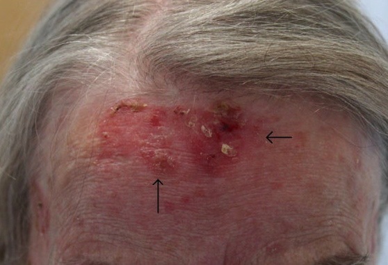
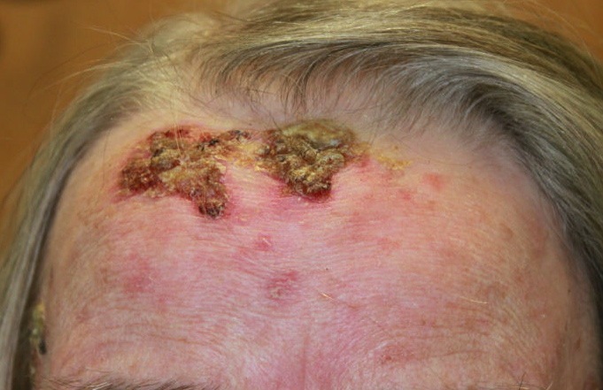
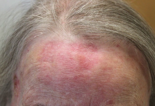
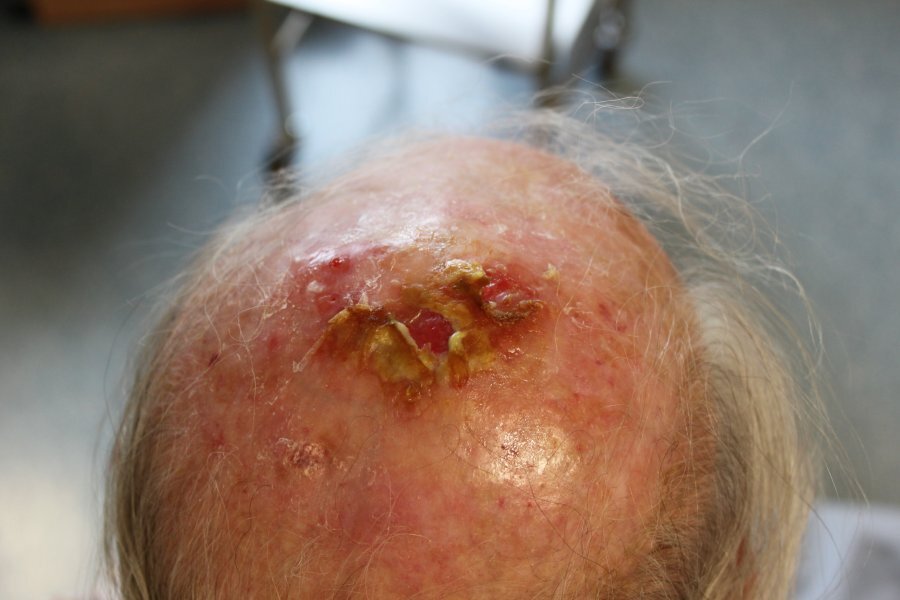
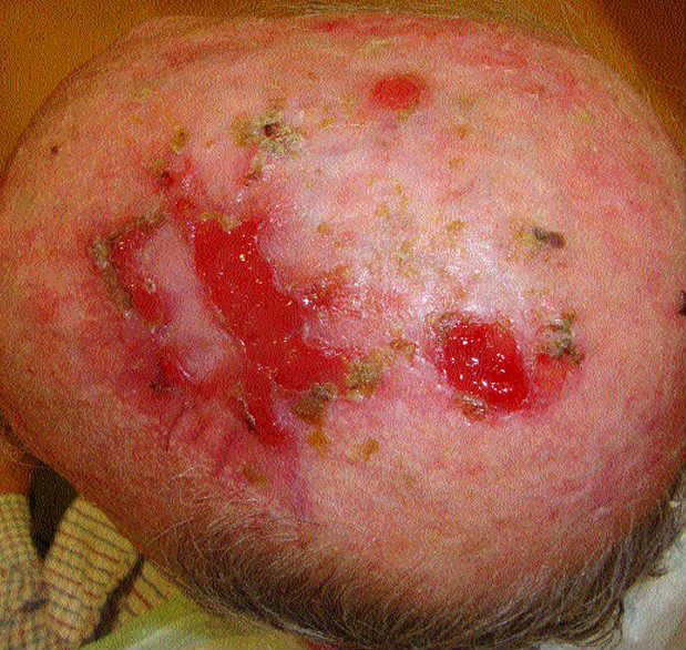
Erosive pustular dermatosis of the scalp

  • Is an uncommon condition affecting UV-damaged areas of the scalp in older patients. The risk appears to be increased with the subsequent treatment of actinic keratoses, especially with cryotherapy
  • Clinically there is varying degrees of scarring associated with yellow-brown crusts, pustules, lakes of pus, erosions and ulceration (refer to figures 11 & 12 below)
  • The primary treatment is the use of super-potent topical steroids

Other resources


Additional images


Disclaimer - the author PCDS cannot accept responsibility for any misleading or incorrect statements, and the management of individual patients remains the direct responsibility of the individual doctor. We do however hope that visitors to this site can contact us regarding comments that are considered misleading or incorrect so that we can continue to improve the site.

Image Rights - The PCDS would like to thank Dermatoweb, DermQuest (Galderma), and others who have contributed images. All named individuals and organisations maintain copyright for the relevant images.

Quick Links

The following pharmaceutical companies have had no involvement in the content of this website or in our conference programmes


Almirall
Galderma
Glenmark
Johnson & Johnson
La Roche-Posay
LEO Pharma
Pierre Fabre
Schuco