Bowen’s disease
LAST UPDATED: Jun 05, 2026
Introduction
Bowen’s disease is an area of UV-damaged skin, which if left untreated can very occasionally turn into squamous cell carcinoma, the rate of transformation is approximately 3%.
This chapter is set out as follows:
Aetiology
-
The main cause is UV radiation
-
Patients with fair skin, blue eyes and blonde hair are more at risk
-
It is more common in women
History
-
Lesions can be single or multiple
-
They are often reported as being very slow-growing and generally asymptomatic
Clinical findings
Distribution
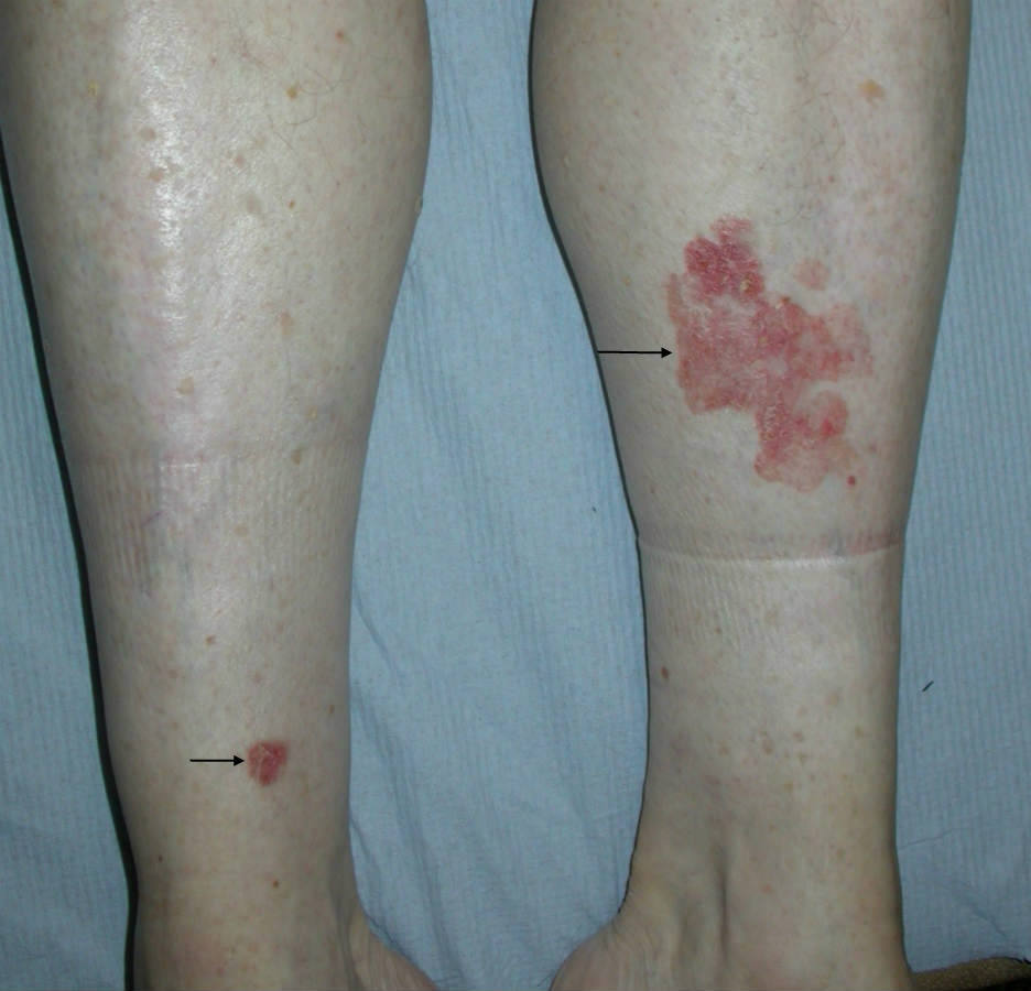
- Sun-exposed sites predominate, especially lower legs in women
Morphology
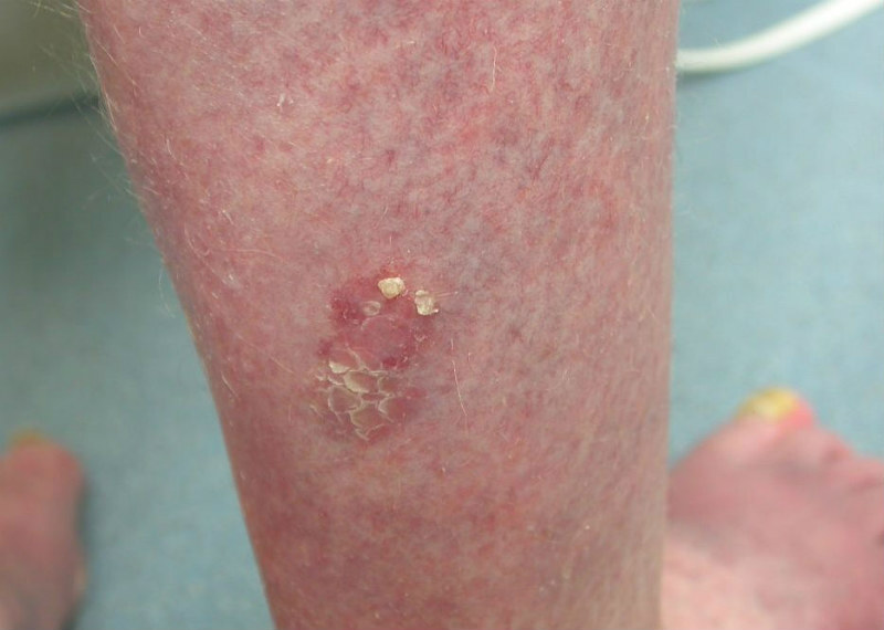
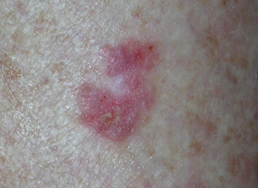
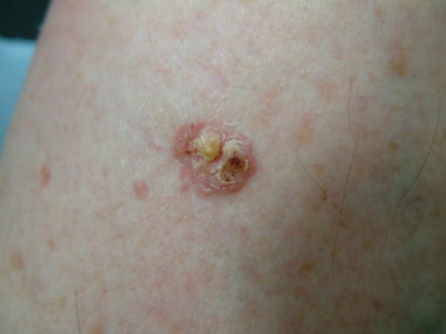
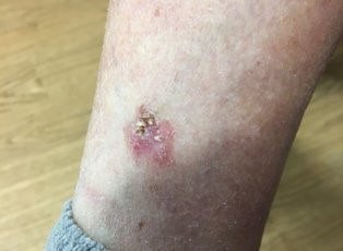
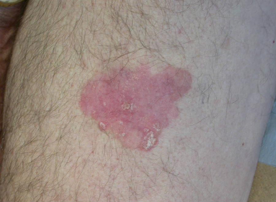
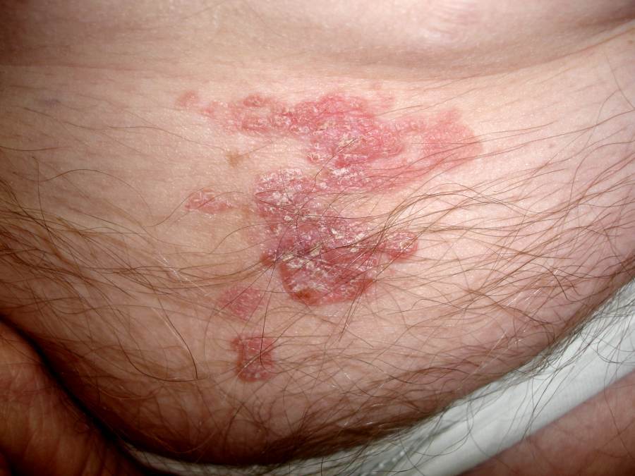
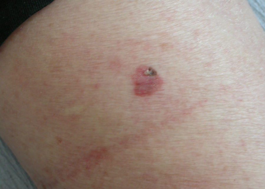
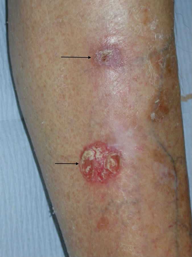
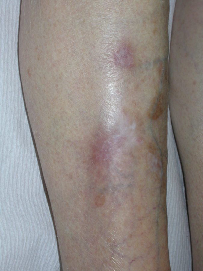
- Well-defined pink and scaly patches or plaques. They tend to have little substance and have finer scale than AK
- As lesions grow they may become crusty, fissured or ulcerated
Dermoscopic features (click or hover over terminologies for description)
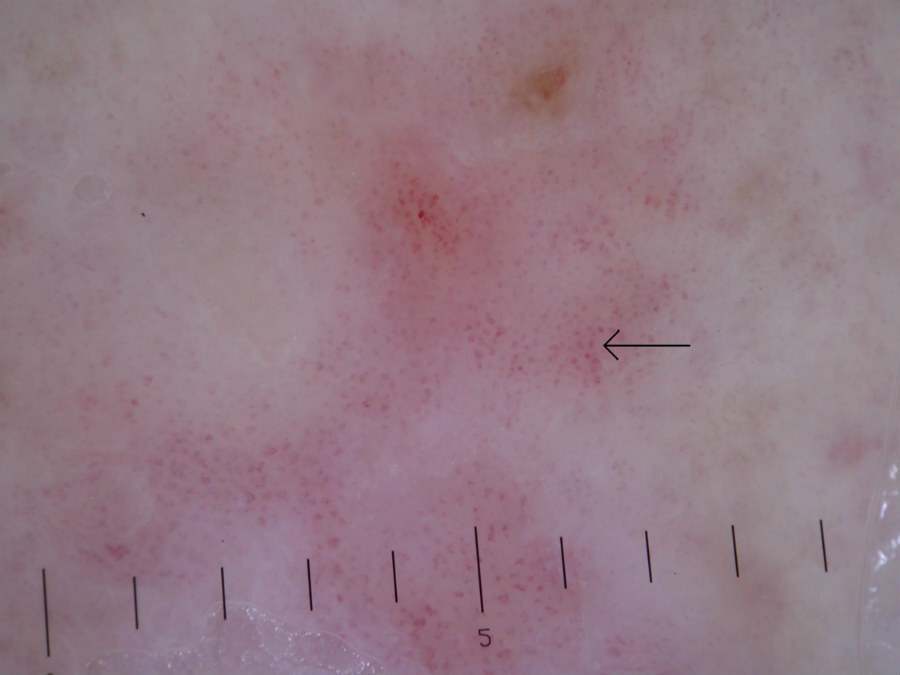
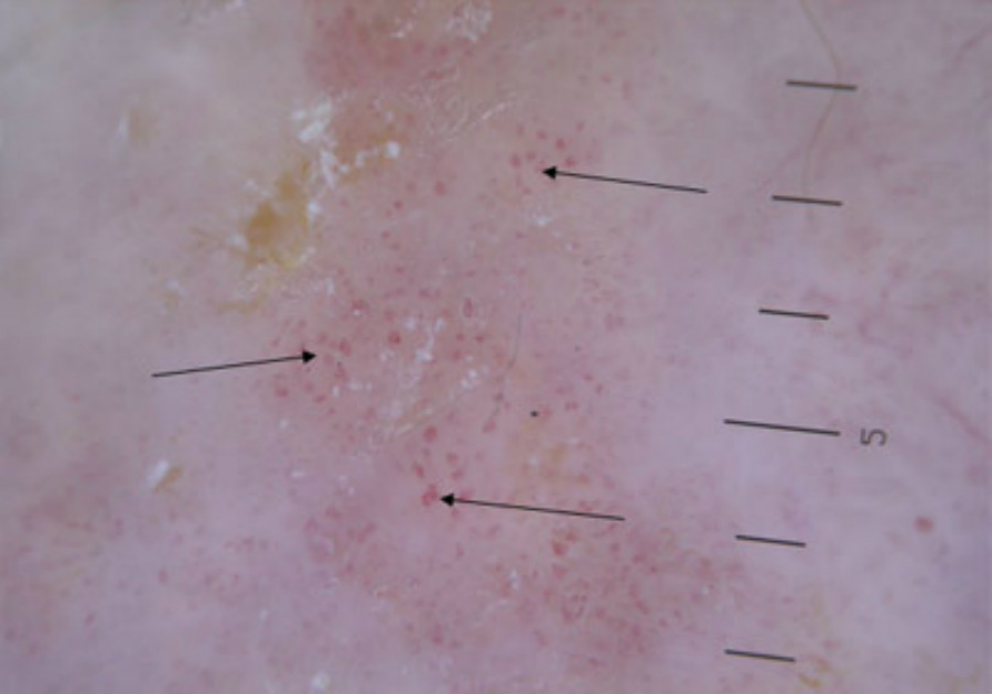
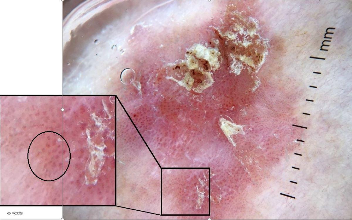
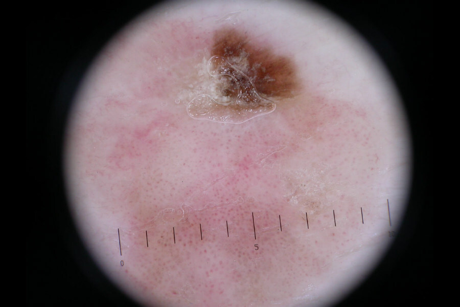
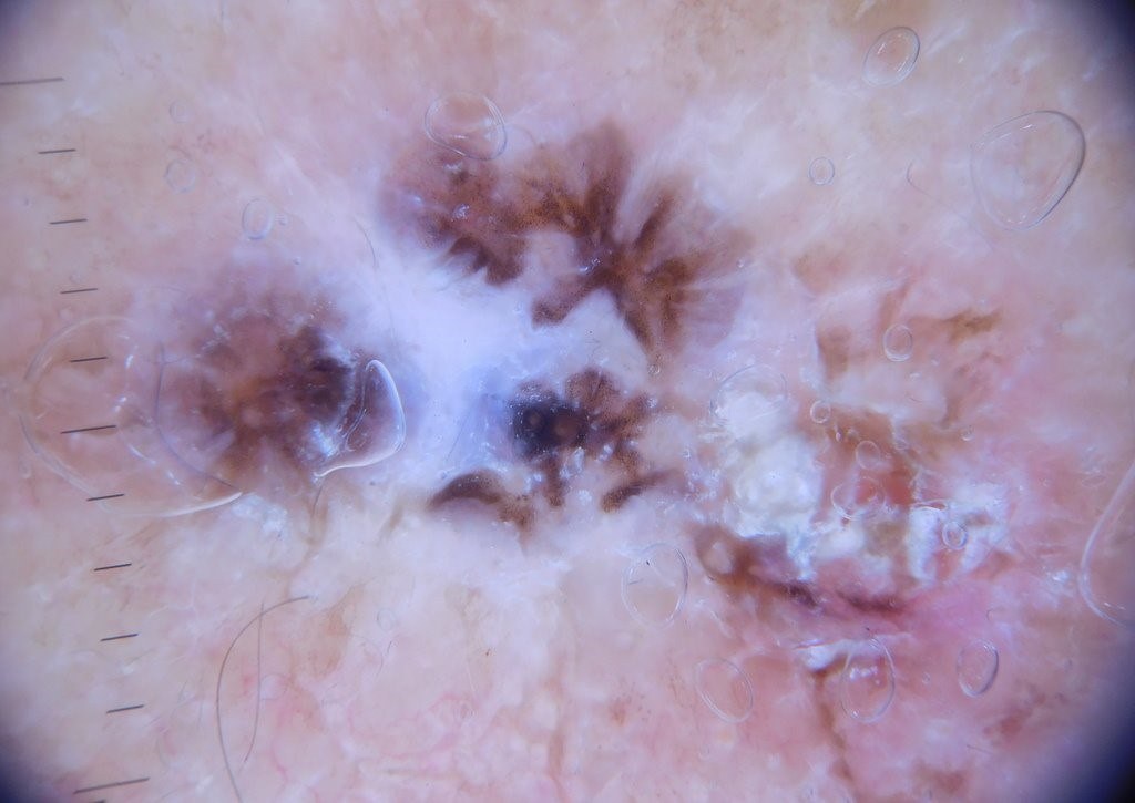
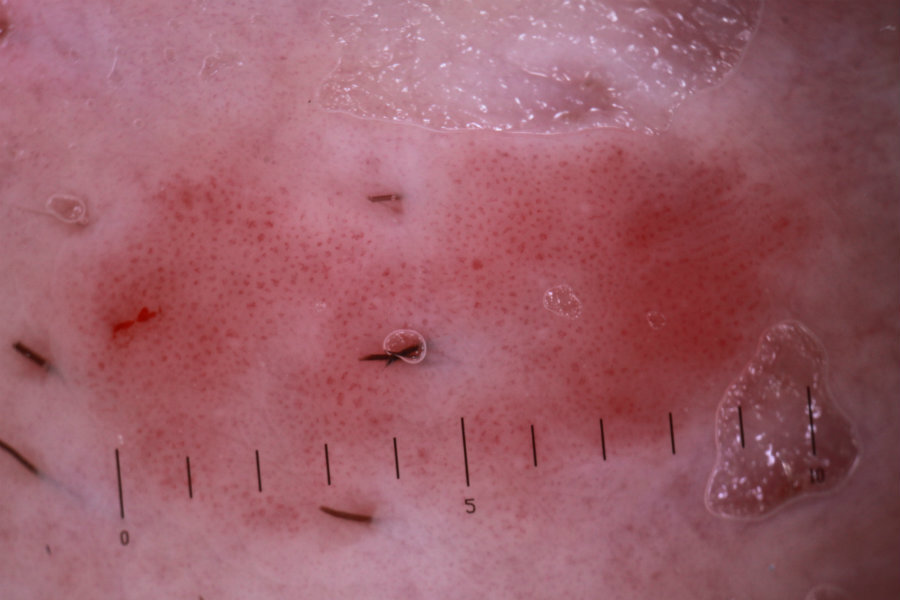
- Typical lesions - erythema, white-yellow keratin, and clusters of highly vascular areas with glomerular or dotted vessels, occasionally can be arranged in a linear fashion (refer here for more information as vascular patterns in dermoscopy)
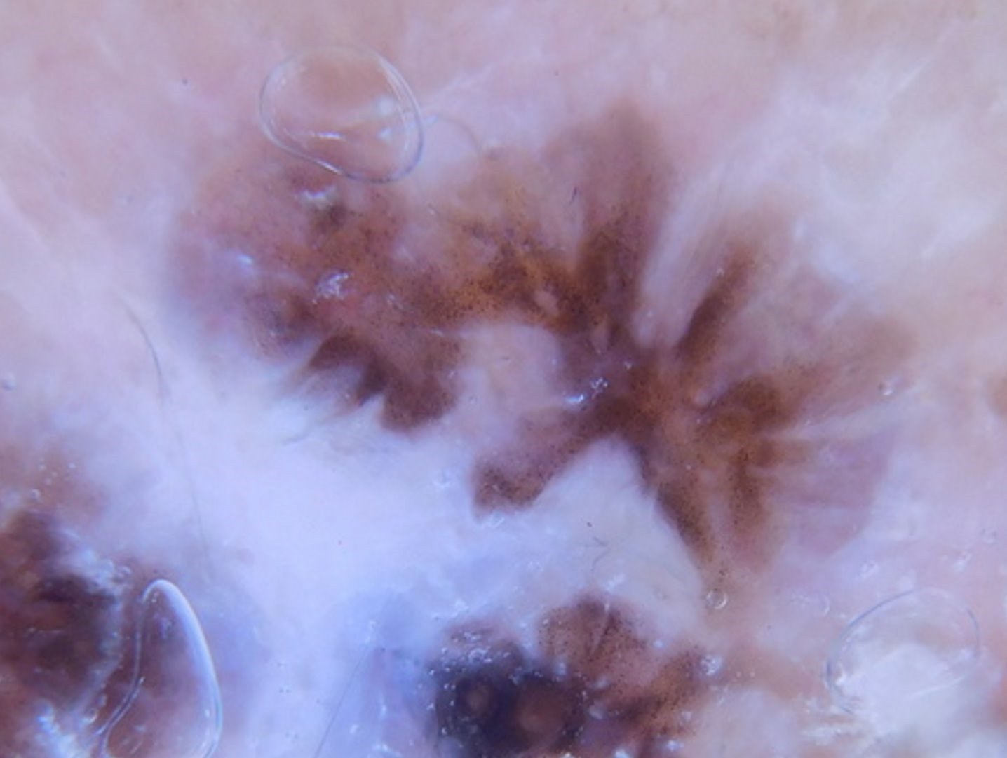
- Pigmented Bowen's - in addition to the above, pigmented Bowen's is typified by a pattern of brown/grey dots and/or structureless areas, sometimes the dots are arranged in a linear and radial fashion. Pigmented Bowen's can mimic melanoma and is appropriate for an urgent suspected cancer referral
For carcinoma in situ of the genitalia please refer to the following:
Clinical Images
Please refer to notes on image rights at bottom of the page with regards to individual image
ownership.
1
2
3
4
5
6
7
8
9
10
11
12
13
14
Investigations
- A biopsy is only needed if there is diagnostic uncertainty, although if a squamous cell carcinoma (SCC) is suspected the patient should be referred urgently to Secondary Care (urgent suspected skin cancer pathway) without a biopsy. Features suggestive of transformation into an SCC include an elevated palpable component, induration, ulceration, tenderness, and surrounding inflammation
- Bowen disease is characterised by full‐thickness epidermal dysplasia and disordered differentiation with loss of epithelial polarity. The intraepidermal portion of cutaneous adnexae is generally affected. Parakeratosis and acanthosis are usually present and keratinocytes show variable pleomorphism, nuclear hyperchromasia and nuclear enlargement
Management
Step 1: general
Step 2: first-line treatments
- A single freeze-thaw cycle with cryosurgery for 20-30 seconds - avoid in the gaiter area of the leg and other areas of poor skin healing. For larger patches treatment may be better tolerated if half of the lesion is treated initially and the other half six weeks later. For more information refer to the chapter Cryosurgery
- 5% 5-fluorouracil cream OD for four weeks (or occasionally BD, which can be considered for larger/thicker patches of Bowen's that are away from poor wound healing sites such as the lower legs). Hands should be washed thoroughly after application. The treated area must be left uncovered and the cream washed off approximately 8 hours after application. Warn the patient to expect some redness, crusting and mild discomfort. After four weeks stop the treatment and consider using a mild topical steroid eg 1% Hydrocortisone or Eumovate ® cream BD for two weeks to help settle down any inflammation.
- Although the risk of leg ulceration is greater with cryosurgery, it can also happen with 5% 5-fluorouracil cream, as such a watch and wait policy can be offered for lesions on the gaiter area of the legs in patients with a reduced life expectancy
Step 3: other treatments
- Photodynamic therapy (where available) - can be very useful for large and multiple lesions, and can be used on almost any body site
Step 4: follow-up
- Patients should be followed up at three months
- The presence of any remaining rough scale would suggest that the lesion has not fully responded to treatment and that more is required, whereas the presence of smooth skin, sometimes with associated post-inflammatory hyperpigmentation (especially on the lower legs), suggests that the lesion has responded well, in which case further follow-up is not required
Other resources
Additional images
Disclaimer - the author PCDS cannot accept responsibility for any misleading or incorrect statements, and the management of individual patients remains the direct responsibility of the individual doctor. We do however hope that visitors to this site can contact us regarding comments that are considered misleading or incorrect so that we can continue to improve the site.
Image Rights - The PCDS would like to thank Dermatoweb, DermQuest (Galderma), and others who have contributed images. All named individuals and organisations maintain copyright for the relevant images.