Vulval intraepithelial neoplasia (VIN) and squamous cell carcinoma
LAST UPDATED: Aug 02, 2021
Introduction
Vulval intraepithelial neoplasia (VIN) is a pre‐malignant condition that may progress to vulval squamous cell carcinoma. There are two forms of VIN, undifferentiated and differentiated, with different aetiologies and prognostic factors.
This chapter is set out as follows:
Aetiology
-
The incidence of VIN is increasing
-
The estimated incidence in undifferentiated VIN is 2 per 100 000 population
History
-
The main symptom of VIN is pruritus but some patients are asymptomatic
-
Squamous cell carcinoma (SCC) may be asymptomatic, or associated with itch, soreness and/or bleeding
Clinical findings
Undifferentiated VIN (uVIN)
-
Mainly affects younger women
-
Associations
-
Smoking - occurs almost exclusively in smokers
-
HPV types 16 and 18
-
Occasionally HIV infection
-
Distribution
-
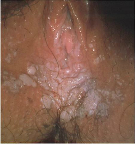
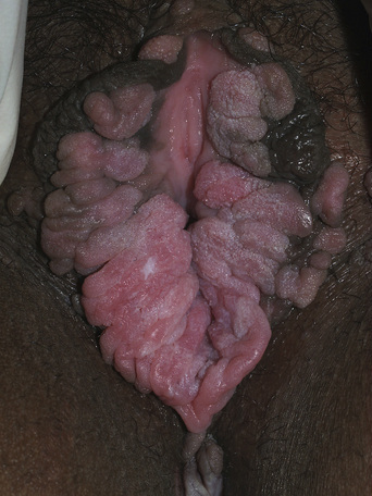
Mutifocal
-
Often associated with multicentric disease, with lesions of intraepithelial neoplasia involving the cervix, vagina and perianal skin
-
Morphology
-
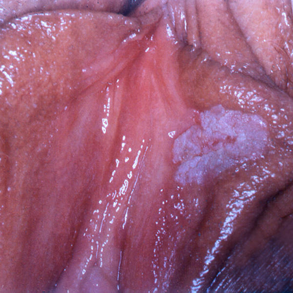
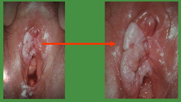
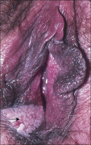
Lesions can be solitary or multiple
-
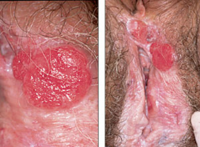
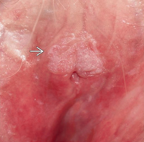
Diverse appearance from plaques to lesions that resemble warts or seborrhoeic keratoses
Differentiated VIN
-
Mainly affects older women
-
Associations
- Lichen sclerosus, and other long-term inflammatory dermatoses such as erosive lichen planus and bullous conditions - differentiated VIN remains as the term reserved for the severe atypia confined to the basal layers, most often seen on a background of a chronic scarring dermatosis
-
Morphology
-
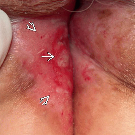
Often subtle, but erythematous, hyperkeratotic or irregular lesions on a background of chronic inflammatory dermatoses should be biopsied early and subjected to good clinicopathological correlation to confirm the diagnosis
Squamous cell carcinoma
-
SCC is the most common malignancy occurring on the female genitalia
-
Distribution
-
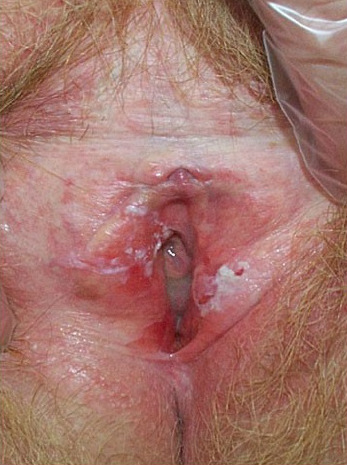
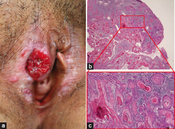
Lesions can occur on any area of the vulva but common sites are the labia majora, clitoris, perineal body and fourchette
-
Morphology
-
Lesions are most commonly nodular, ulcerated, or warty
Clinical Images
Please refer to notes on image rights at bottom of the page with regards to individual image
ownership.
Investigations
-
Good clinicopathological correlation is needed to make a diagnosis
-
Undifferentiated VIN is a multicentric problem, and other sites that need to be monitored are the cervix, vagina and perianal area. If there is perianal disease, anoscopy should be performed to exclude involvement of the anal canal
Management
Undifferentiated VIN
The risk of progression to invasive disease (SCC) is estimated as 10% or less in multifocal uVIN, but this risk is likely to be higher in immunocompromised patients, those with perianal disease and in the older woman with a solitary plaque
Treatment options include topical treatments, cryosurgery and surgical excision. Imiquimod cream has proved very effective in the treatment of multifocal uVIN in younger patients and recurrent disease after surgical excision
Disclaimer - the author PCDS cannot accept responsibility for any misleading or incorrect statements, and the management of individual patients remains the direct responsibility of the individual doctor. We do however hope that visitors to this site can contact us regarding comments that are considered misleading or incorrect so that we can continue to improve the site.
Image Rights - The PCDS would like to thank Dermatoweb, DermQuest (Galderma), and others who have contributed images. All named individuals and organisations maintain copyright for the relevant images.