Hyperpigmentation - of the face and neck
LAST UPDATED: Feb 15, 2023
Introduction
Facial hyperpigmentation is common, and can cause significant cosmetic disfigurement with subsequent emotional impact.
This chapter is set out as follows:
History
It is important to approach facial hyperpigmentation, which is much more common in patients with skin of colour, in a logical way. As such the conditions have been grouped in this chapter as follows:
-
Group 1: Serious causes of hyperpigmentation (eg Addison's disease, haemochromatosis)
-
Group 2: Post-inflammatory hyperpigmentation (eg acne, eczema)
-
Group 4: Melasma and other causes of more diffuse facial hyperpigmentation (eg matural dyschromia, exogenous ochronosis)
-
Group 5: Localised hyperpigmentation of the face (eg periorbital hyperpigmentation, naevus of Ota)
-
Group 6: Hyperpigmentation of the neck (eg poikiloderma of Civatte, acanthosis nigricans, erythema dyschromicum perstans)
-
Group 7: Lesional hyperpigmentation (eg dermatosis papulosa nigra, Hori naevi, actinic lichen planus)
-
Group 8: Others - very rare causes of facial hyperpigmentation
For further information on some of these conditions refer to the related chapters.
Clinical findings
Group 1: Serious causes of hyperpigmentation
The following are rare, but must not be missed:
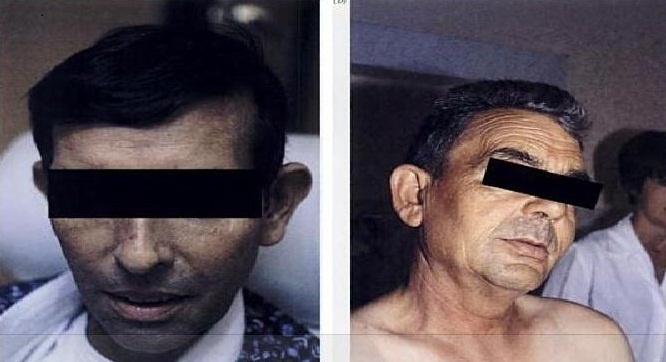
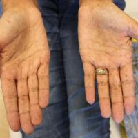
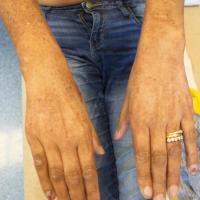
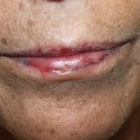
Addison's disease
-
Systemic features include fatigue, weight loss, dizziness on standing, abdominal pain, vomiting and psychiatric symptoms
-
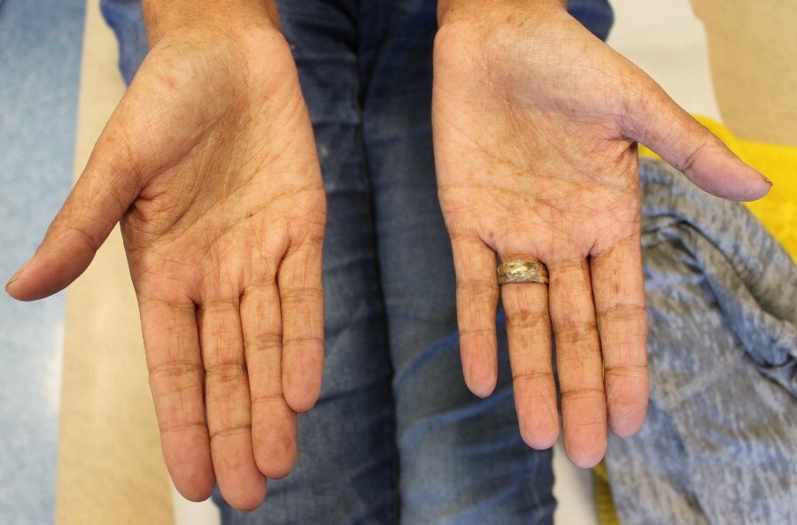
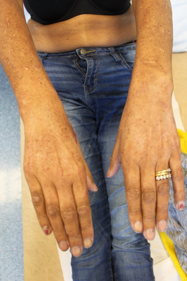
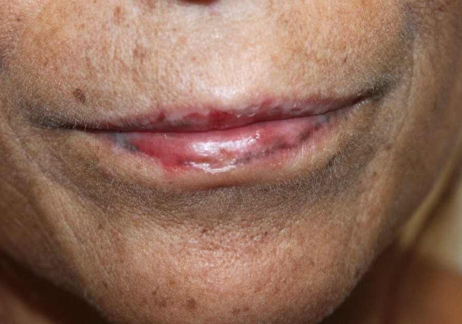
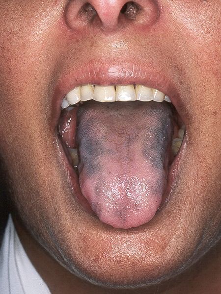
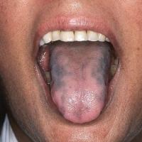
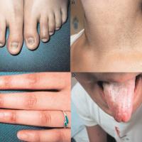
Hyperpigmentation affects UV-exposed sites, palmar creases, buccal mucosa, gums, scars, hair and nails, areas subject to friction. There is accentuation of normally high pigmentation areas such as the areolae, axillae, genital skin and umbilicus
-
Investigations:
-
Low serum sodium and raised serum potassium
-
Serum urea and albumin are raised because of dehydration
-
Serum cortisol level taken ideally between 8-9 am (random measurements have a low sensitivity for Addison's disease due to the pulsatile nature and diurnal variation of cortisol secretion). If the level of serum cortisol is:
-
< 100 nanomol/L - adrenal insufficiency is highly likely (if the patient is not on oral or inhaled steroids)
-
> 400 nanomol/L - adrenal insufficiency is unlikely (diagnosis not excluded if the patient is acutely unwell at the time since cortisol values may increase during illness)
-
Between 100 and 400 nanomol/L - refer to a specialist for further investigations eg synacthen test
Haemochromatosis
-
Systemic features - diabetes, cirrhosis and cardiac failure
-
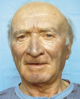
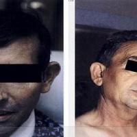
Hyperpigmentation - slate grey or brown-bronze with a predominance for the face and other UV-exposed sites
-
Investigations:
-
Iron levels - most people with haemochromatosis have elevated levels of iron in the blood
-
Transferrin saturation - transferrin is a protein that binds iron and transports it between the tissues. This test is one of the most sensitive tests for detecting early haemochromatosis. A transferrin saturation greater than 45% should be investigated further
-
Ferritin levels - ferritin is a protein that reflects the body's iron stores. Blood ferritin levels increase when the body's iron stores increase; however, levels of ferritin usually do not rise until iron stores are high. Therefore, ferritin levels may be normal early in the course of haemochromatosis. Ferritin levels greater than 400 ng/mL support a diagnosis of haemochromatosis, however, ferritin levels can also be increased in other conditions
Group 2: Postinflammatory hyperpigmentation (PIH)
-
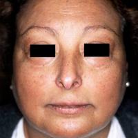
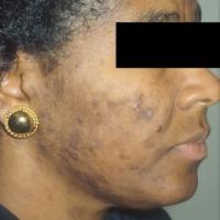
PIH refers to darkening of the skin that occurs after an inflammatory eruption such as acne, eczema, lupus or following cutaneous injury. There may be a history of itch and / or signs of active skin disease such as erythema and scale. On occasions the skin can become hypopigmented
-
As with many of the causes of facial hyperpigmentation, the incidence and severity of PIH is much greater in skin of colour
-
Management:
-
Treatment is of the primary disorder - the patient will develop less postinflammatory hyperpigmentation if the skin condition is treated promptly and effectively
-
As with other causes of facial hyperpigmentation it is important to consider the possibility of contact allergic dermatitis or a photocontact allergic dermatitis, referred to as Riehl melanosis. Affected individuals usually have pruritus with mild erythema and scale. Patients should be referred for patch testing and sometimes photopatch testing
-
Once the hyperpigmentation has developed the time taken for the affected skin colour to lighten is highly variable and can take several years
Group 3: Drug reactions
-
Several medications can cause hyperpigmentation including the phenothiazines (especially chlorpromazine), minocycline, phenytoin, antimalarial drugs eg chloroquine and hydroxychloroquine, busulfan and other cytotoxic drugs, amiodarone, anti-retroviral drugs and tricyclic anti-depressants (especially imipramine) - the colour change seen is likely to have a grey tone
-
Lichenoid drug eruptions, eg caused by gold, antimalarials, thiazides, ACEI, betablockers and quinine, can also affect the face
-
After discontinuing the drug it can take many months, sometimes years, for symptoms to improve
Group 4: Melasma and other causes of more diffuse facial hyperpigmentation
Melasma (syn. chloasma)
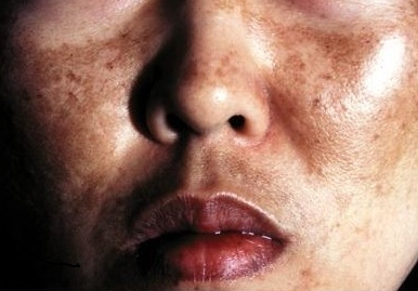
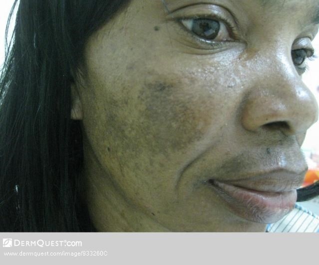
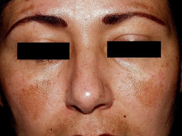
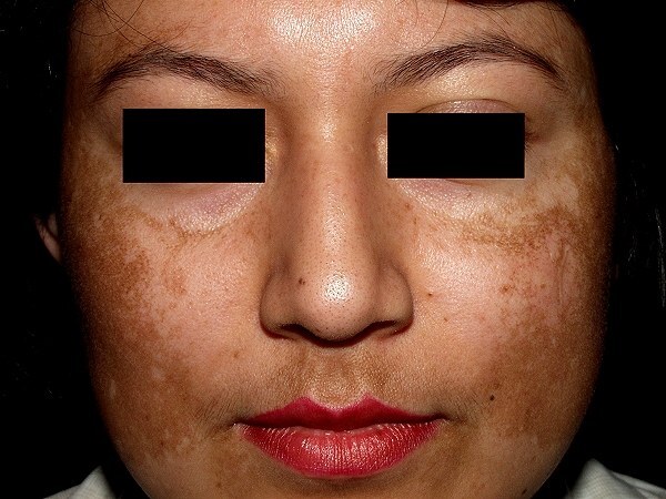
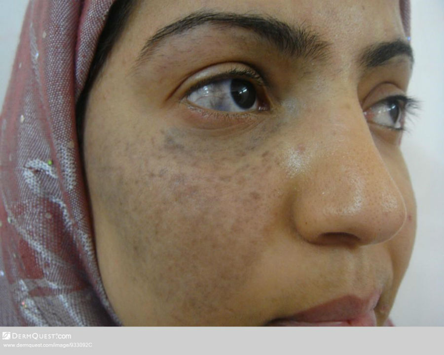
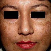
- Symmetrical involvement, most commonly of the centrofacial, malar and mandibular regions. The forearms can also be affected
- Light to dark brown patches - if the excess melanin is in the epidermis the patches are brown and more well-defined, whereas, if the excess melanin is in the dermis the patches are more grey-brown with less well-defined margins. Mixed types occur. A Wood's light may be helpful as it will enhance the colour if the pigment is mainly epidermal (eg some cases of melasma, and post-inflammatory hyperpigmentation), but not if the pigment is in the dermis. Epidermal melasma is more likely to respond to treatment
- For more information refer to the chapter Melasma
Maturational dyschromia
-
Dyschromia refers to skin discolouration or patches of uneven colour resulting in temporary or permanent hyperpigmentation (increase in pigment production) and / or hypopigmentation (decrease in pigment production). Mottling, or mottled skin, is another type of dyschromia in which changes in the blood vessels cause a patchy appearance
-
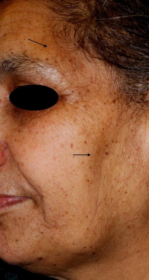
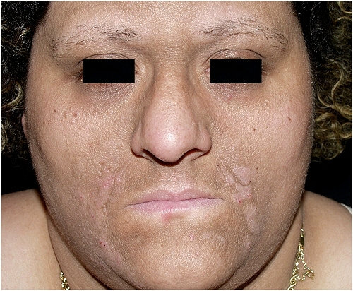
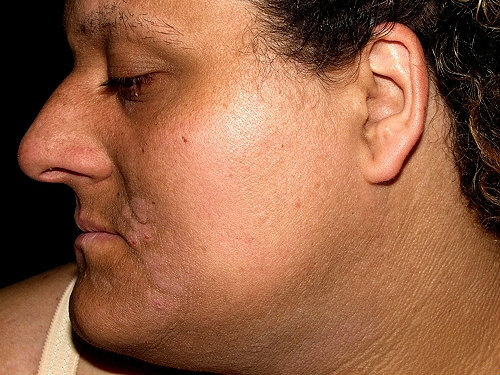
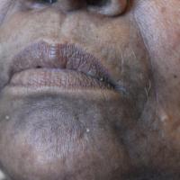
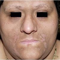
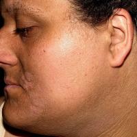
Maturational dyschromia is common. It is mainly seen in mature patients with skin of colour. Maturational dyschromia causes a diffuse hyperpigmentation predominantly affecting the lateral forehead, temples, and cheekbones, the margins are often ill-defined and the texture can be velvety. Most patients are overweight and 70% have the metabolic syndrome so it is worth testing for diabetes etc. Pressure or trauma may contribute (eg lying on one side in bed) but not UV-light
-
Refer to the section on management for more information
Lichen planus pigmentosus (LPP)
- An uncommon variant of lichen planus, the cause in unknown
- Affects predominantly young to middle-aged patients with skin of colour
- Unlike classical lichen planus, LPP does not tend to itch
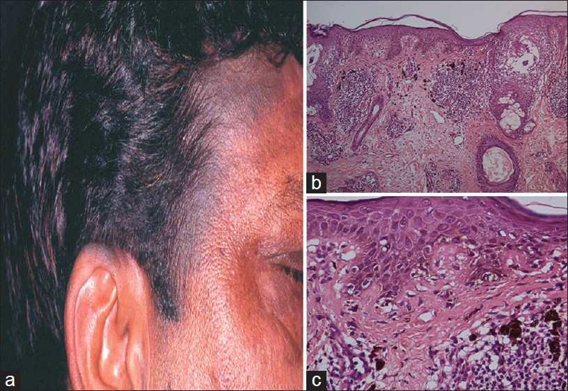
- Oval shaped grey-brown to brown macules and patches mainly on the face, neck and intertriginous areas, although can be more widespread. A minority of patients also have classical lesions of lichen planus
- Lesions can persist for many years
- Refer to the chapter Lichen planus
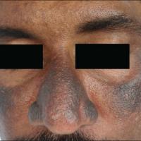
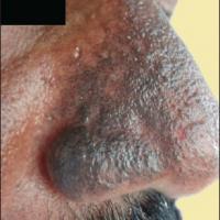
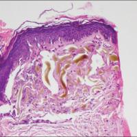
Exogenous ochronosis
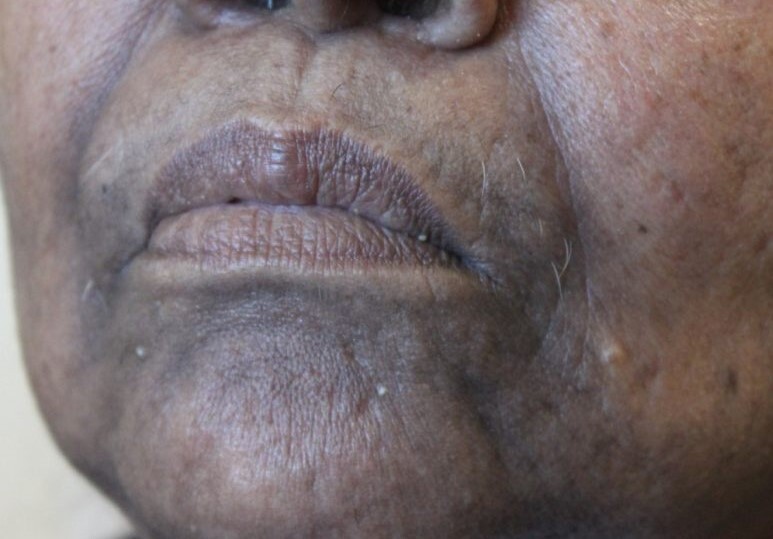
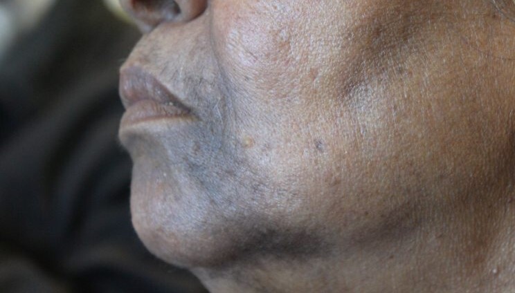
- Occurs secondary to the use of chemical substances applied to the face eg hydroquinone or resorcinol, often used by the patient to treat one of the conditions listed above. Such treatment may have been prescribed or used without the knowledge of a health professional
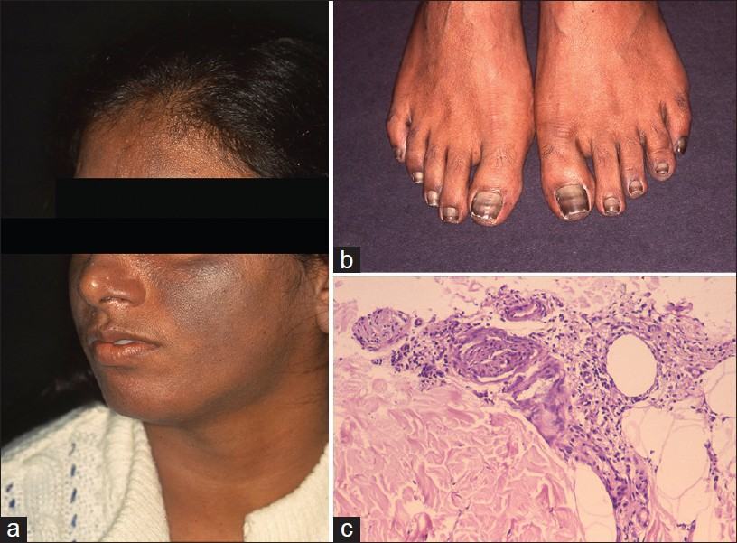
- Blue-black pigmentation of the face, sides and back of the neck. It can also affect extensor surfaces
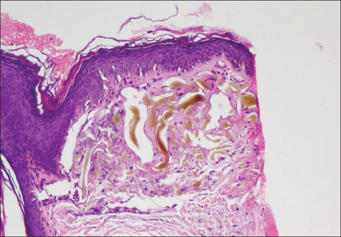
- The appearances can be similar to those of PIH or melasma and sometimes a biopsy is needed to differentiate. In exogenous ochronosis a microscopic deposition of ochre-coloured pigment can be found in the dermis
Group 5: Localised facial hyperpigmentation
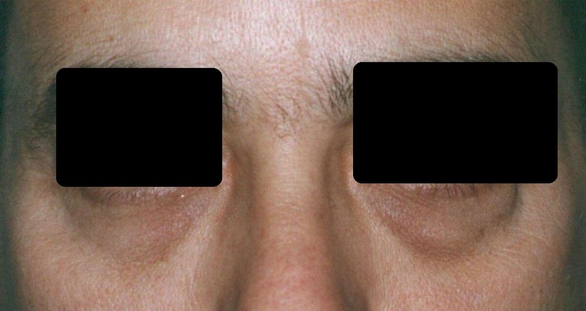
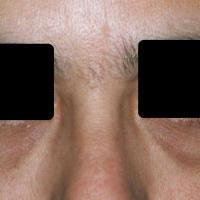
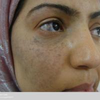
Periorbital hyperpigmentation (syn. dark circles; periorbital hypermelanosis)
-
A common condition
- The cause may be multifactorial and include both genetic and environmental factors
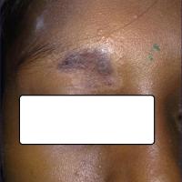
- Bilateral darkening of the orbital skin and eyelid
- The skin should be asymptomatic. If itch is present, or there are other skin signs, consider a diagnosis of eczema / contact allergic dermatitis
Naevus of Ota
-
Predominantly affects patients with skin of colour, although it can arise in any skin type. The majority present either at birth, infancy or around puberty
- Blue/grey to slate-brown patches of hyperpigmentation. The distribution of the pigment is usually unilateral affecting:
- Skin - the ophthalmic and maxillary divisions of the ophthalmic nerve
- The sclera can be involved, and sometimes other parts of the eye
- Occasionally the hard palate
Group 6: Hyperpigmentation of the neck
Poikiloderma of Civatte (POC)
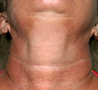
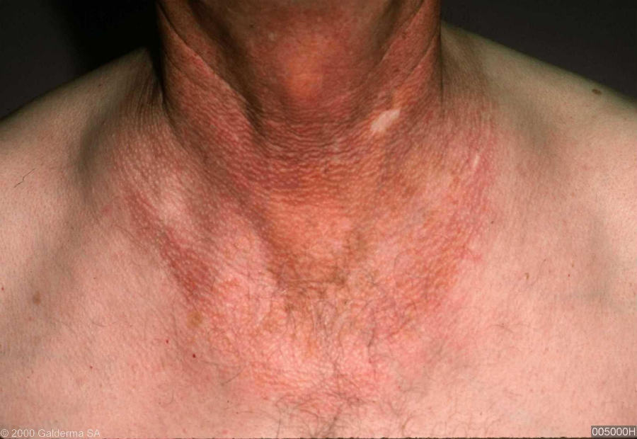
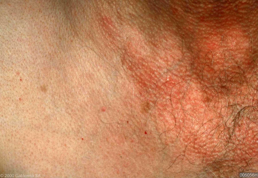
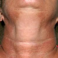
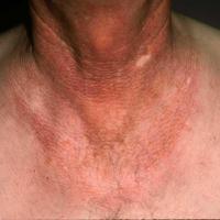
- Refers to skin changes with thinning, increased pigmentation and dilation of the small blood vessels (telangiectasia)
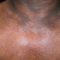
- The skin is red-brown with prominent hair follicles, affecting the neck and lateral cheeks, characteristically with sparing of the shaded area under the chin
- The cause is unknown, however, there is an association with UV exposure and it is more common in patients with fair skin. An additional theory is the photosensitising components of cosmetics and toiletries especially perfumes, although many doubt this link
- Refer to the section on management for more information
Acanthosis nigricans (AN)
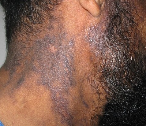
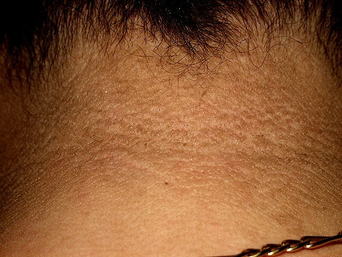
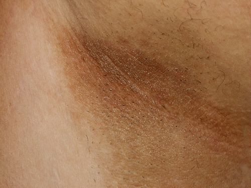
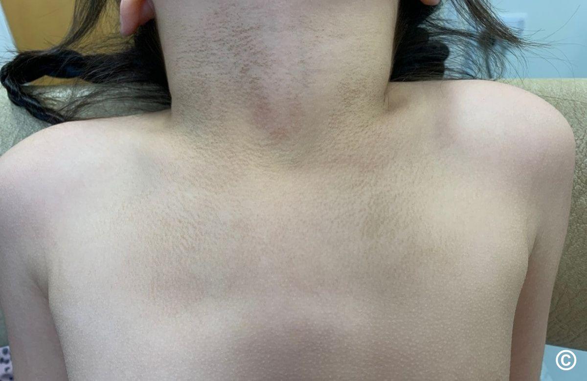
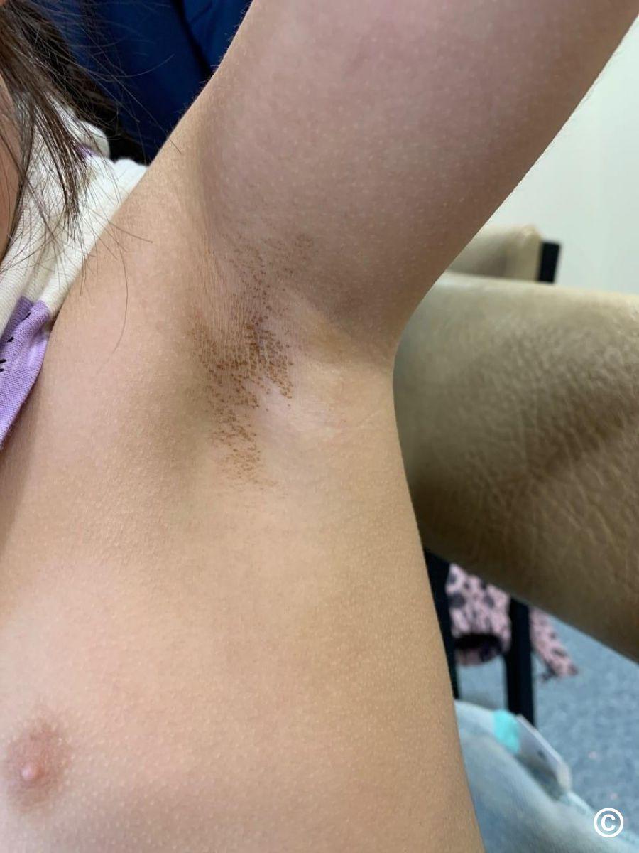
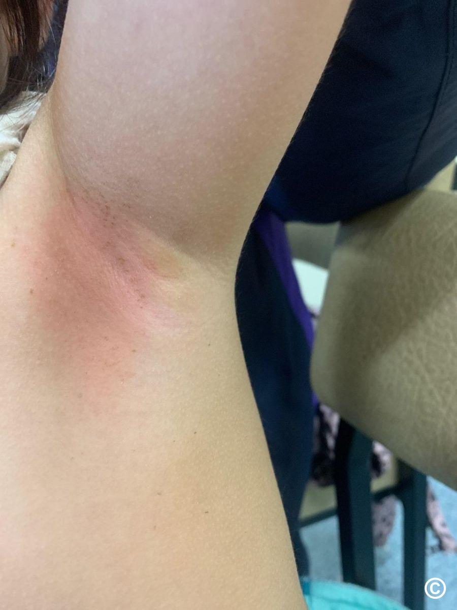
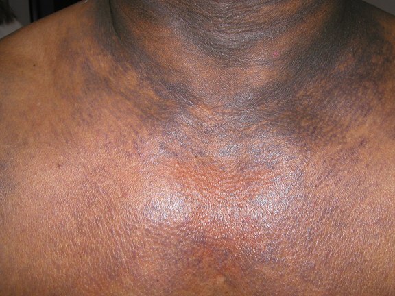
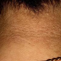
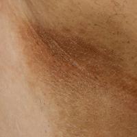
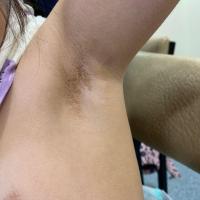
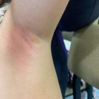
- Characterised by darkening, thickening and hyperpigmentation of the skin, occurring mainly in the folds of the skin in the axilla, groin and back of the neck. The face and other sites can be affected
-
Most cases of AN are related to insulin resistance. Rarely is it associated with underlying malignancy, such patients have much more extensive skin thickening including of the palms and soles
- Refer to the chapter Acanthosis nigricans
Terra firma-forme dermatosis
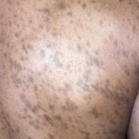
- Brown-black macules, papules, or plaques resembling dirty skin
- Lesion may also have a papillomatous, verrucous, or reticulate appearance
- The neck and other flexural sites, along with the face, trunk, and ankles, are the most commonly affected areas, although any area of the body can be affected
- The scale can be removed with alcohol wipes
- Refer to the chapter Terra firma-forme dermatosis
Dyskeratosis congenita (syn. the Zinsser-Engman-Cole syndrome)
- A rare group of genetic diseases that most commonly manifest with mucocutaneous signs, bone marrow failure and / or lung or liver fibrosis
- There is considerable variability in the severity, age at onset and organ involvement, even within individual families
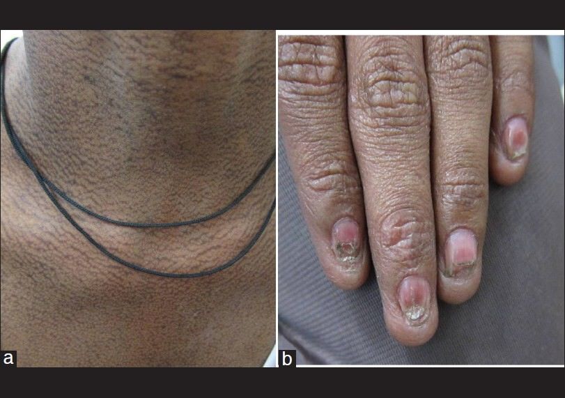
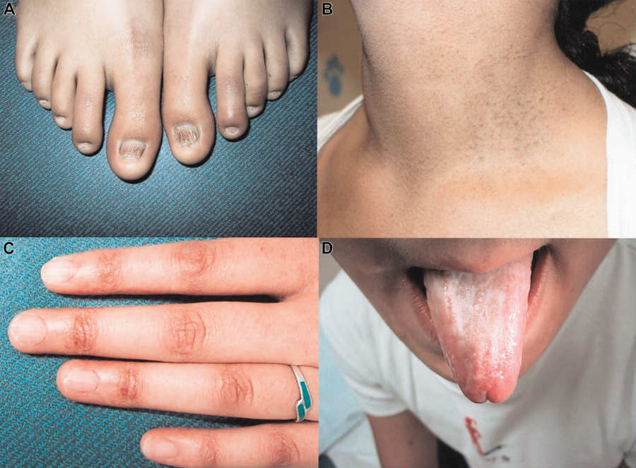
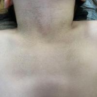
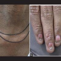
- The skin looks prematurely aged and photodamaged with dyspigmentation and atrophy. There is reticulate (lace-like) hyperpigmentation of skin creases, described as a 'dirty neck'
- Other mucocutaneous features include early hair greying or hair loss, sparse eyelashes, hyperhidrosis, squamous cell carcinomas often arising at a young age (50% by 40 years), nail dystrophy (longitudinal ridging, loss of nails, koilonychia), and oral leukoplakia
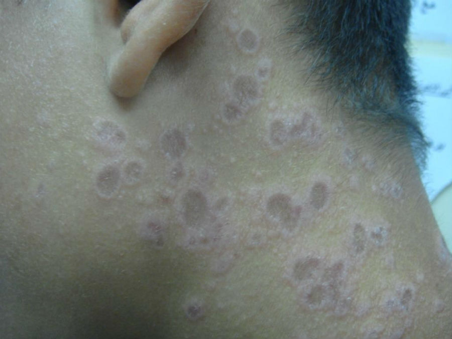
Erythema dyschromicum perstans (syn. ashy dermatosis)
- An asymptomatic, slowly progressive eruption usually presenting during the second and third decade of life
- The majority of the patients are from Latin America. Males and females are equally affected
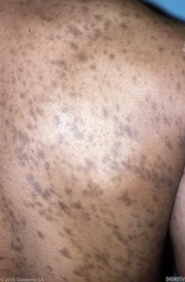
- A symmetrical involvement of the trunk, neck, upper extremities and sometimes the face with slate-grey to blue-brown oval, irregularly shaped macules and patches. In the early stages lesions may have thin, raised and erythematous borders
Group 7: Lesional hyperpigmentation
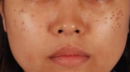
Ephelides (freckles) and lentigines
-
Are a common finding in white patients and less so in skin of colour
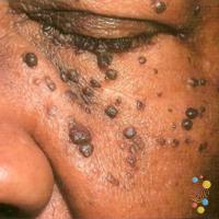
Dermatosis papulosa nigra (DPN) and seborrhoeic keratosis
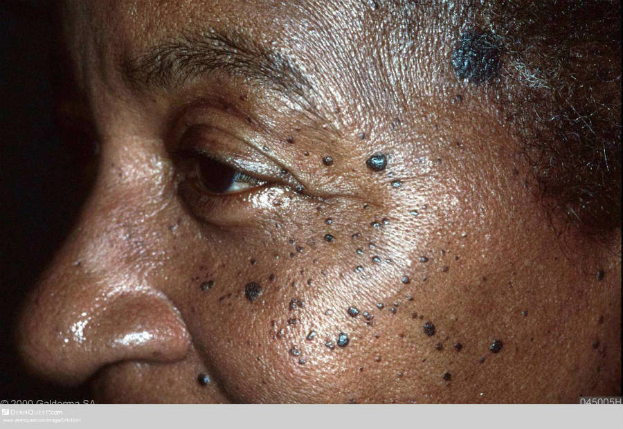
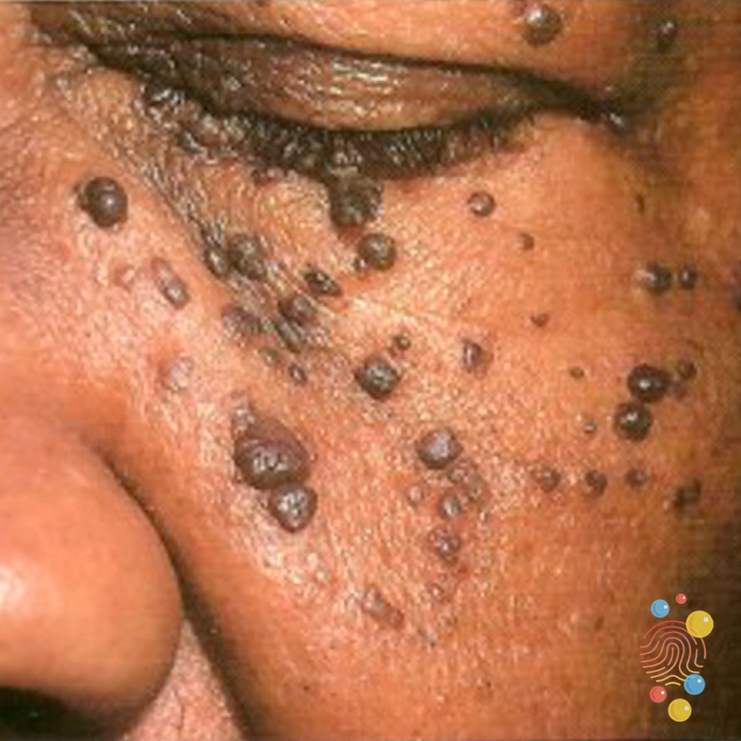
- A common finding in African-American, Afro-Caribbean and sub-Saharan black patient
- Considered a variant of seborrhoeic keratoses, with an earlier age of onset in many cases, arising often in adolescence with lesions increasing in number and size after that
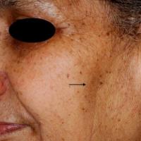
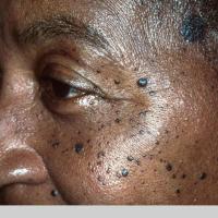
- Multiple 1-5 mm pigmented papules - lesions are distributed symmetrically across the malar eminences, forehead and less often the neck, chest and back
Hori's naevus
- A common finding in the Asian population, especially Chinese and Japanese patients aged 20-70 years
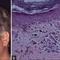
- Symmetrical blue-grey or grey-brown macules primarily on the zygomatic area, although can become more extensive
Actinic lichen planus
- A rare photodistributed variant of lichen planus, of unknown cause
- The majority of patients are of a Middle Eastern extraction. It mainly occurs in children and young adults
- Tends to be worse in the summer and improves over the winter
- Lesions are photodistributed affecting the face, neck and dorsal aspects of the arms and present as annular, red-brown patches with a striking hypopigmented zone and fine scale
Group 8: Others - very rare causes of hyperpigmentation
Argyria
- Characterised by a metallic, slate-grey or blue-grey pigmentation. The hyperpigmentation is most apparent in the UV-exposed areas of skin, especially the forehead, nose, and hands, as well as mucous membranes. Argyria results from prolonged contact with or ingestion of silver salts
Erythromelanosis follicularis faciei et colli
- Characterised by well-demarcated erythema, hyperpigmentation and follicular papules. There are fewer than 50 reported cases, mostly from Japan
Post-chikungunya pigmentation
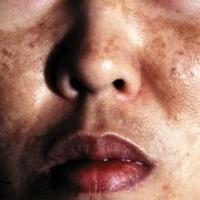
- Characterised by brown-black freckle-like macules, or less commonly slate pigmentation, mainly on the centrofacial skin. It occurs as part of Chikungunya fever, which is a febrile, mosquito-borne viral illness
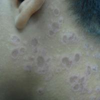
Clinical Images
Please refer to notes on image rights at bottom of the page with regards to individual image
ownership.
1
2
3
4
5
6
7
8
9
10
11
12
13
14
15
16
17
18
19
20
21
22
23
24
25
26
27
28
29
30
31
32
33
34
35
36
37
38
39
40
41
42
43
44
45
46
47
48
Management
UV protection
-
Some conditions e.g. melasma and poikiloderma of Civatte benefit from UV protection - mineral suncreens (protect against UVA, UBV, and visible light) are most effective
-
Patients adhering to strict UV protection of the face may require vitamin D supplements - refer to the patient information leaflet on UV protection and vitamin D
Camouflage clinics
-
Can advise on the best ways of covering up affected areas
-
Such clinics may be found in local out-patient departments or under the name of Changing Faces
Melasma
-
Refer to the related chapter Melasma
Management of other causes of facial hyperpigmentation
-
Maturational dyschromia - responds well to laser treatment
-
Poikiloderma of Civatte - the redness can be reduced by a pulsed-dye laser
-
Exogenous ochronosis - stop any creams causing the problem. The best policy is then to wait, although the pigmentation is likely to fade very gradually over a long period of time. An Nd:Yag laser may be of benefit although evidence is limited
-
Dark circles - an Nd:Yag laser may benefit a few patients although evidence is limited
-
With regards to all of the above, patients must be informed of the following:
-
The main reason for referring to a specialist is if there are diagnostic uncertainties
-
Treatments are likely to be provided only on a private basis (refer to local guidelines)
-
Some patients, eg those with dark circles, are unlikely to get significant benefit from treatment
Disclaimer - the author PCDS cannot accept responsibility for any misleading or incorrect statements, and the management of individual patients remains the direct responsibility of the individual doctor. We do however hope that visitors to this site can contact us regarding comments that are considered misleading or incorrect so that we can continue to improve the site.
Image Rights - The PCDS would like to thank Dermatoweb, DermQuest (Galderma), and others who have contributed images. All named individuals and organisations maintain copyright for the relevant images.