
Vascular patterns are most easily identified in pink and other pale skin lesions ie those with limited (or no) pigment. To see vessels properly it is important not to put too much pressure through the dermatoscope, this can be achieved by only resting the scope gently on the lesion and using a contact fluid or using a non-contact method. In relation to terminology, a monomorphous vascular pattern refers to one pattern of vessels, a polymorphous pattern refers to more than two pattern types.
Please click on the links below to go to the most relevant vessel/vascular group:
Reticular vessels - a network-like appearance


Dot vessels


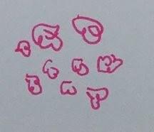
Bowen’s (often grouped), psoriasis (and other inflammatory dermatoses), warts (red or thrombosed), porokeratosis, benign melanocytic naevi.
Dotted vessels can also be a feature of early melanoma. Clues to melanoma include any of - areas of brown pigment, lack of rough white surface scale (the presence of which is much more suggestive of Bowen's), or a clinical ugly duckling ie the only pink ''naevus'' that the patient has.
Coiled vessels - a variation of dotted vessels, larger in size as a result of tortuous capillaries. Sometimes referred to as glomerular as reminiscent of the histological appearance of the renal glomerular apparatus


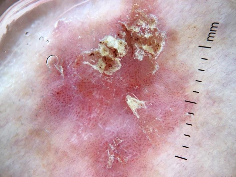

Bowen’s (vessels often grouped, sometimes seen in addition to dotted vessels), porokeratosis, psoriasis, stasis dermatitis (and other inflammatory dermatoses).
Clods - larger than a dot

Capillaritis (pigmented purpuric dermatosis) - red dots and clods. Can have additional colours of yellow and brown.
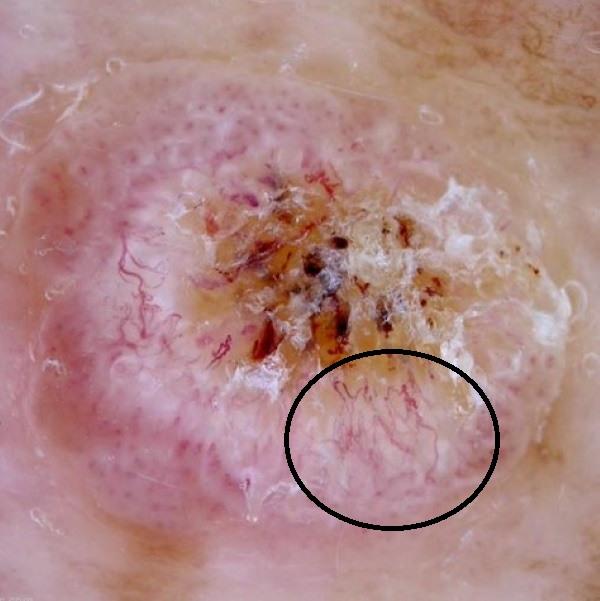
Radial vessels - radial infers pointing to the centre

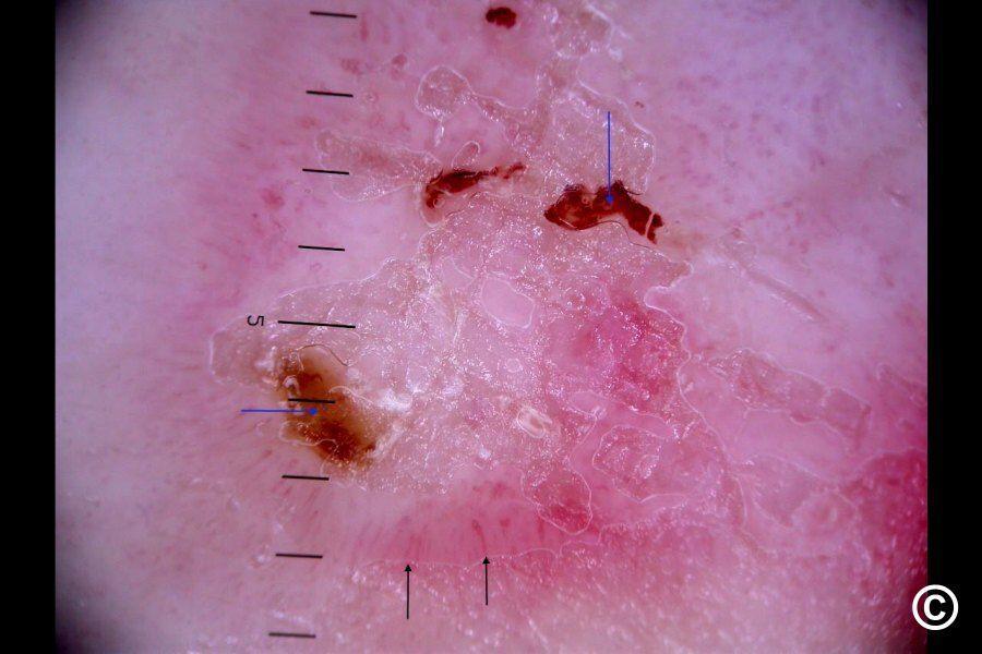
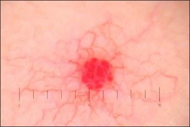
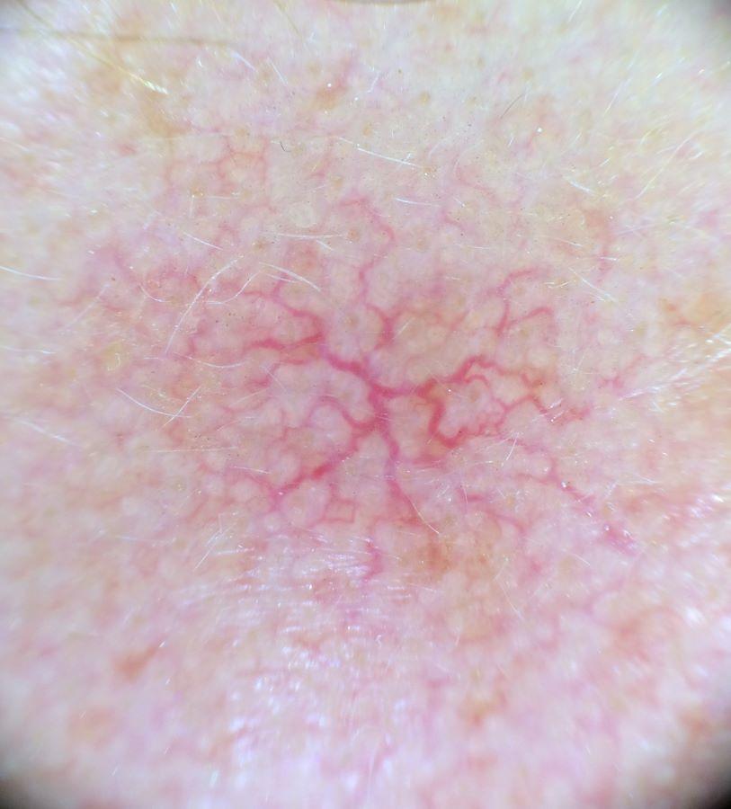
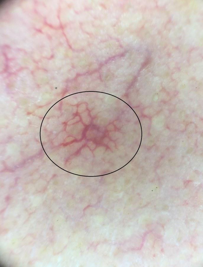
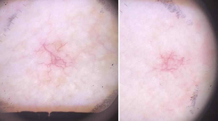
Superficial BCC - peripheral hairpin (thin and looped) vessels, often sit parallel to each other. Spider naevi have a dilated central arteriole with radiating radial vessels
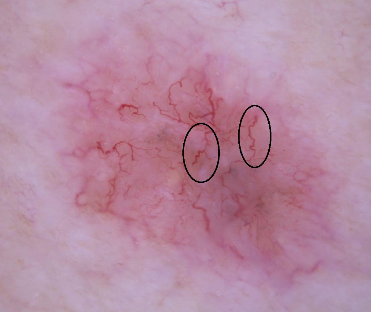
Serpentine (snake-like) vessels - refers to linear irregular vessels with more than one bend/kink
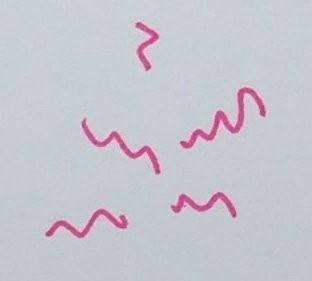


Polymorphous vessels


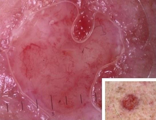


If the pattern includes dots then melanoma and other skin cancers such as SCC are differentials. If there are no dots then consider BCC, lichenoid keratosis, or porokeratosis.
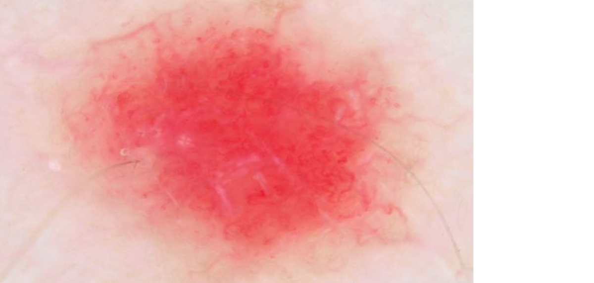
No pattern - featureless milky red
Radial vessels - radial infers pointing to the centre


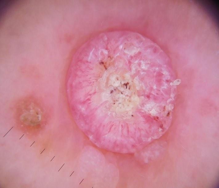

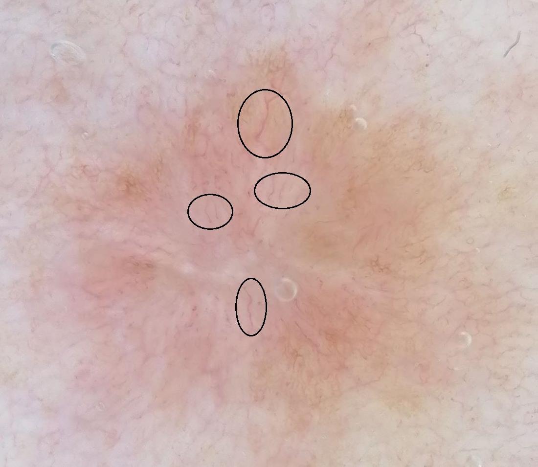
Peripheral:
BCC - thin looped vessels often described as hairpin-like, commonly sit parallel to each other.
SCC - usually more irregular in shape.
Radial vessels occasionally found in sebaceous gland hyperplasia and molluscum contagiosum.
Any site:
Dermatofibroma - most commonly outside the central white structures.
Crown vessels - consist of linear, curved vessels with minimal branching, situated along the periphery and extending toward the center without crossing it, giving the appearance of a crown


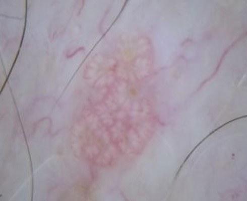


Other peripheral vessels


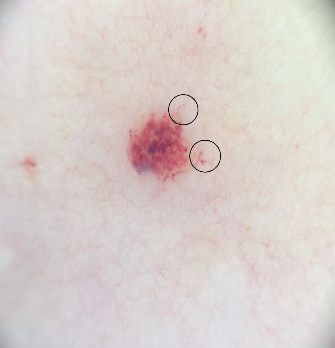
Clods


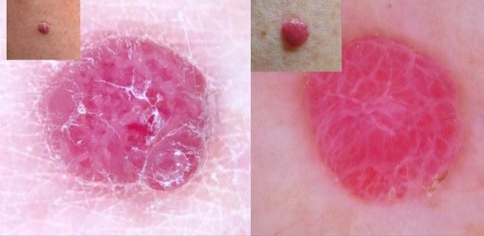
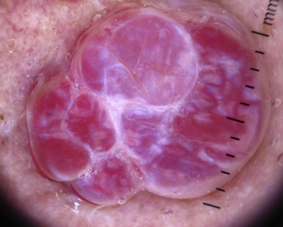

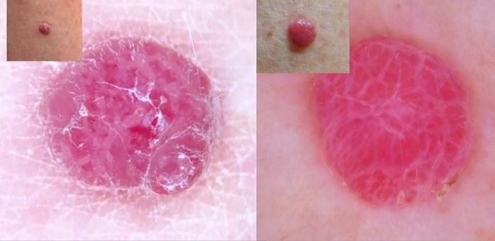
Angioma - well-demarcated red/purple/black clods (referred to as lacunae in angioma) separated by white fibrous stroma. Individual vessels often absent; occasionally seen at the periphery. Individual vessels running over the lesion and/or brown pigment suggest melanoma.
Pyogenic granuloma - largely red and homogenous, often intersected with white lines. Dermoscopcially difficult to differentiate from amelanotic melanoma. If vessels present, unlike in melanoma these tend to be regular and do not cross septal lines.
Lymphangioma - typical clods have the hypopyon sign with a two-tone appearance; darker red-blue at the base and lighter yellow or pink at the top. The clods are separated by pale septae. Sometimes individual vessels can be seen feeding the clods.
Looped vessels
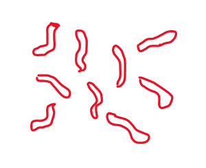


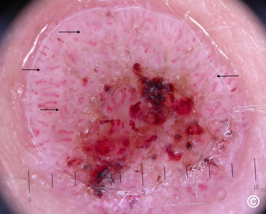


In seborrhoeic keratoses vessels are relatively uniform, sometimes sitting in a white halo, usually scattered throughout the lesions unless obscured by keratin.
More variable looped vessels can indicate an irritated seborrhoeic keratosis (should revert back to normal within 3 weeks), viral warts, or malignancy including melanoma and SCC/KA. In melanoma, the looped vessels do not have a white halo, which instead may be pink.
Frogspawn-like vessels - central vessel inside skin-coloured or pink clods



Arborising vessels - have the appearance of tree branches

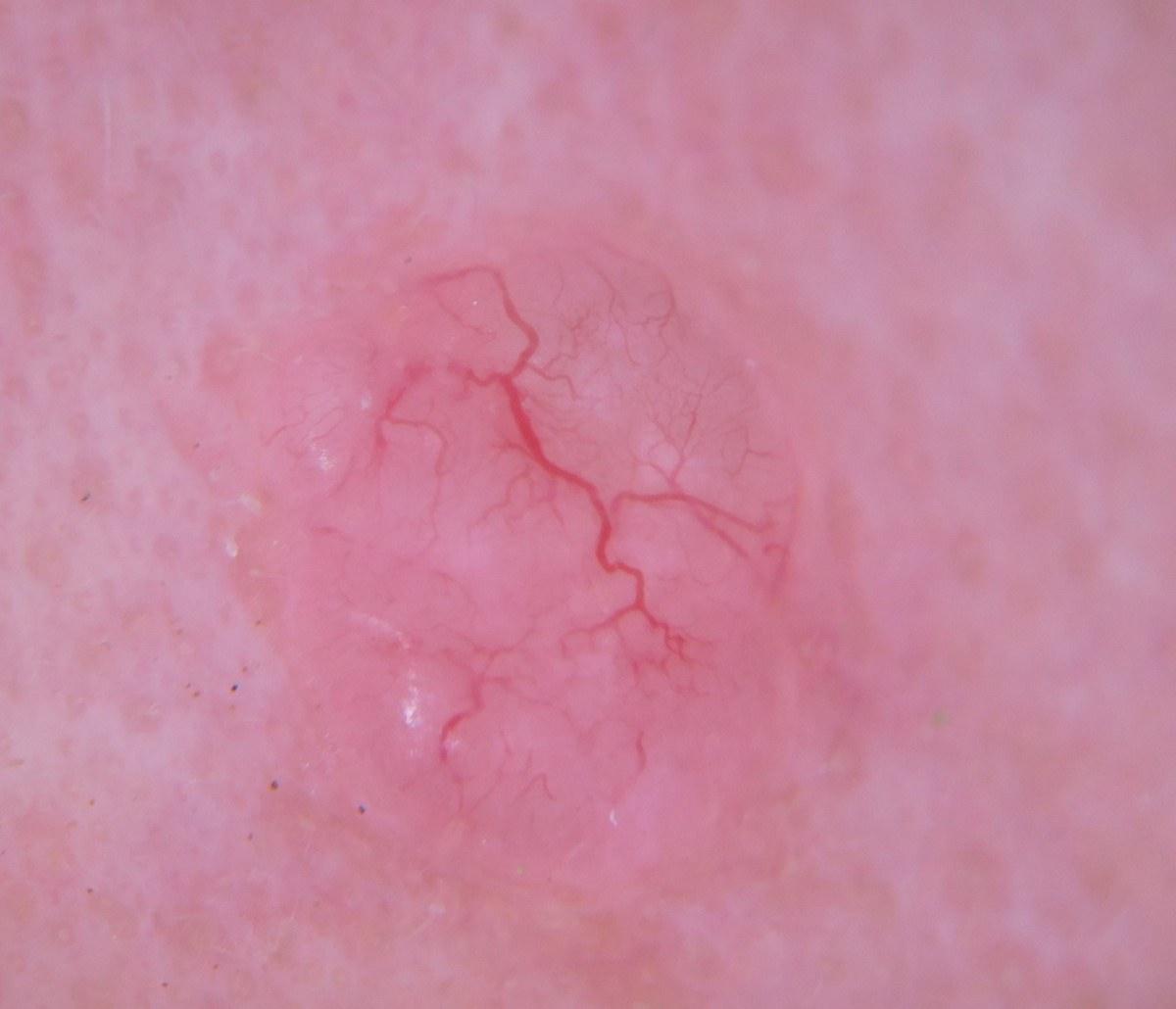

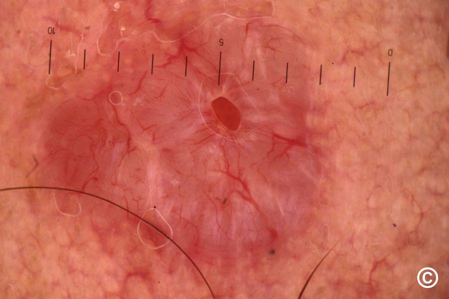

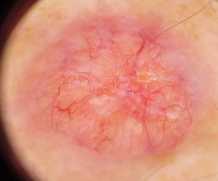
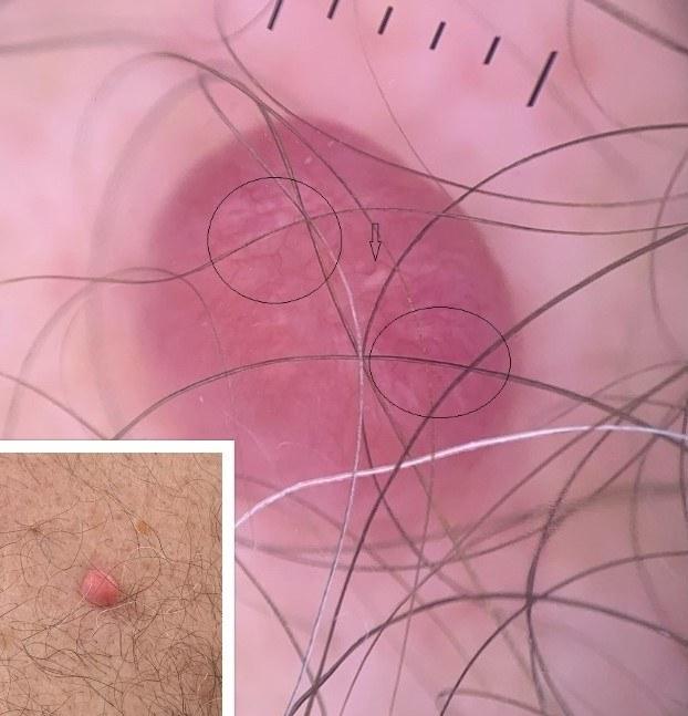

Sharply focused arborising vessels are commonly found in solid BCC, typical vessels start with a thicker calibre, which get thinner as they branch. They are not usually a feature of superficial BCC. Similar vessels can be found in benign adnexal tumours.
Arborising vessels can be found in any tumour under the superficial vascular plexus including dermal naevi, epidermoid cysts, fibrous papules, pilomatrixoma, juvenile xanthogranuloma, keloid/hypertrophic scars. Generally, in these other lesions vessels tend to keep the same calibre and may be less well focused.
Rarely, arborising vessels can be seen in melanoma and Merkel cell carcinoma.
Comma vessels (c-shaped) - slightly curved vessels that appear to take the shape of a comma. Vessels slightly out of focus


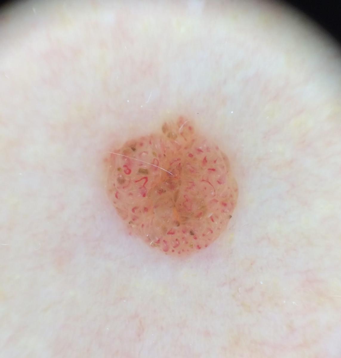

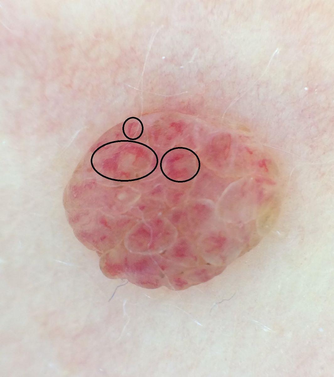

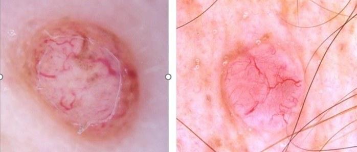
The typical comma vessel is broad and shaped like a comma; however, can can be of variable thickness and some less curved. Mainly found in benign melanocytic dermal naevi; occasionally fibrous papules (of the nose).
Dot vessels
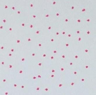

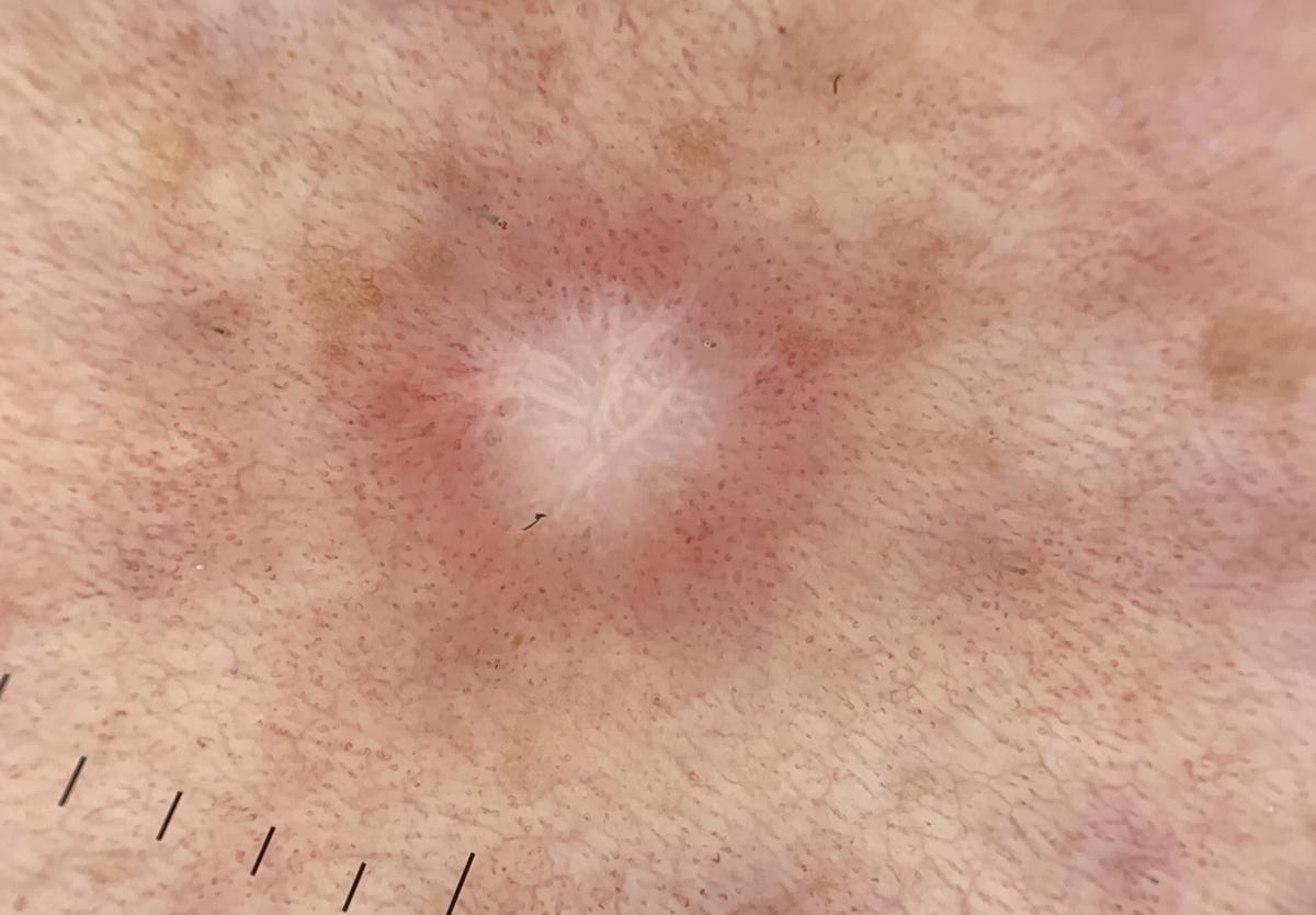
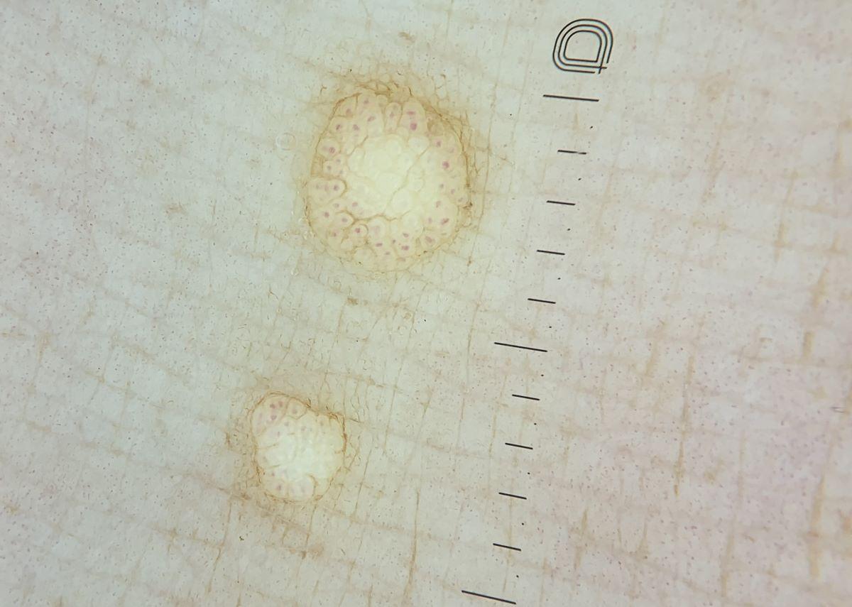
Benign melanocytic naevi, dermatofibromas, viral warts, molluscum contagiosum (a punctate pattern), Spitz naevi, melanoma (dots plus irregular linear vessels is the most common pattern in melanoma), inflammatory skin conditions.
Coiled (glomerular) vessels
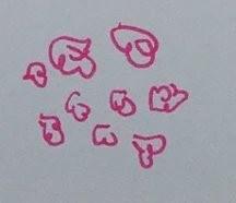

Thicker forms of clear cell acanthoma have a coiled rather than dotted appearance that form a serpentine pattern (see below). Eccrine poroma is usually a glomerular pattern (often in a halo) on a ''branch'', sometimes described as looking like cherry blossom.
Serpentine pattern - sometimes multiple vessels join up to form a serpentine, sometimes known as a ''string of pearls''


Polymorphous vessels / random vascular pattern / featureless milky red


Melanoma, SCC/KA, BCC, various uncommon malignancies, seborrhoeic keratoses, pyogenic granuloma.
Polymorphous vessels refers to 3 or more vessels types - in such cases the lesion is very likely to be malignant. One or two vessel types can be found in both benign and malignant lesions.
The most common vascular combination for melanoma is dot vessels plus linear irregular vessels. The presence of corkscrew vessels usually indicates melanoma. Areas of tan pigmentation can also be a clue.
Sometimes melanoma / SCC have no specific vascular clues and are just a pink EFG (all of elevated, firm, and growing), standing out as the ugly duckling. From a dermoscopic perspective such lesions may have a milky red featureless appearance.
The following pharmaceutical companies have had no involvement in the content of this website or in our conference programmes







