Cellulitis, erysipelas, and necrotising fasciitis
LAST UPDATED: Dec 13, 2024
Introduction
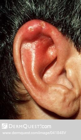
Cellulitis is a common bacterial infection of the dermal and subcutaneous tissue. Erysipelas is best regarded as a more superficial form of cellulitis. Cellulitis / erysipelas usually follow a breach in the skin, although a portal of entry may not be obvious. If treated promptly the infection is usually confined to the affected area, however, more severe episodes can lead to septicaemia.
Necrotising fasciitis (NF) is an uncommon but rapidly progressive and life-threatening infection of the deep dermis, adipose tissue and subcutaneous fascia.
This chapter is set out as follows:
Aetiology
Cellulitis / erysipelas
-
Streptococcal infection, especially group A (Streptococcus pyogenes), is the most common cause of cellulitis and erysipelas. Staphylococcus aureus can occasionally cause cellulitis
-
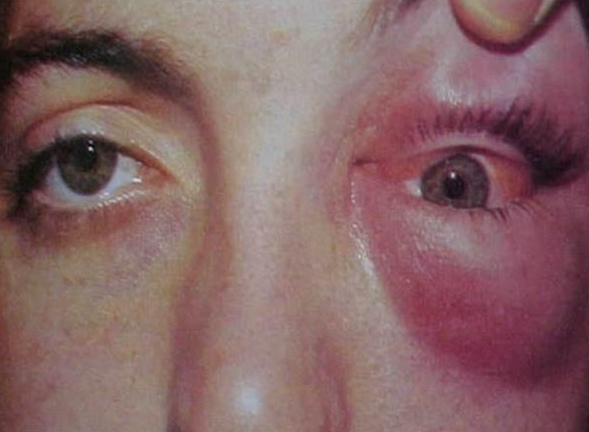
Other infections are rarely associated, except in certain groups eg immunocompromised patients (a range of organisms), unilateral childhood facial cellulitis can be due to Haemophilus influenza type b (although less so now since the introduction of the Hib vaccination), and orbital cellulitis (as opposed to the more common periorbital cellulitis) is often caused by Streptococcus pneumonia or one of the other sinus pathogens
- Risk factors for cellulitis / erysipelas
- Skin conditions of the lower legs such as gravitational eczema, leg ulcers, lymphoedema, tinea, trauma
- Previous episodes of cellulitis
- Obesity
- Immunocompromised patients
- Chronic disease such as diabetes mellitus, chronic liver or renal disease
- Pregnancy
Necrotising fasciitis
-
The most widely broadcast cases of necrotising fasciitis are caused predominantly by group A streptococci, and sometimes by staphylococcus aureus and other organisms. While the portal of entry is thought to be an open wound, such a finding may not be apparent
-
Other forms of necrotising subcutaneous infections include clostridial cellulitis (gangrene), and those resulting from multiple organisms, one of which is usually an anaerobe. Clostridial infections usually follow significant injury or surgery and result in gas under the skin, which clinically is felt as crepitus
History
-
Any age can be affected
-
Pain is a common feature - in necrotising fasciitis, pain is severe and often out of context to the clinical findings
-
Systemic features - depending on the severity can include malaise, fever, and vomiting
Clinical findings
Cellulitis and erysipelas
-
Distribution
-
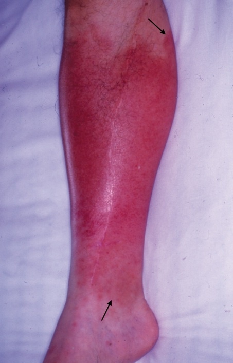
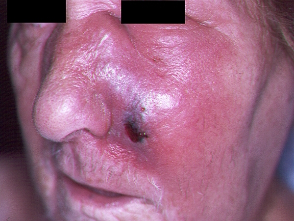
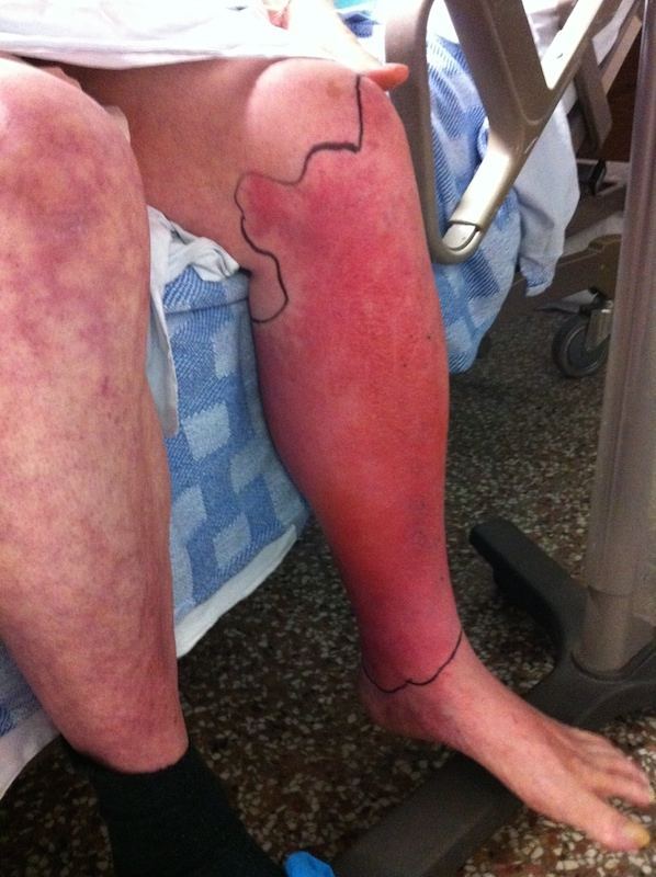
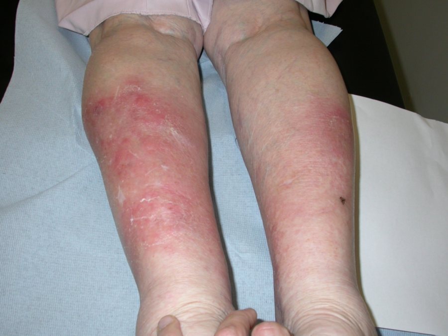
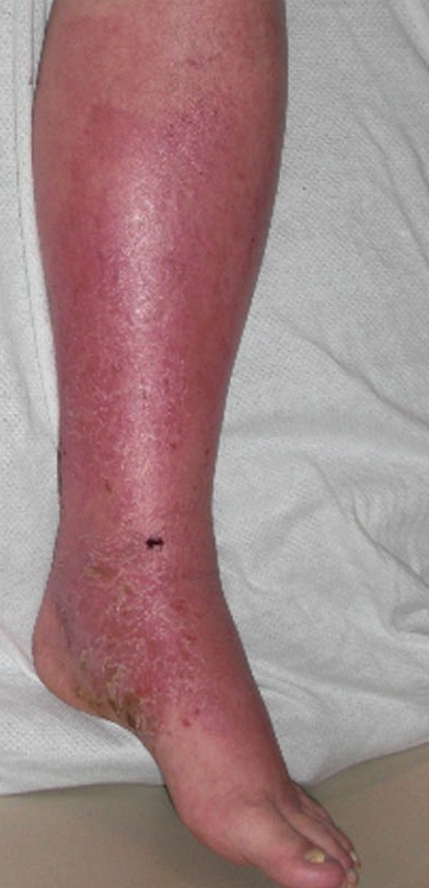
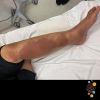
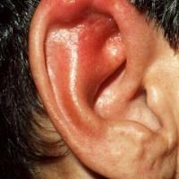
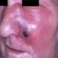
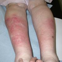
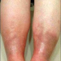
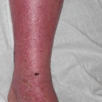
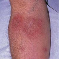
Although any site can be affected, the legs followed by the face are the most common sites
-
Facial involvement tends to be more superficial ie erysipelas
-
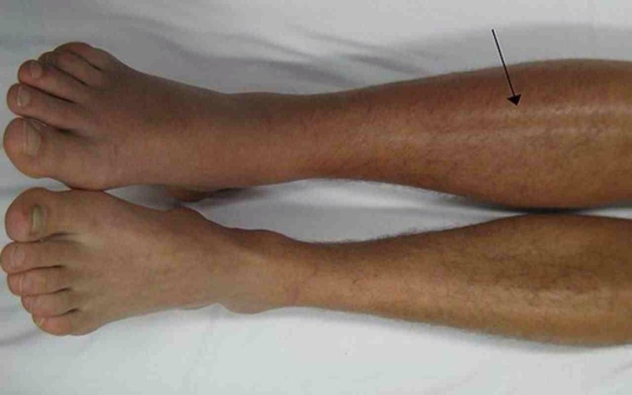
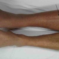
The rash is normally unilateral, except on the face where it is occasionally bilateral
-
Morphology
-
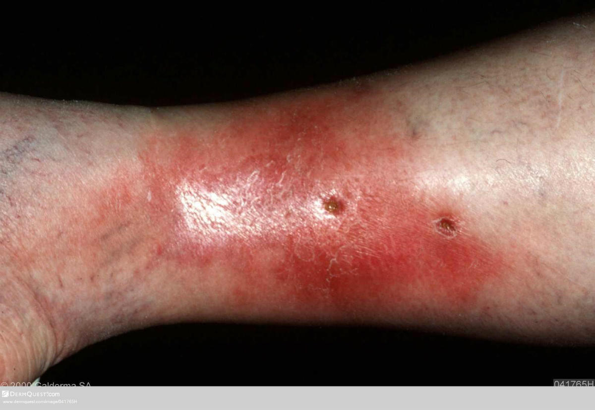
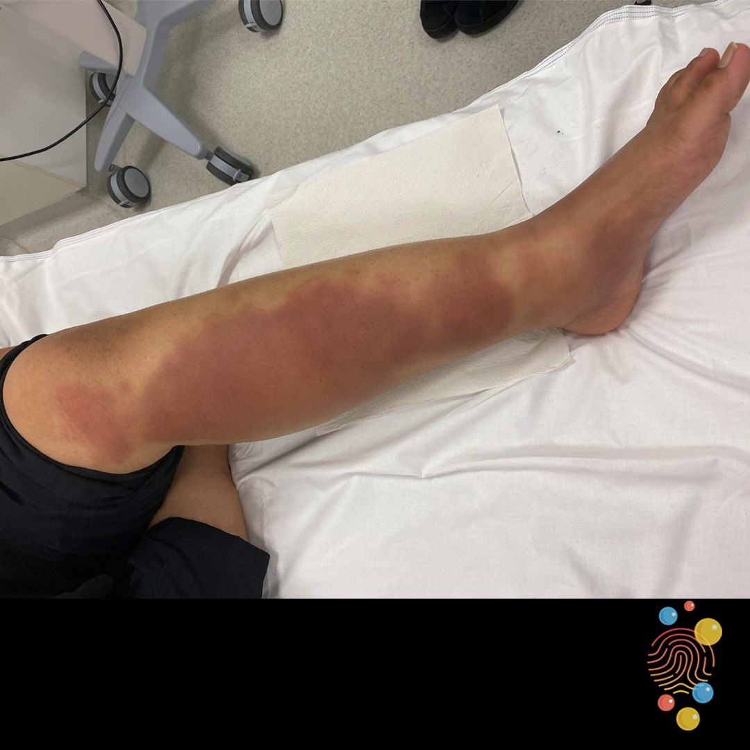
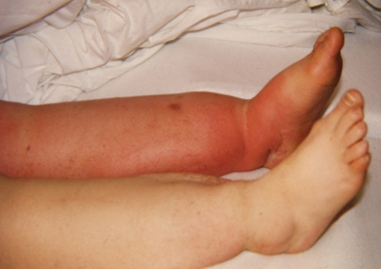
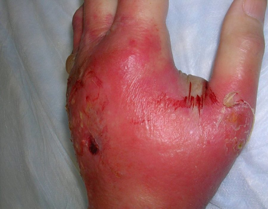
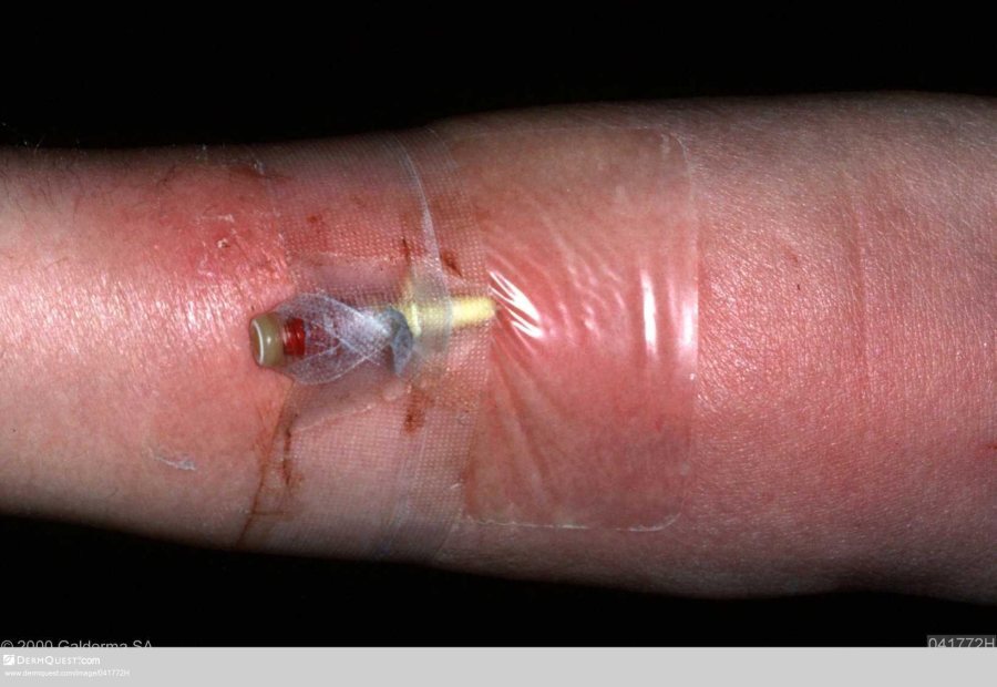
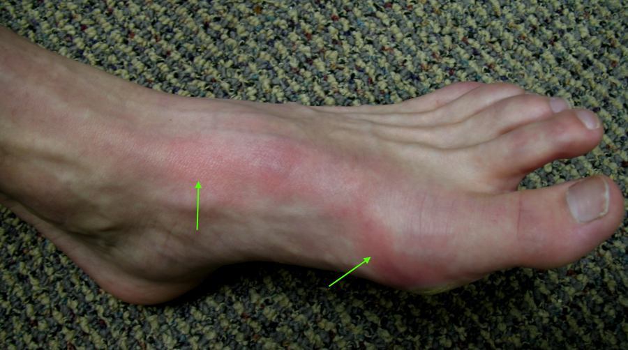
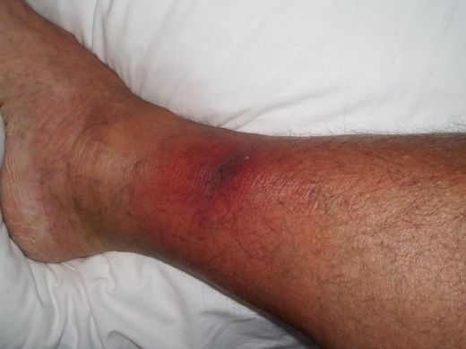
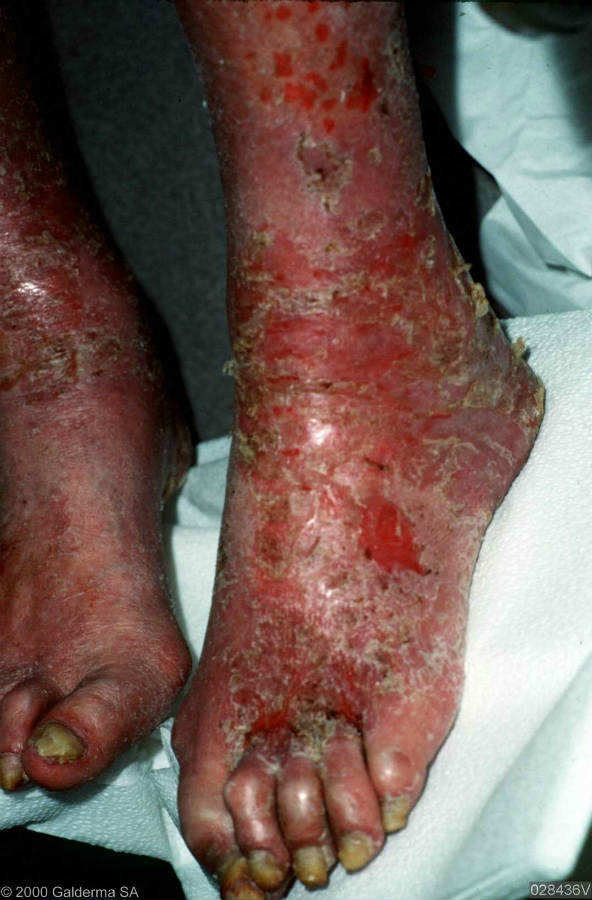
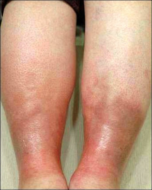
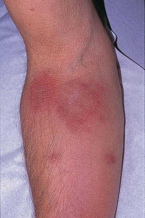
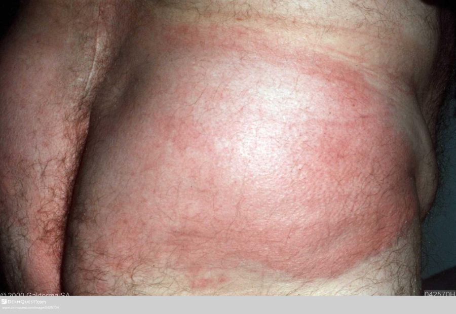
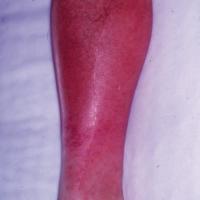
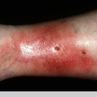
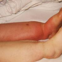
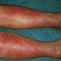
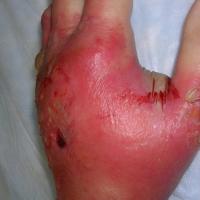
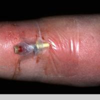
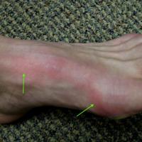
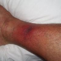
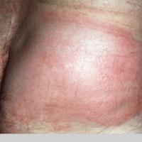
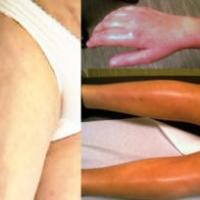
Erythema - the edge is more well-demarcated in erysipelas than in cellulitis
-
Oedema is common
-
Hot and tender to touch
-
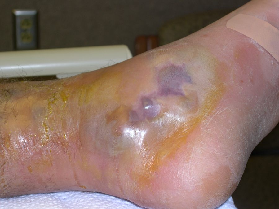
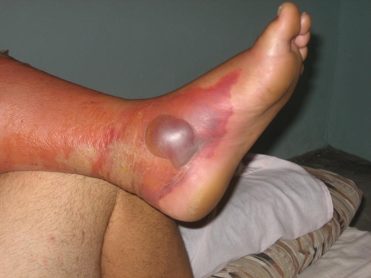
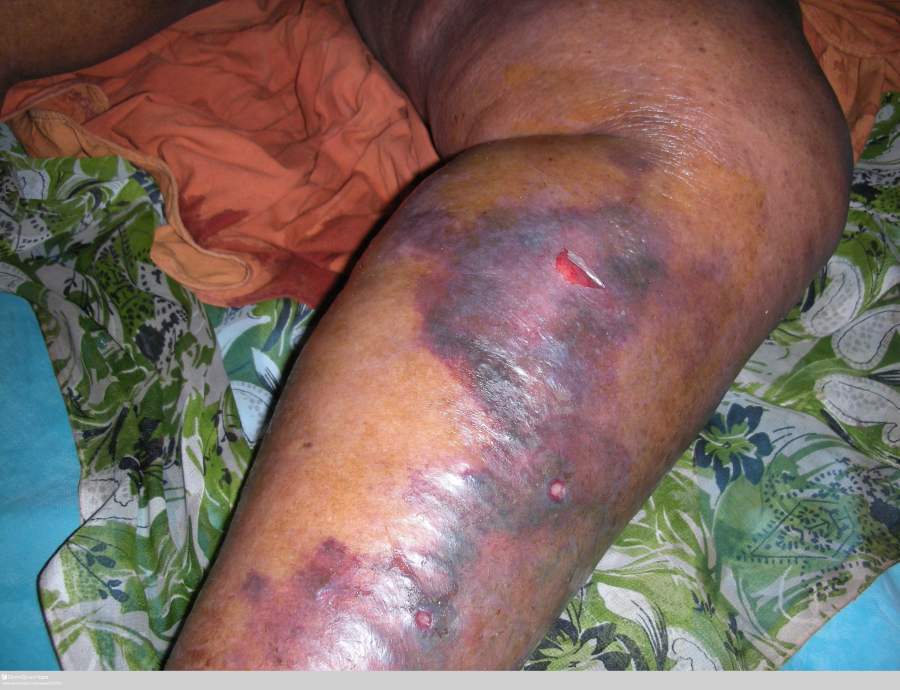
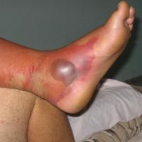
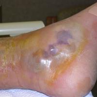
The presence of haemorrhage, bullae, or lymphangitis suggest streptococcal infection as opposed to staphylococcal infection
-
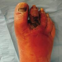
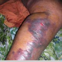
Severe cellulitis can lead to ulceration and more deep-seated tissue damage
-
Complications
-
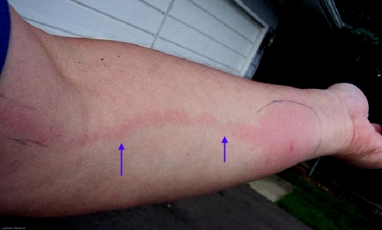
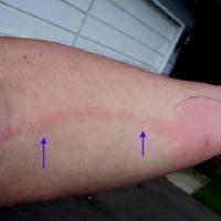
Lymphangitis presents with a red line originating from the cellulitis and spreading proximally to lymph nodes along the lymphatic vessels
-
Deep-seated infection eg fasciitis and myositis
-
Septicaemia, which can occasionally be fatal
-
Nephritis
Necrotising fasciitis
Early diagnosis of NF is vital in terms of reducing morbidity and mortality, and should be suspected in the following scenarios:
- If the level of pain and tenderness, or systemic upset, is out of proportion to the physical signs
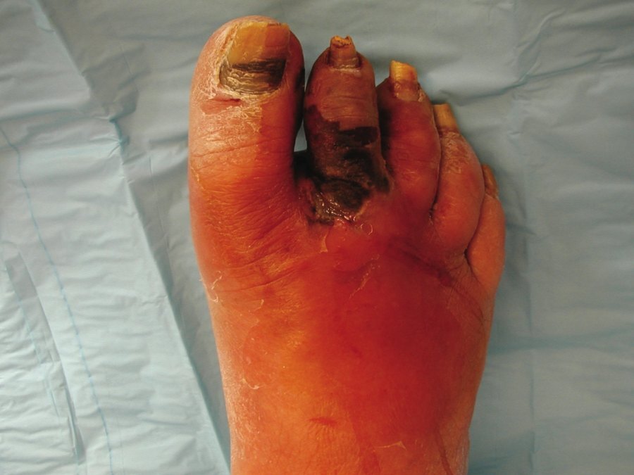
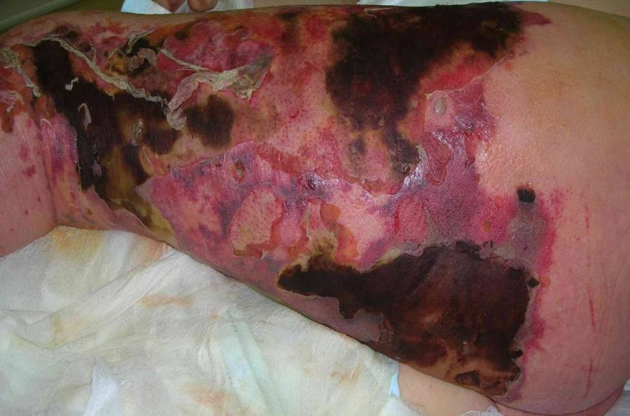
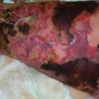
- Dusky-violaceous areas along with erythema
- Crepitus
- Over a short period of time blisters develop, the affected area becomes necrotic, and the patient very toxic
Differential diagnosis
-
Deep vein thrombosis
-
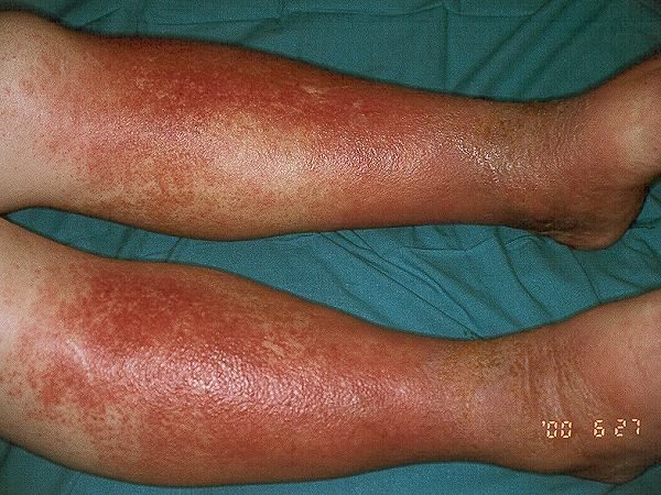
Gravitational eczema
-
Many patients with gravitational eczema are incorrectly diagnosed of having bilateral cellulitis, which is extremely rare, and are inappropriately given systemic antibiotics over several months
-
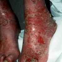
Patients with gravitational eczema will have itch, non-tender erythema and sometimes areas of brown discoloration
-
Gravitational eczema can become infected, which occasionally evolves in to a secondary cellulitis
-
Refer to the chapter Gravitational eczema
-
Contact allergic dermatitis
-
Can present acutely as erythematous, sore and tender areas of skin, sometimes with blisters
-
Refer to the chapter Contact allergic dermatitis
-
Panniculitis, including sclerosing panniculitis (syn. acute lipodermatosclerosis)
-
Causes multiple tender nodules and plaques
-
The acute stage of lipodermatosclerosis causes tender erythema of the lower legs. However, compared to cellulitis, lipodermatosclerosis is usually bilateral and patients are afebrile
-
Refer to the chapter Panniculitis
-
Eosinophilic cellulitis (syn. Wells syndrome)
-
Is rare
-
Clinically the condition presents with large, indurated erythematous plaques, and less commonly nodules, that evolve over several weeks
-
The majority of patients have a blood eosinophilia
-
Refer to the chapter Eosinophilic cellulitis
-
Eosinophilic fasciitis
-
Is also rare
-
The cutaneous manifestations of eosinophilic fasciitis evolve as the condition progresses. The acute inflammatory stage consists of pain, swelling and tenderness of the distal limbs. These findings are later replaced by induration, and eventually fibrosis with limitation of the movement of the hands and feet. The affected skin is taut and firmly adherent to underlying tissue with dimpling and a peau d'orange appearance
-
The condition has a symmetrical distribution
-
There is a blood eosinophilia in 70% of cases
-
Refer to the chapter Eosinophilic fasciitis
Clinical Images
Please refer to notes on image rights at bottom of the page with regards to individual image
ownership.
1
2
3
4
5
6
7
8
9
10
11
12
13
14
15
16
17
18
19
20
21
22
23
24
25
26
27
Investigations
-
Skin swabs should be taken, and if present from open areas / areas of exudate, although in many cases results are negative. If bullae or abscesses form, culturing the fluid from inside these lesions yields an organism in more than 90% of cases
-
Blood cultures will be needed for inpatients
Management
Patients needing admission
-
Necrotising fasciitis
-
Have a low threshold for admitting patients with possible necrotising fasciitis, which should be suspected at an early stage if the level of pain and tenderness, or systemic upset, is out of proportion to the physical signs
-
Patients require urgent surgery to remove affected tissue, and high dose antibiotics
-
If diagnosed and treated early, most patients will survive with minimal scarring. However if there is significant tissue loss, later skin grafting will be necessary and in some patients amputation of limbs is required. Up to 25% of patients will die from the disease
-
The following cases of cellulitis:
-
Associated with marked systemic upset eg fever, vomiting, tachycardia, hypotension
-
Spreading cellulitis that is not responding to oral medication
-
Lymphangitis
-
Have a lower threshold for admitting those more at risk eg infants and very young children, the very elderly, immunocompromised patients, and those with significant comorbidities
-
Orbital cellulitis
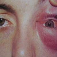
- Features of which include the sudden onset of unilateral swelling of conjunctiva and lids, painful or restricted eye movements, proptosis, diplopia, reduced visual acuity, and abnormal pupil reactions
Management of uncomplicated cellulitis (and erysipelas)
Initial treatment of cellulitis
-
Antibiotic
-
First line treatment - flucloxacillin 500 mg QDS (adjust doses accordingly in children), which is bactericidal on streptococci and staphylococci
-
For penicillin allergy use clarithromycin 500 mg BD (adjust doses accordingly in children)
-
For patients on statin therapy who are penicillin allergic, clarithromycin can be used if the statin is withheld during treatment. Alternatively use doxycycline 200 mg taken for the first day and then 100 mg daily
-
Facial cellulitis - consider co-amoxiclav if sinus pathogens are a possible cause
- Duration of treatment - in terms of antibiotic stewardship, NICE recommend a minimum of 5 days treatment; however, some patients may need longer. We also recommend referring to local guidance. Ultimately, treatment should be tailored to the individual patient
-
For the lower leg - rest and elevate the affected area where possible to reduce pain, swelling and damage to the venous system
-
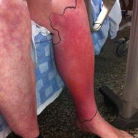
Mark out the extent of the cellulitis and review the patient while on treatment. Give advice and ask the patient to report immediately if the condition deteriorates (spreading infection / systemic symptoms), or if the antibiotics are not tolerated
Post-treatment of cellulitis
Identify and manage any causal / risk factors, which may include:
-
Treatment of lower leg skin conditions such as gravitational eczema, leg ulcers, and oedema. If tinea is present, treat accordingly, and then consider prophylactic treatment with terbinafine cream once or twice a week to the plantar surface of the feet and in-between the toes
-
Weight control and adequate glycaemic control if diabetic
Treatment of recurrent cellulitis
Antibiotic prophylaxis should be offered to patients who have two or more attacks of cellulitis per year:
-
Penicillin V 250 mg BD (1g if weight >75kg) should be the first choice. The dose may be reduced to 250 mg OD after one year of successful prophylaxis. For those allergic to penicillin, clarithromycin 250 mg daily (OR erythromycin 250 mg BD) is recommended. For those both allergic to penicillin and on statins use doxycycline 50 mg OD
-
Prophylaxis may need to be life-long if relapse occurs when antibiotics are discontinued after a two year period of successful prophylaxis
Other resources
Disclaimer - the author PCDS cannot accept responsibility for any misleading or incorrect statements, and the management of individual patients remains the direct responsibility of the individual doctor. We do however hope that visitors to this site can contact us regarding comments that are considered misleading or incorrect so that we can continue to improve the site.
Image Rights - The PCDS would like to thank Dermatoweb, DermQuest (Galderma), and others who have contributed images. All named individuals and organisations maintain copyright for the relevant images.