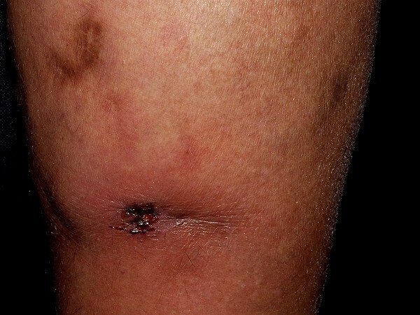
Panniculitis
LAST UPDATED: Aug 09, 2021
Introduction
Panniculitis refers to a group of conditions with inflammation of the adipose tissue, the most well-known of which is erythema nodosum. The detail of panniculitis lies outside the remit of this website, consequently this chapter provides only an overview and is set out as follows:
Aetiology
Panniculitis may be classified histologically according to whether it is septal, lobular, mixed or associated with vasculitis, within each group there are multiple aetiologies. The list that follows gives a flavour of the different causes, for more information further reading will be required (eg Rook's textbook of dermatology).
Septal panniculitis
-
Eosinophilic panniculitis
-
Is characterised by a prominently eosinophilic infiltration of subcutaneous fat
-
It is not a disease entity but a reactive process, associated with patients who have a variety of associated clinical conditions. Once a diagnosis of eosinophilic panniculitis has been established, appropriate evaluation for an associated clinical condition should be performed
Lobular panniculitis
-
Nodular panniculitis
-
Most commonly seen in young women
-
Dull red tender nodules measuring 1-2 cm in diameter
-
Mainly affects lower limbs but other sites can be affected
-
Often resolves with hyperpigmentation or atrophy
-
Sometimes accompanied by fever and arthralgia, known as relapsing febrile nodular panniculitis
-
Lipoatrophic panniculitis
-
A rare disease that usually affects children and characterised by eruption of tender erythematous nodules and plaques principally of the arms and legs that leave residual circumferential bands of lipoatrophy
-
Associated with crystal deposition
-
Examples include gout and renal failure
-
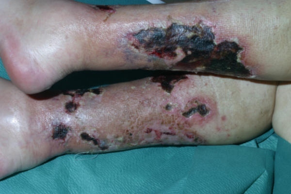
With renal failure the changes are part of a process known as calciphylaxis, a condition which remains a highly morbid and poorly understood disease of vascular calcification and tissue necrosis, most frequently of the skin, but also affecting other organs such as the heart and gastrointestinal tract. While most frequently described in patients with end-stage renal disease (1-4% of this population), calciphylaxis is seen in other settings, such as multiple myeloma, polyneuropathy, organomegaly, endocrinopathy, cirrhosis, and rheumatoid arthritis
-
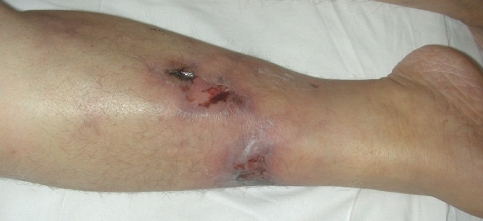
Clinically, nodules or plaques tend to progress quickly to livedo-like areas and then ulcerate. The thighs, abdomen and buttocks are the most commonly affected sites. Ulceration is associated with severe pain
-
In patients with renal failure, lesions on the trunk are associated with a much higher mortality rate than lesions on the legs
-
Enzymic panniculitis - this has been associated with acute / chronic pancreatitis, as well as pancreatic carcinoma
-
Fat necrosis - cold injury
-
This is a localised reaction occuring secondary to cold injury
-
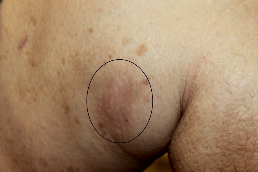
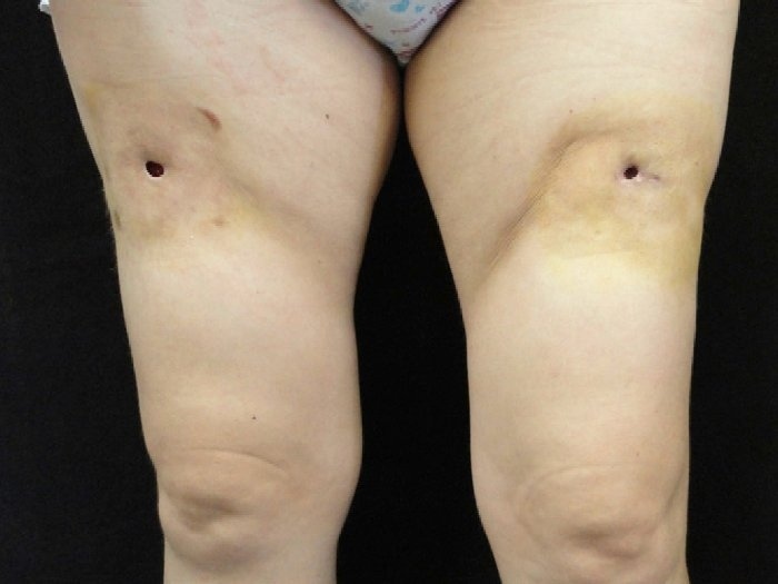
Cold panniculitis of the thighs or buttocks may occur in horse riders or skiers wearing inadequate clothing, especially if tight fitting. Wearing adequate clothing will often lead to an improvement of symptoms
-
The affected skin is ill-defined, red-blue and cold to touch
-
Alpha-1-antitrypsin deficiency
-
Tends to be severe
-
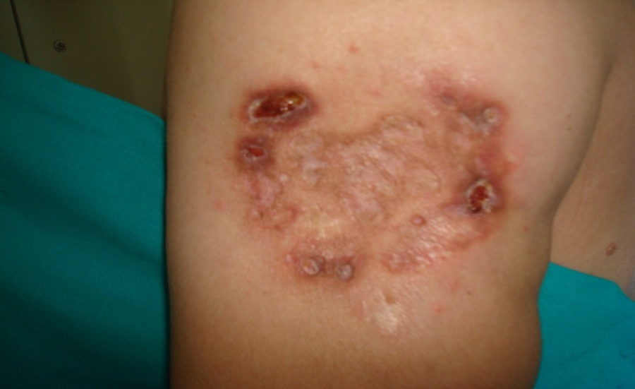
Lesions, often with ulceration, occur predominantly on the trunk and proximal limbs
-
Subcutaneous t-cell lymphoma - a rare form of cutaneous lymphoma, which can for long periods of time mimic benign forms of panniculitis
Mixed panniculitis
-
Lipodermatosclerosis (syn. sclerosing panniculitis)
-
This typically arises in patients with venous insufficiency. Obesity is a common factor
-
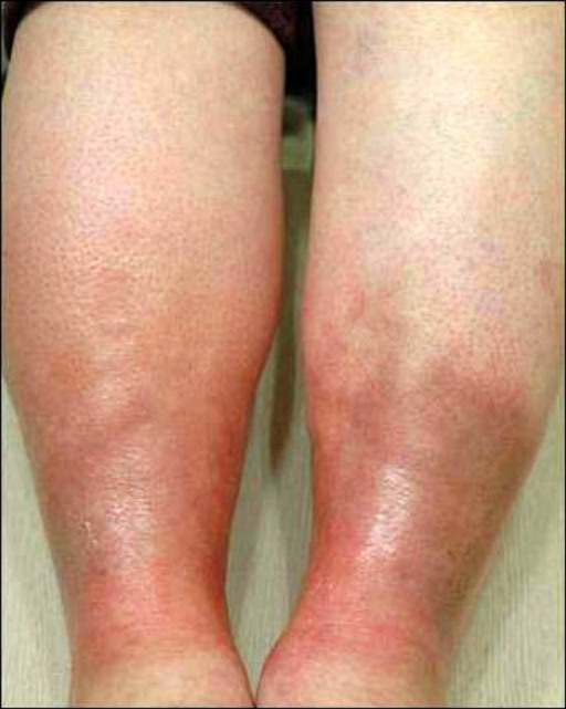
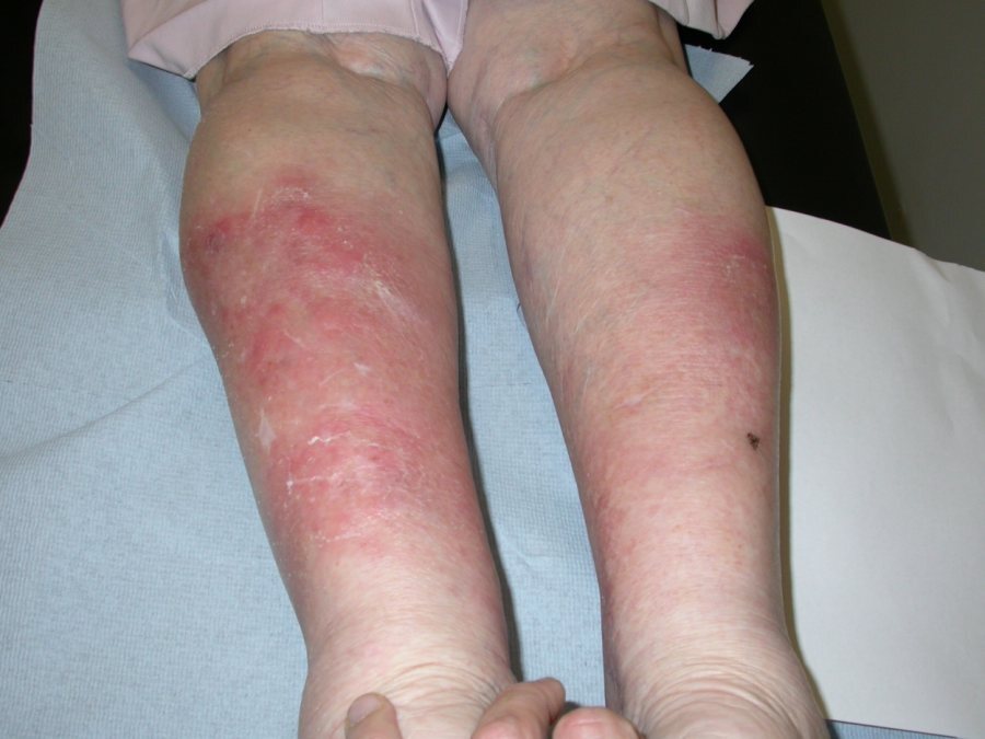
The acute form presents as episodes of painful inflammation in the lower legs, above the ankles, resembling cellulitis. It can affect one or both legs
-
Chronic lipodermatosclerosis may follow an acute episode or develop gradually. Features of chronic lipodermatosclerosis include pain, hardening of the skin, colour change (red / brown), atrophie blanche (localised white areas), an ''inverted champagne bottle” or “bowling pin” appearance, and leg ulcers
-
For more information refer to the chapter Leg ulcers (and disorders of venous insufficiency)
-
Lupus profundus
-
A rare entity with deep-seated inflammation
-
More common in young women
-
The overlying skin may be associated with discoid lupus in 20% of cases
-
Any site can be affected. Lesions may ulcerate and scar
-
Other connective tissue panniculitis
-
Panniculitis associated with complement deficiency
-
Infective panniculitis
-
This is becoming more of a problem in patients with immunodeficiency
-
Hepatitis has been linked
-
Factitial panniculitis - this is self-inflicted and may be caused by injection or less commonly blunt trauma
Panniculitis with vasculitis
-
Most deep-seated vasculitis syndromes, especially the large vessel vasculitis eg polyarteritis nodosa, begin as a septal panniculitis and involve the fat lobules later
-
Nodular vasculitis (syn. erythema induratum of Bazin and Whitfield)
-
This is a specific cause of panniculitis
-
It is a chronic relapsing lobular panniculitis with septal vasculitis
-
The condition is typically seen in healthy, middle-aged, sometimes obese women with venous stasis
-
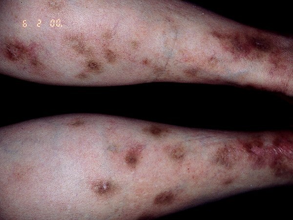
Distribution - often affects the posterolateral aspects of the lower third of the legs. Can affect one or both legs
-
Morphology - tender, dusky nodules or plaques. Although in most cases no cause will be identified, it is important to exclude tuberculosis, which is more likely if lesions are ulcerated. Deep incisional biopsies should be taken for both histopathological examination, and tissue culture to look for either mycobacterium tuberculosis, or atypical mycobacteria. The use of PCR (polymerase chain reaction) testing aids in diagnosis and significantly reduces the number of false negative results
- For more information refer to the chapter Vasculitis
Clinical findings
-
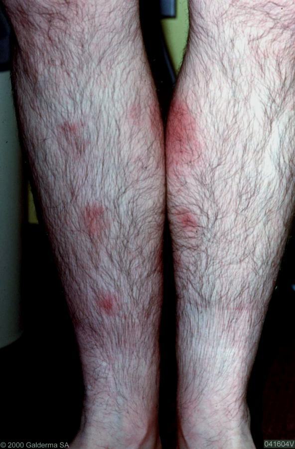
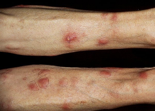
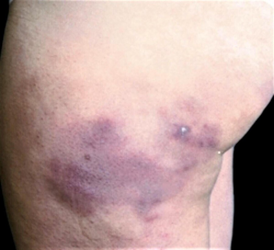
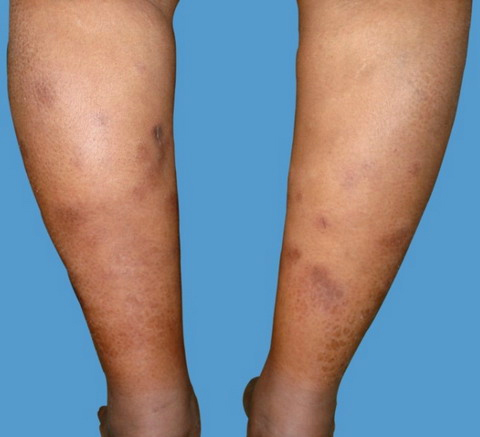
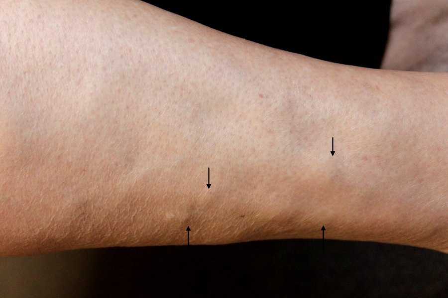
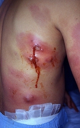
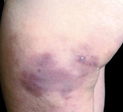
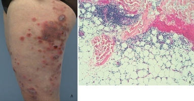
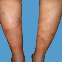
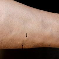
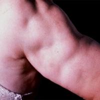
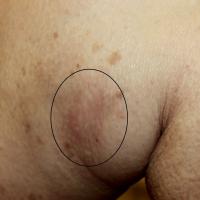
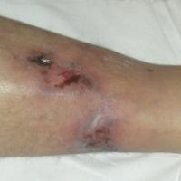
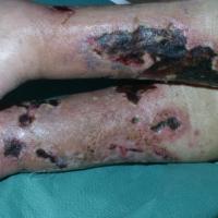
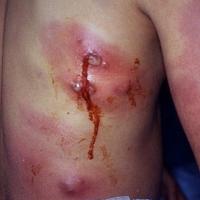
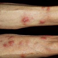
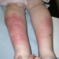
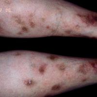
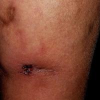
In general, lesions are nodular, plaque-like or indurated, and tender to touch
-
Depending on the type there may be ulceration, atrophy and scaring
Clinical Images
Please refer to notes on image rights at bottom of the page with regards to individual image
ownership.
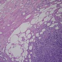
1
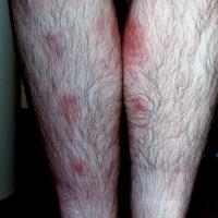
2
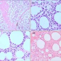
3
4
5
6
7
8
9
10
11
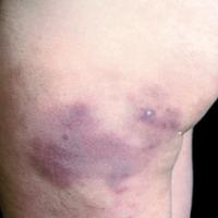
12
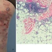
13

14
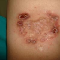
15
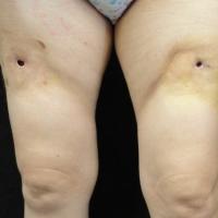
16
17
18
19
Investigations
-
Refer to section on management
Management
Step 1 - erythema nodosum / other uncomplicated cases
- Refer to the chapter Erythema nodosum if typical features
- Many cases of cold panniculitis can be improved by wearing adequate clothing, which is not tight fitting
-
For lipodermatosclerosis refer to the chapter Leg ulcers (and disorders of venous insufficiency)
-
If the features are atypical (atypical site ie away from lower legs, ulceration, not settling within six weeks) referral is indicated
- Conservative measures - rest and elevate the affected area. NSAID can help with pain
Step 2 - referral
At the same time as referring it can be helpful to organise investigations:
-
Those as for erythema nodosum (FBC, throat swab, ASO titre, CXR)
-
In addition - routine biochemistry screen, autoimmune screen (including ENA autoantibodies), complement levels, immunoglobulins with plasma electrophoresis, alpha-1-antitrypsin levels. If the patient is potentially immunocompromised also consider HIV / hepatitis screen
Step 3: management in Secondary Care
-
One or more deep incisional biopsies is usually required to ascertain the diagnosis
-
Treatment depends on the cause
-
If no cause is found treatment options include hydroxychloroquine with or without mepacrine, tetracyclines, dapsone, corticosteroids (intra-lesional or systemic) and potassium iodide
Disclaimer - the author PCDS cannot accept responsibility for any misleading or incorrect statements, and the management of individual patients remains the direct responsibility of the individual doctor. We do however hope that visitors to this site can contact us regarding comments that are considered misleading or incorrect so that we can continue to improve the site.
Image Rights - The PCDS would like to thank Dermatoweb, DermQuest (Galderma), and others who have contributed images. All named individuals and organisations maintain copyright for the relevant images.