Eosinophilic fasciits
LAST UPDATED: Jul 01, 2023
Introduction
Eosinophilic fasciitis is a rare scleroderma-like condition characterised by an acute presentation of pain, swelling and tenderness of the distal limbs, progressing to fibrosis, which can result in reduced mobility of the hands, feet and other joints.
This chapter is set out as follows:
Aetiology
History
-
Eosinophilic fasciitis predominantly occurs in Caucasians, with females and males equally affected
-
It may occur at any age, although most cases arise between the ages of 30 and 60 years
-
It has an acute, painful onset
-
Malaise, weakness, and fever are frequently present
Clinical findings
-
There are three cutaneous stages
-
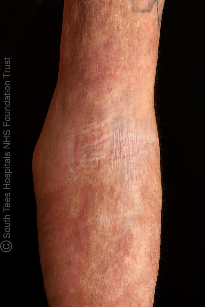
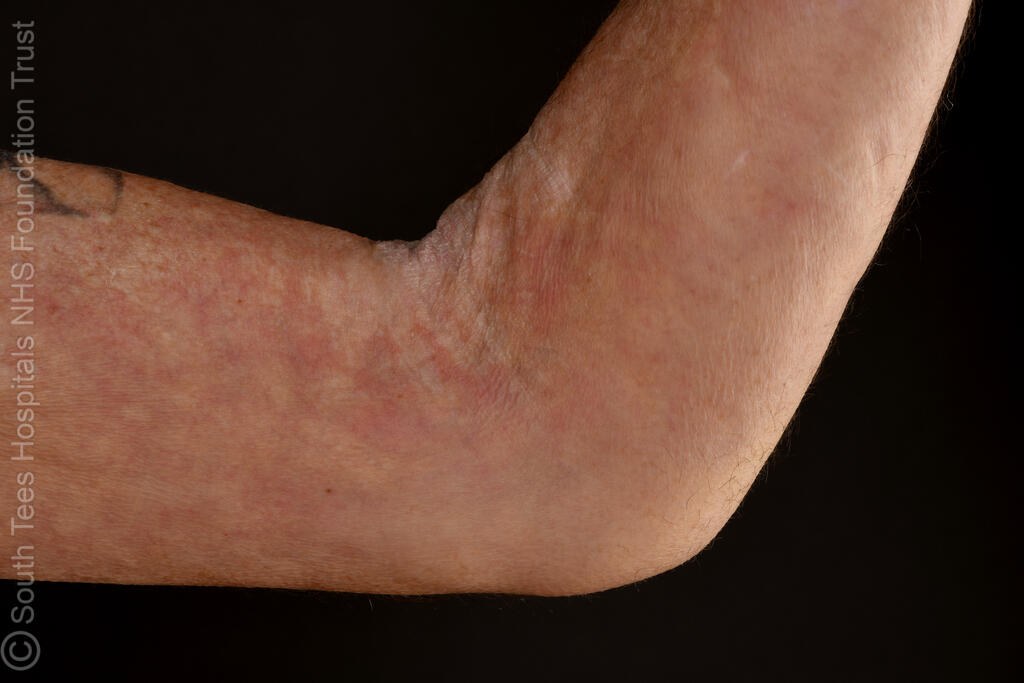
The initial phase is a symmetric, diffuse, erythematous tenderness of the distal limbs. The trunk may be involved, but the hands, feet, and the face are spared
-
The skin then becomes oedematous with a characteristic orange-peel appearance
-
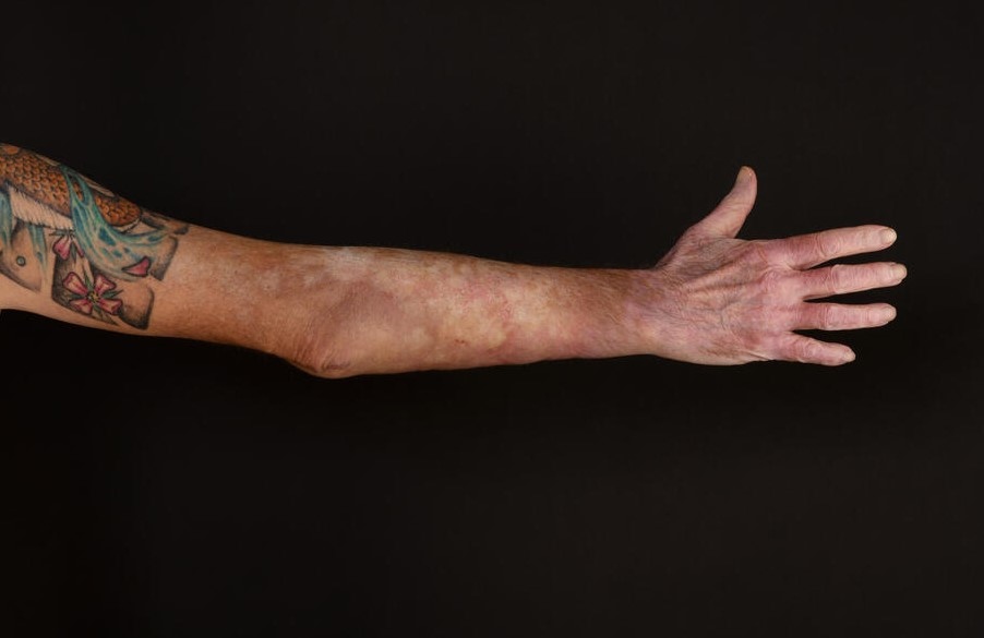
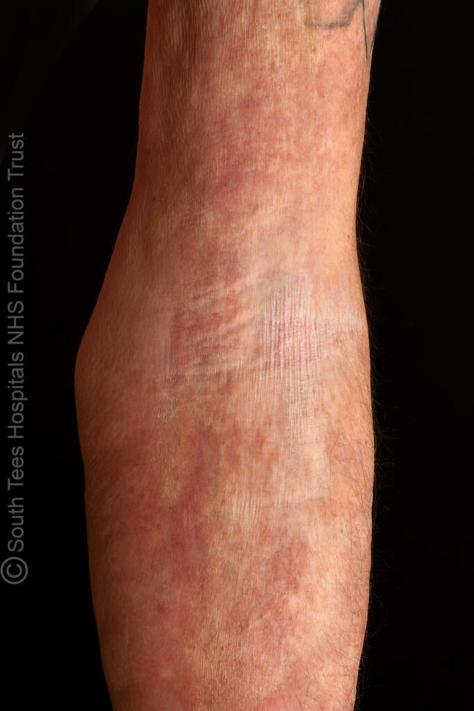
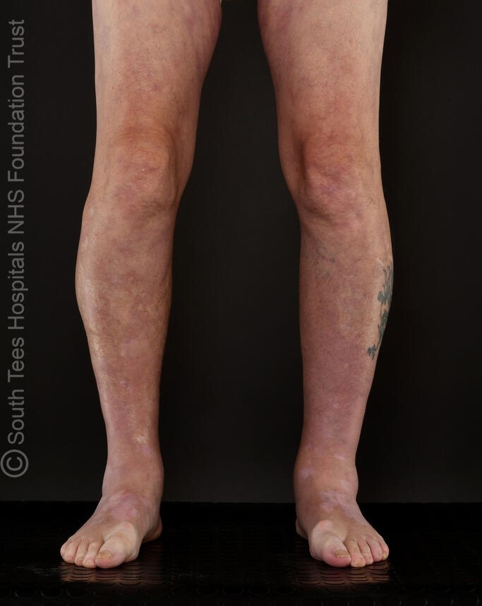
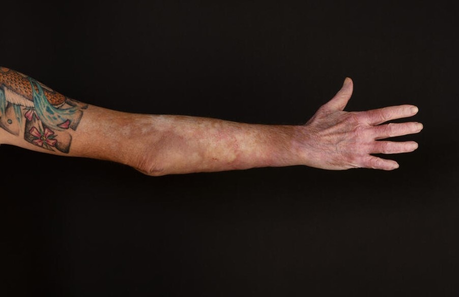
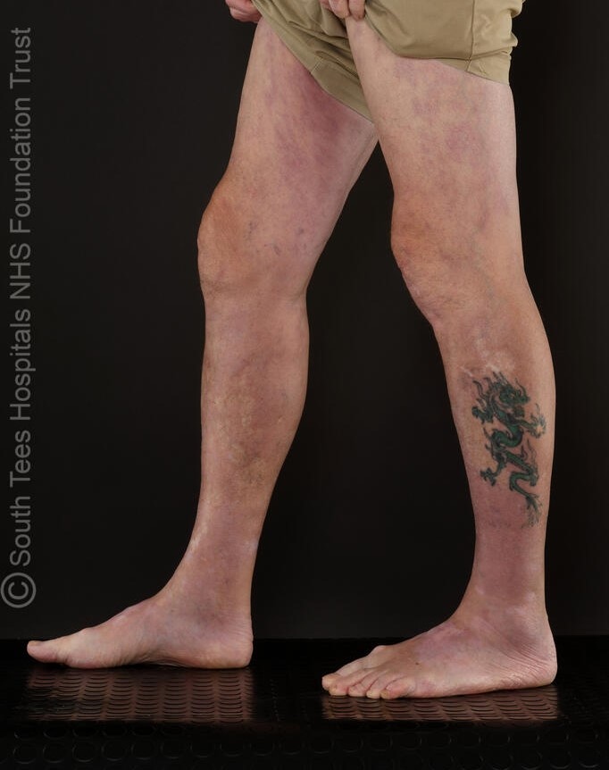
The final phase involves fibrotic change with areas of hypopigmentation, induration, and skin tightness of the ankles and wrists
-
In severely affected cases both the skin and subcutaneous tissues are bound-down to the underlying muscle, with a woody-like appearance. In such cases joint contractures can be more extensive and involve the knees, elbows, and shoulders
-
Other clinical features sometimes reported include the Carpal Tunnel syndrome, and inflammatory arthritis
- Differential
- Pansclerotic morphoea has a more insidious onset and is characterised by near total body surface involvement, with circumferential lesions, sparing the fingers and toes, which usually extends into the subcutaneous tissue, and may affect the fascia, muscle, and bone
- Unlike in systemic sclerosis, Raynaud's phenomenon, abnormal nailfold capillaries, and sclerodactyly are absent
Clinical Images
Please refer to notes on image rights at bottom of the page with regards to individual image
ownership.
Investigations
-
A FBC reveals eosinophilia in 70% of cases. Some patients have an elevated ESR and hyperglobulinaemia
-
A full-thickness skin biopsy that includes the dermis, subcutaneous fat and fascia is necessary to confirm a diagnosis of eosinophilic fasciitis, and shows dermal sclerosis, with inflammation and fibrosis of the fat and deep fascia. The fascia is thickened and infiltrated with several cell types including lymphocytes and eosinophils (reference: Rook's Textbook of Dermatology)
- If clinically indicated, MRI of the involved areas shows a high-intensity signal in the fascia
Management
-
Oral corticosteroids are the mainstay of treatment, with most patients responding well to moderate-to-high doses, particularly if started early in the course of the condition. Maintenance low-dose treatment may be required for 2-5 years
-
Steroid-sparing drugs such as hydroxychloroquine, methotrexate, and ciclosporin are sometimes required. More recently there has been success with the biologics
-
Physiotherapy is required to prevent and treat joint contractures
-
In some cases the disease resolves spontaneously
Disclaimer - the author PCDS cannot accept responsibility for any misleading or incorrect statements, and the management of individual patients remains the direct responsibility of the individual doctor. We do however hope that visitors to this site can contact us regarding comments that are considered misleading or incorrect so that we can continue to improve the site.
Image Rights - The PCDS would like to thank Dermatoweb, DermQuest (Galderma), and others who have contributed images. All named individuals and organisations maintain copyright for the relevant images.