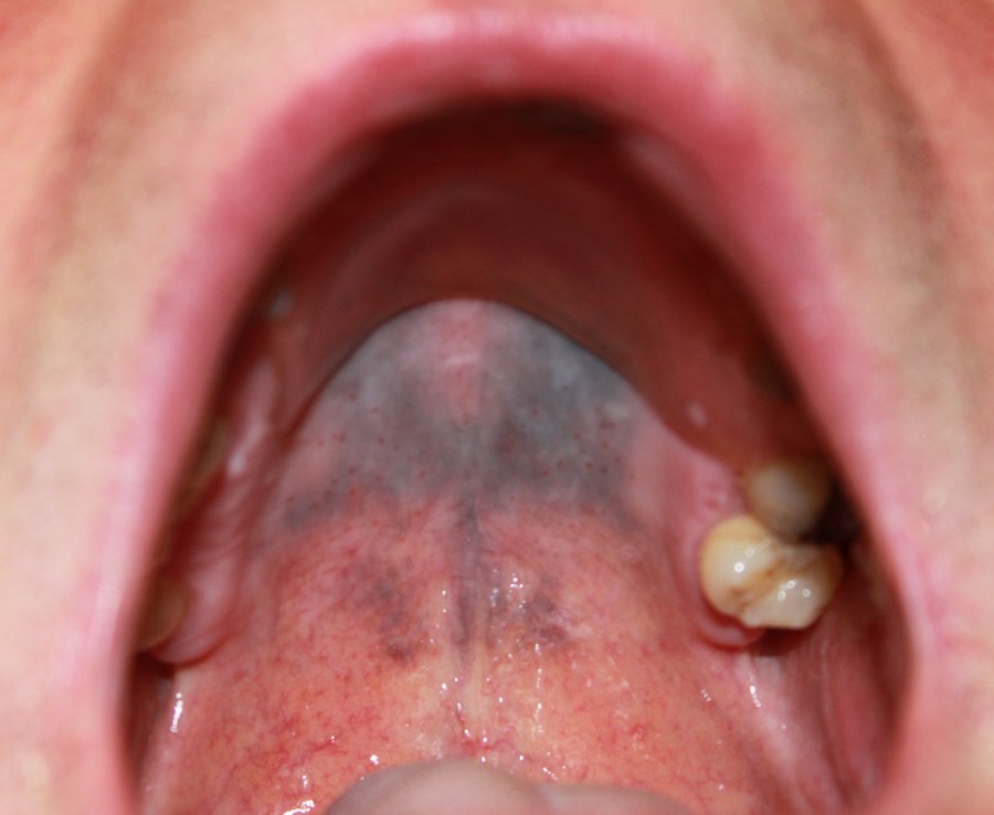
Cutaneous lupus erythematosus
LAST UPDATED: Jan 25, 2024
Introduction
Cutaneous lupus erythematosus is a diverse group of autoimmune connective tissue disorders localised to the skin that can be associated with systemic lupus erythematosus (SLE) to varying degrees. Cutaneous lupus erythematosus (CLE) is classified as:
- Acute (ACLE)
- Subacute (SCLE)
- Intermittent (lupus tumidus)
- Chronic (CCLE) - discoid lupus (DLE), lupus profundus, chilblain lupus erythematosus
The chapter, which is set out as below, focuses on the key features of the most common and/or important presentations including discoid lupus erythematosus, subacute cutaneous lupus erythematosus, and acute cutaneous lupus erythematosus including its relationship to systemic lupus erythematosus.
Aetiology
-
Lupus erythematosus is an autoimmune condition
- It is thought that a combination of environmental factors and genetics most likely contribute to the development of CLE
- Genetic factors - SCLE is associated with the human leukocyte antigen (HLA)
- Environmental factors
- All types of CLE are aggravated by UV-radiation (UVR). In SCLE, exposure to UVR results in increased expression of the Ro/SSA antigen on the surface of keratinocytes, binding the anti-Ro/SSA antibody and leading to the disease
- CLE is more severe in smokers, especially DLE
- Medications - approximately one-third of cases of SCLE are caused by medications
Clinical findings
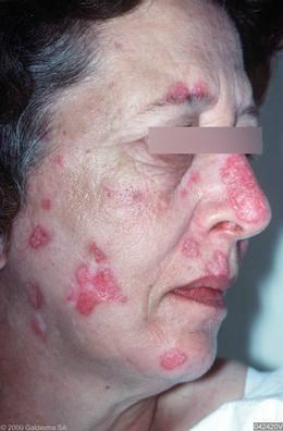
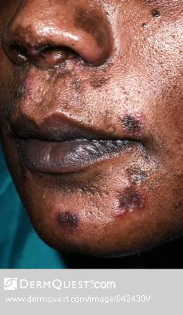
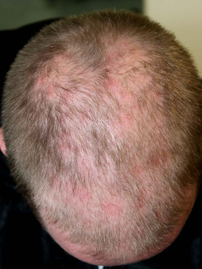
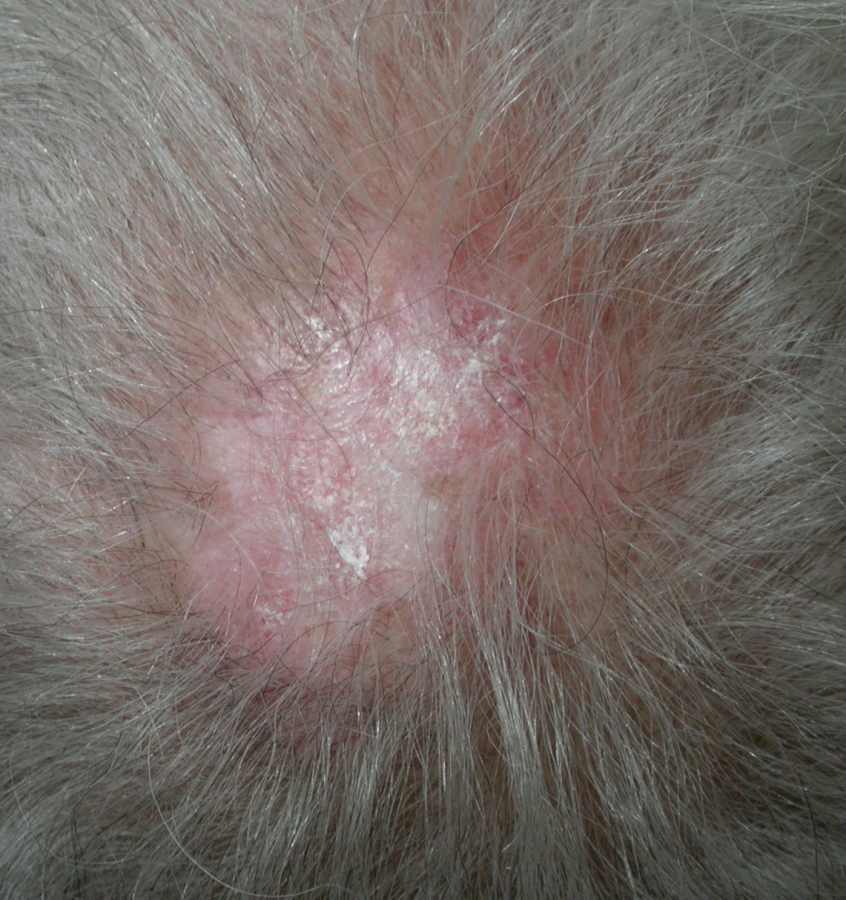
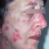
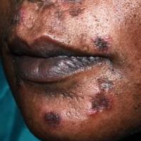
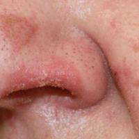
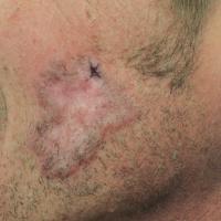
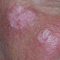
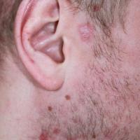
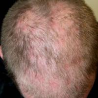
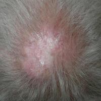
Discoid lupus erythematosus - DLE (figures 1-10)
-
Overview
-
DLE is the most common form of lupus and is responsible for 50-85% of cases of cutaneous lupus
-
It occurs 2-3 times more frequently in women than in men, and is slightly more common in African Americans than in whites or Asians
-
Although DLE may occur at any age it most often develops in patients aged 20-40 years
-
Exacerbation is common with increased UV exposure, particularly in the spring and summer
- Cigarette smoking is more common in patients with cutaneous lupus erythematosus than healthy controls, with a prevalence of 60–80%. Current smokers demonstrate increased disease activity and damage, and a more pronounced effect on QoL. Despite previous reports that smoking interferes with antimalarial efficacy in cutaneous lupus erythematosus, it is more likely that this reflects increased disease activity in this group
- Drug-induced DLE is rare and usually associated with anti-TNF inhibitors and fluorouracil
-
Patients with DLE can be divided into two groups: localised and generalised
-
Localised DLE occurs when the head and neck only are affected, and is nearly always confined to the skin
-
Generalised DLE occurs when other areas are affected, regardless of whether the head and neck are involved. Patients with widespread involvement often have haematological and serological abnormalities, are more difficult to treat, and are more likely to develop SLE, although the overall risk for all cases of cutaneous lupus is somewhere in the region of 1.3-6.5%
-
50% of DLE patients achieve complete resolution over many years. The prognosis is worse if associated with Raynaud’s phenomenon, chilblains and alopecia. Ultimately some patients will be left with scarring
-
Distribution
-
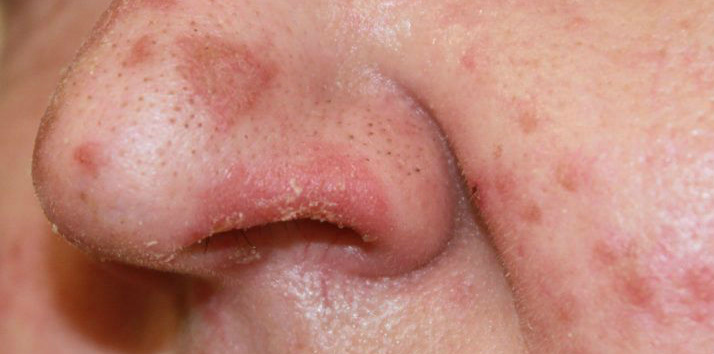
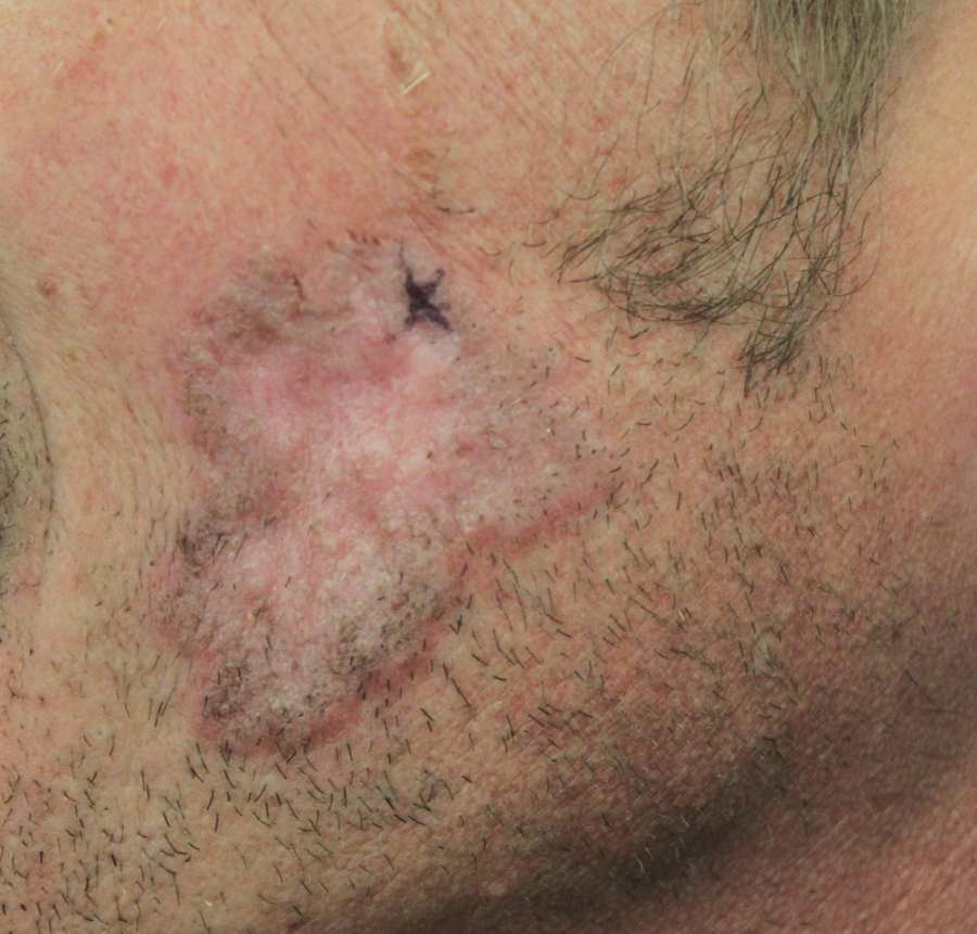
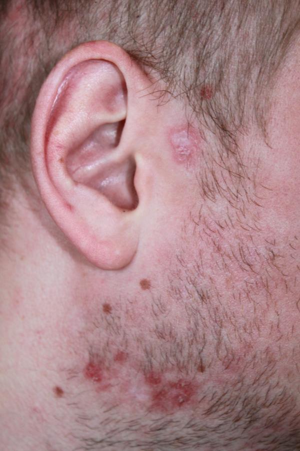
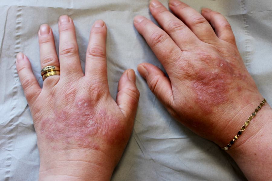
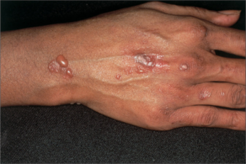
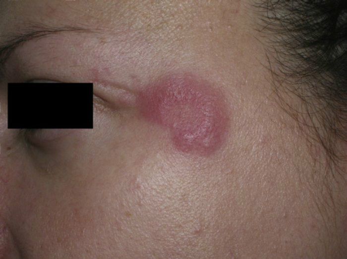
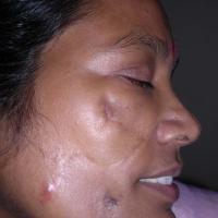
Discoid LE predominantly affects the cheeks, nose and ears, and sometimes the V of the neck, the upper back, and the dorsal hands
-
Occasionally it is more widespread
-
Typical morphology
-
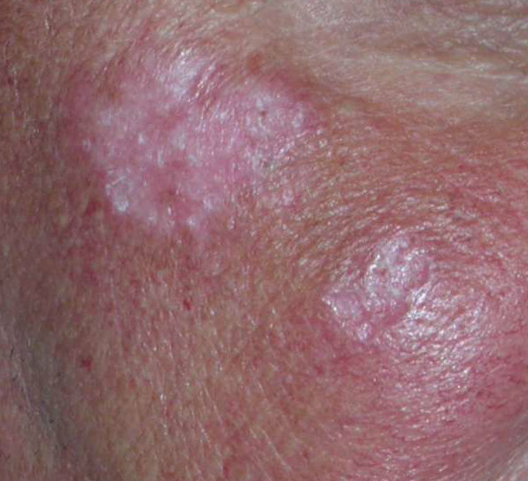
One or several well-defined erythematous patches or plaques with horny plugs
-
Adherent scale is common
-
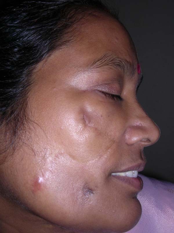
Untreated lesions develop a central white scarred area and areas of cribriform scarring
-
Other cutaneous features
-
Wide follicular pits arise in the concha of the ears
-
Eyebrows may be sparse and erythematous
-
Scarring alopecia in one-third of patients - refer to the chapter Alopecia - an overview for more information
-
Hyperpigmentation is common especially in dark-skinned individuals
-
Lesions are occasionally hyperkeratotic and warty, tumid, or annular atrophic plaques
-
Associations
-
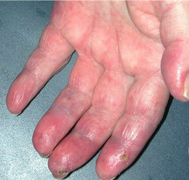
Raynaud’s phenomenon, perniosis (chilblain lupus), and panniculitis
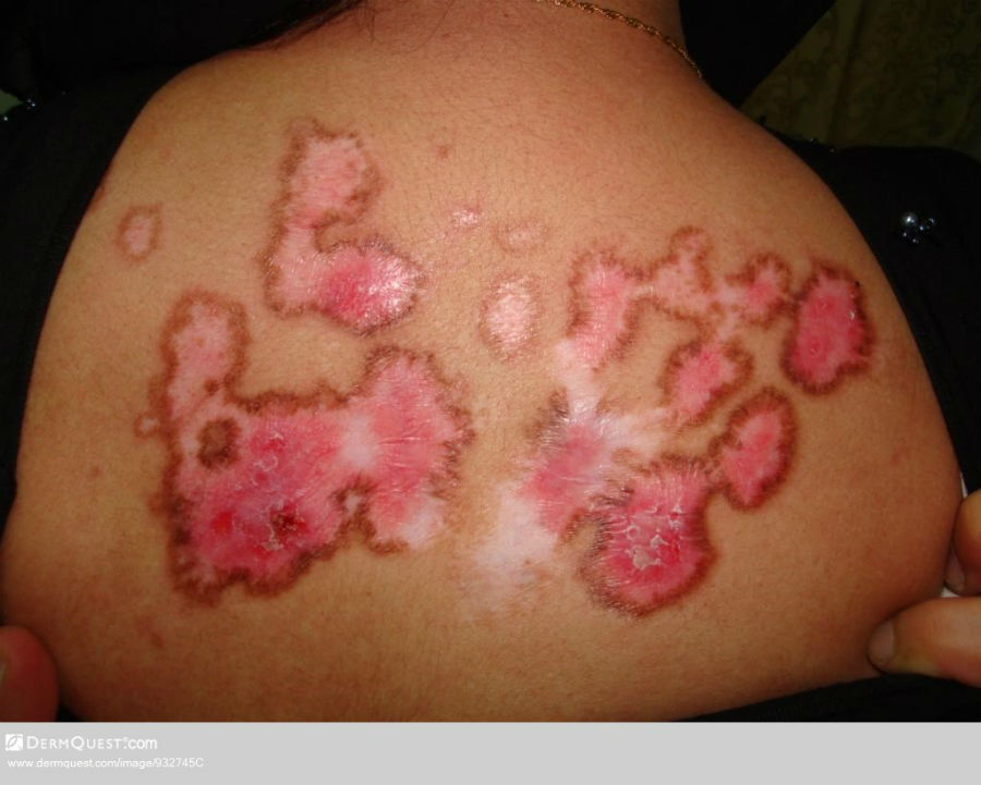
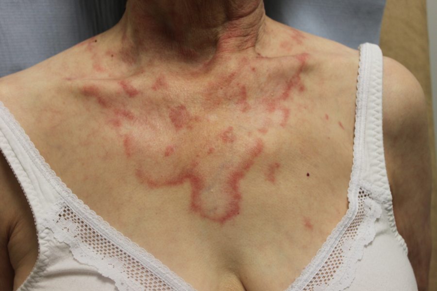
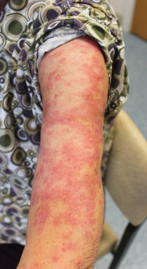
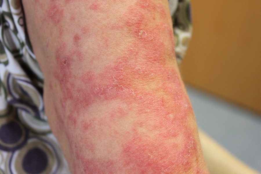
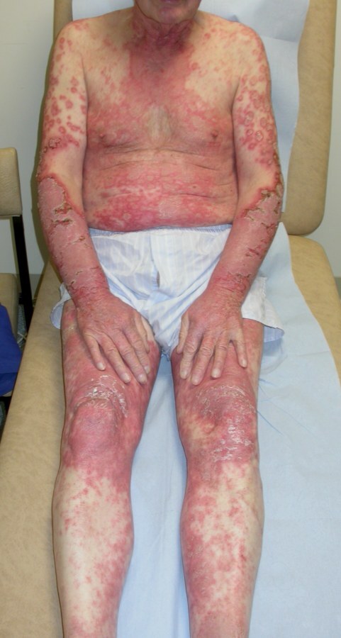
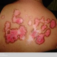
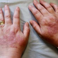
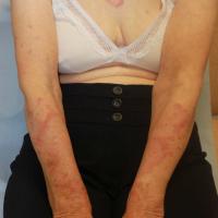
Subacute cutaneous lupus erythematosus - SCLE (figures 11-18)
-
Overview
-
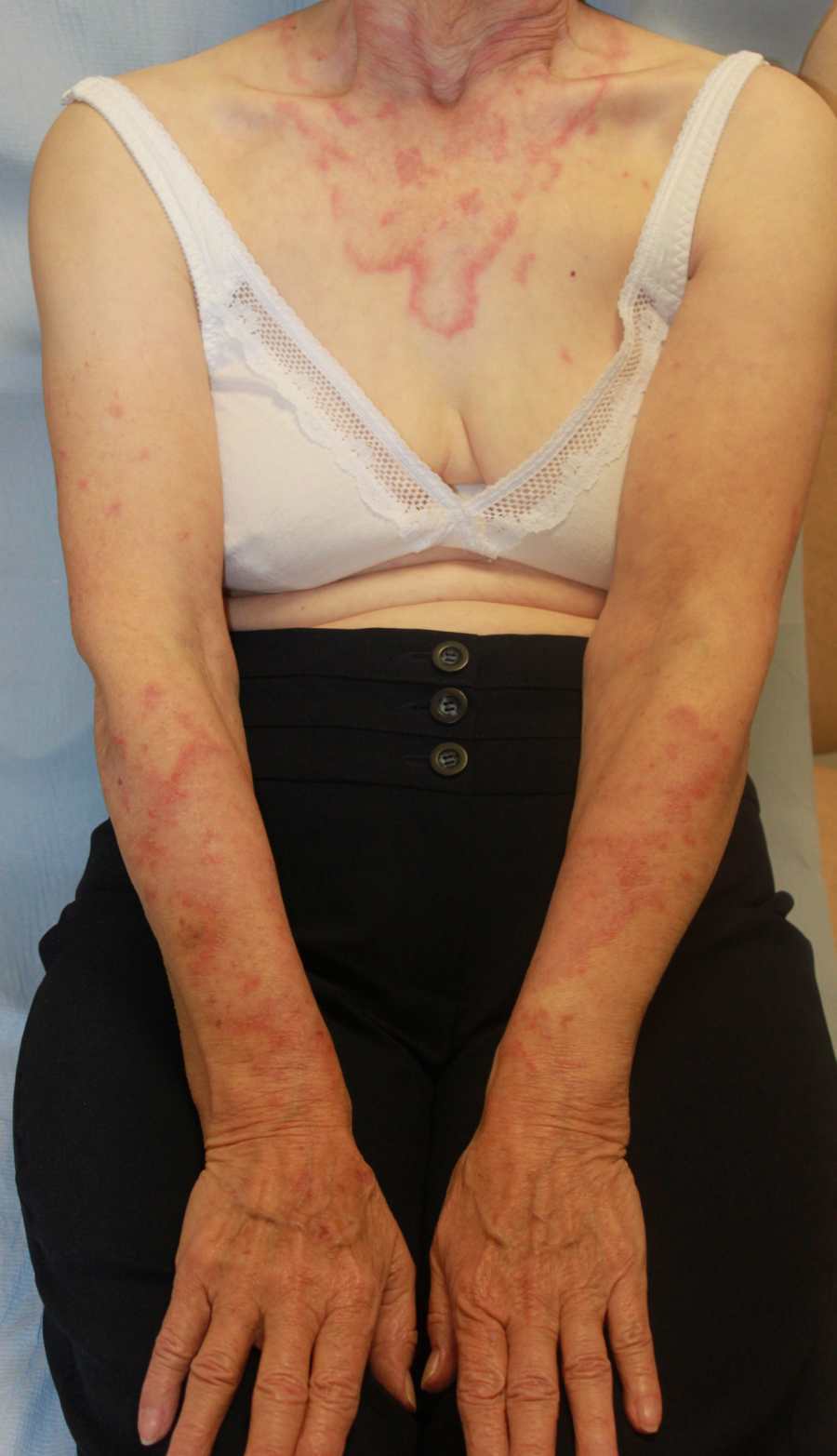
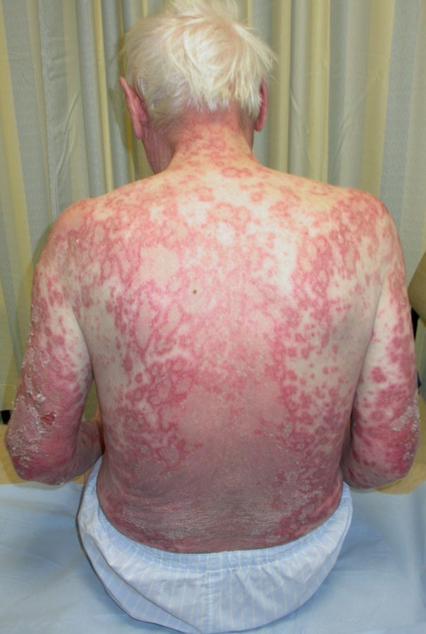
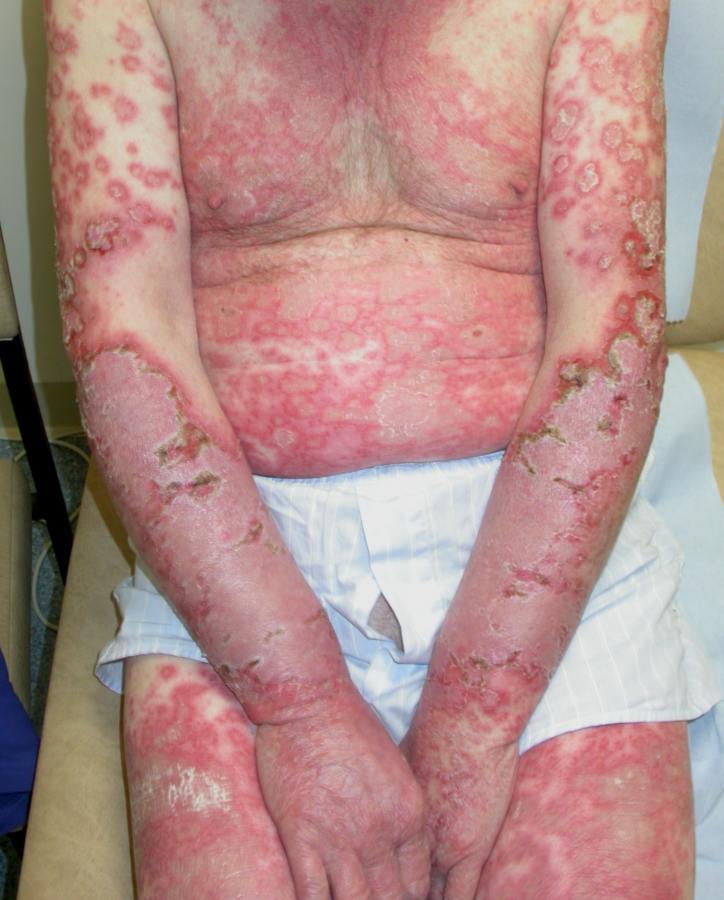
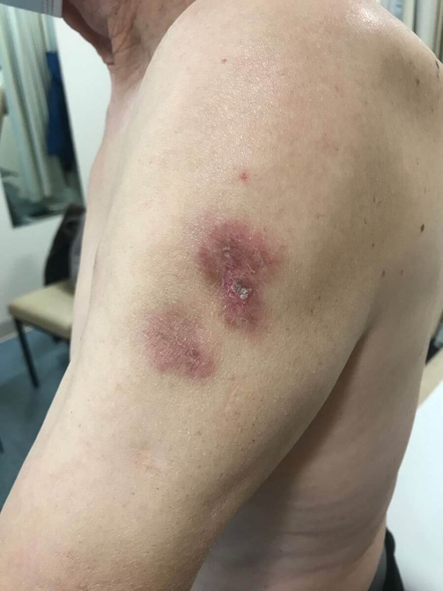
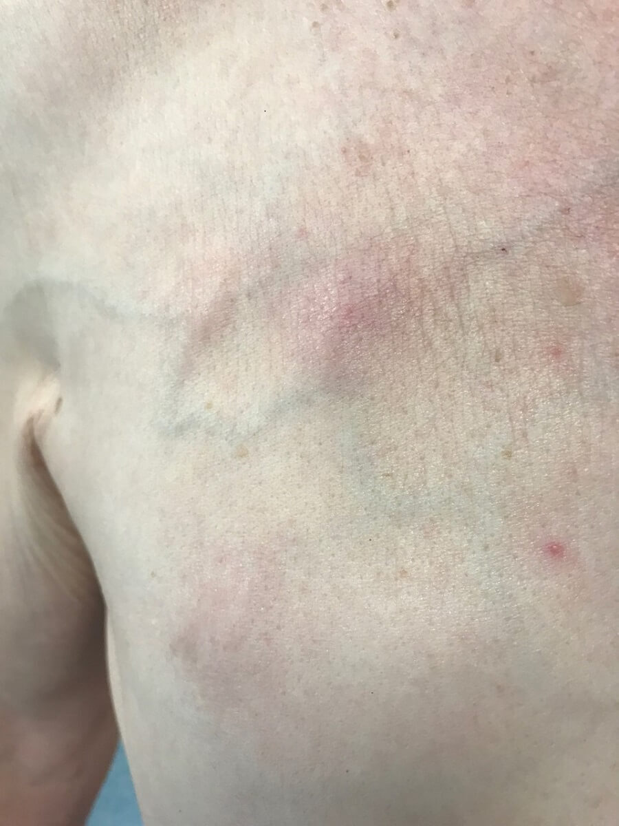
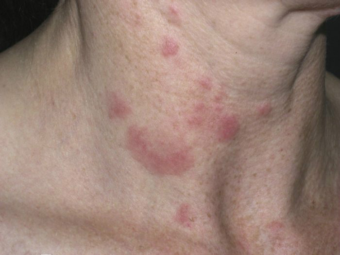
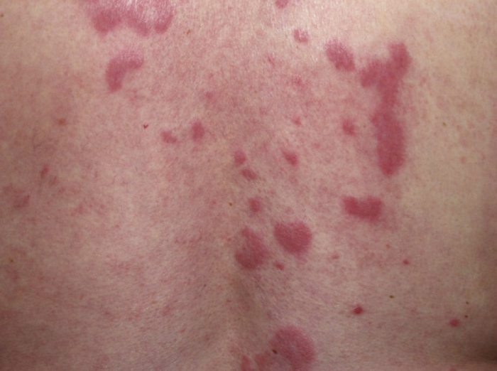
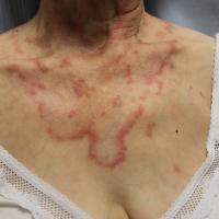
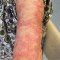
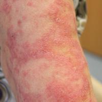
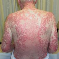
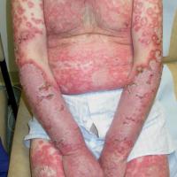
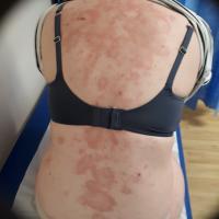
SCLE is a non-scarring, non–atrophic, photosensitive dermatosis. SCLE commonly develops in UV-exposed areas, including the upper back, shoulders, extensor arms, neck, and upper torso, although the face is often spared
-
It is more common in Caucasian's (> 85%) with a female to male ratio of 4:1. The condition typically occurs in patients aged 15-70 years, with the mean age being approximately 43 years
- SCLE may occur in patients with SLE, Sjögren's syndrome, complement deficiency, and it may be drug-induced. Some patients with SCLE may also have the typical lesions of DLE
-
It normally has a good prognosis
-
Distribution
-
Photosensitivity is common
-
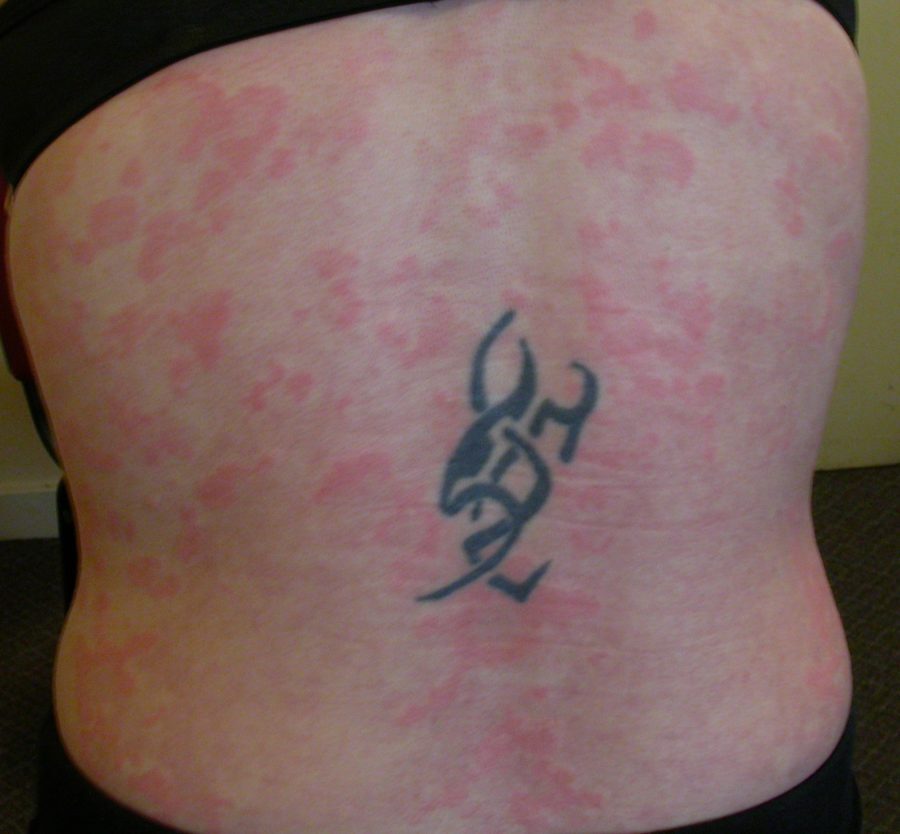
The main areas affected are the neck, trunk and outer arms, although the face is often spared
-
Typical morphology
-
Non-scarring papulo-squamous or annular polycyclic lesions
-
Fine scale is a common feature
-
Other cutaneous features
-
Diffuse non-scarring alopecia
-
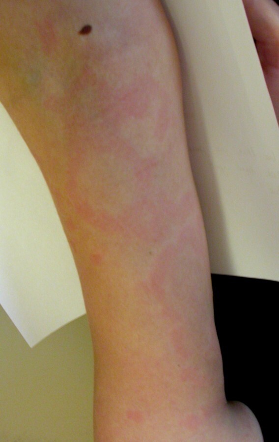
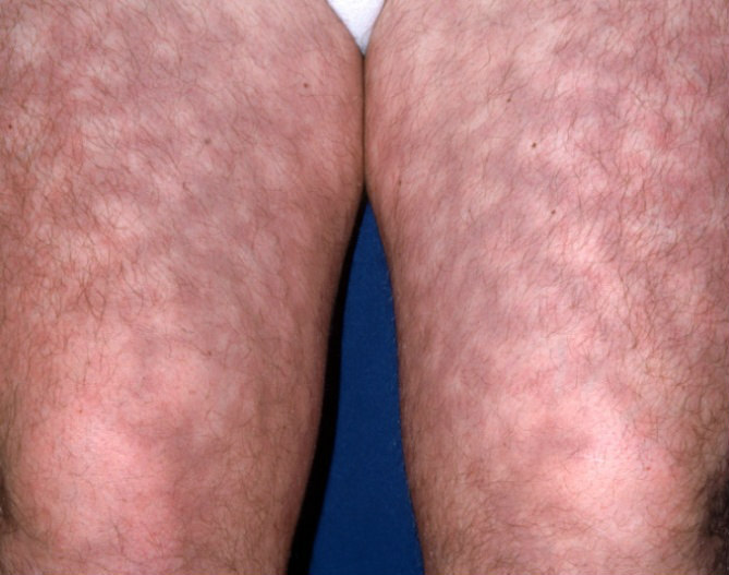
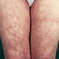
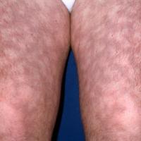
Non-palpable livedo-reticularis
-
Raynaud's phenomenon
-
Urticarial vasculitis
-
Systemic features
-
Arthritis
-
Renal involvement is uncommon, and mild
-
Associations
-
SCLE is the most common subtype of cutaneous lupus erythematous associated with Sjögren's syndrome. It can also be associated with rheumatoid arthritis, small vessel vasculitis, Sweet's syndrome and Crohn's disease
-
Drug-induced (DI-SCLE)
- Approximately one-third of SCLE cases have been attributed to a drug trigger and the frequency of DI-SCLE has increased in recent years as a result of new therapeutics and increased awareness of DI-SCLE
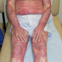
- The level of suspicion should be increased if the rash is widespread ie also involves the trunk and distal extremities, and/or an implicated drug has recently been introduced, although in some cases there can be a long incubation period
-
Autoantibodies - anti-Ro may be positive, and anti-histone antibodies are more likely to be positive compared with idiopathic SCLE
- There are a wide range of drugs implicated - antihypertensives including calcium channel blockers (especially Diltiazem and Verapamil), beta-blockers and angiotensin-converting enzyme inhibitors / proton pump inhibitors (class effect. Usually anti-Ro positive. Delay of several months before cutaneous features) / diuretics (including loop and thiazide) / terbinafine / naproxen / statins / antihistamines (anti-Ro negative) / carbamazepine / cinnarizine / several chemotherapy agents (the taxanes and Tamoxifen) / anti-TNF therapies / leflunomide / monoclonal antibodies
- Most cases of DI-SCLE are reversible on stopping the offending drug, although resolution can take up to 12 months (most improve within 3 months) and concurrent therapies may be required during this time
- SCLE can occasionally be associated with neonatal lupus - refer to the section underneath on SLE
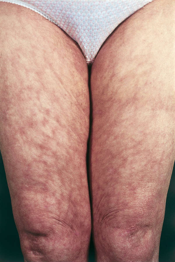
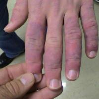
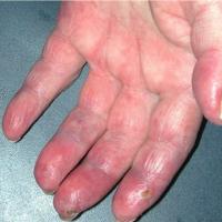
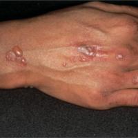
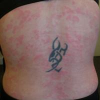
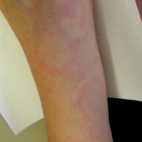
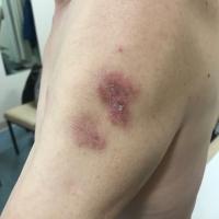
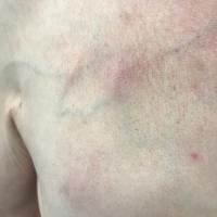
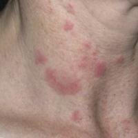
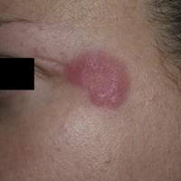
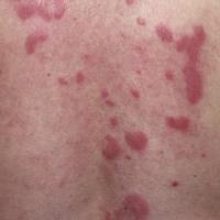
Acute cutaneous lupus erythematosus and systemic lupus erythematosus (figures 19-30)
- Systemic lupus erythematosus (SLE)
- Is a chronic systemic disorder characterised by multisystem organ inflammation, most commonly the skin, joints, and vasculature, and associated immunological abnormalities. The main clinical features include fever, rashes and arthritis, but renal, pulmonary, cardiac, and neurological involvement may occur
- Presents most commonly in young to middle-aged adults, is eight times more common in females, and is three times more common in black people than in white people
- Approximately 80% of cases have cutaneous features, which in approximately 25% of cases are the presenting features
- Mucocutaneous features of SLE
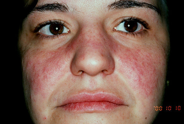
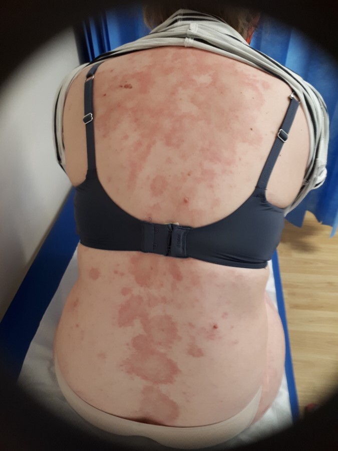
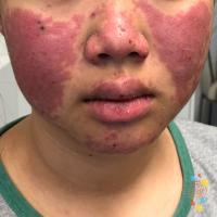
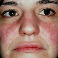
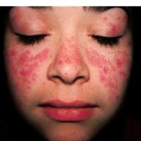
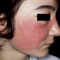
- Acute cutaneous lupus erythematosus typically presents as transient erythematous patches associated with a flare of systemic lupus erythematosus (SLE). It is usually very photosensitive. Lupus-specific skin changes include:
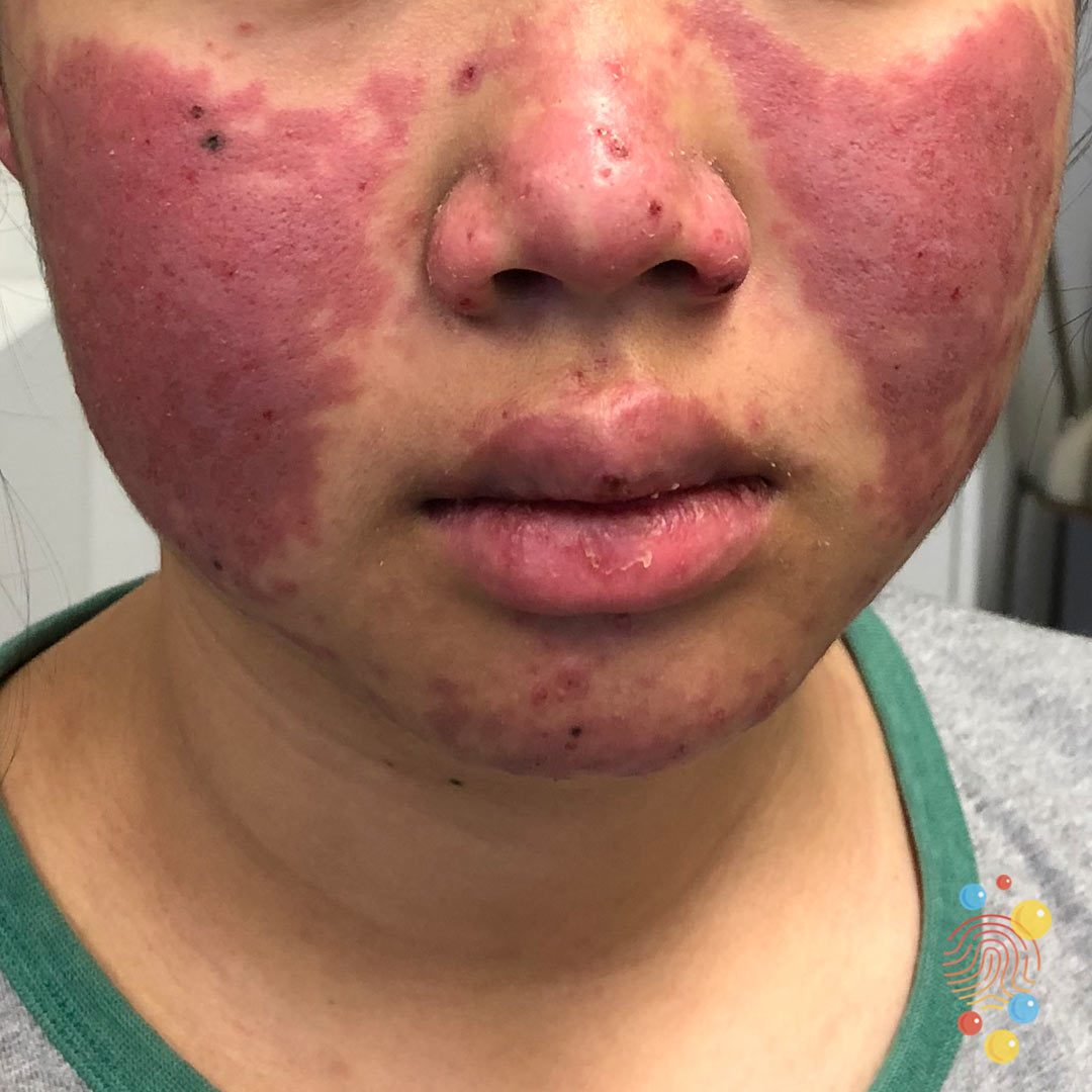
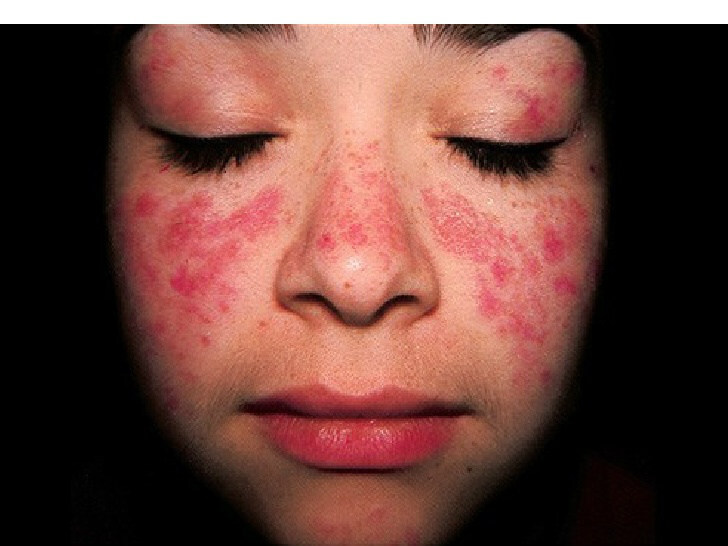
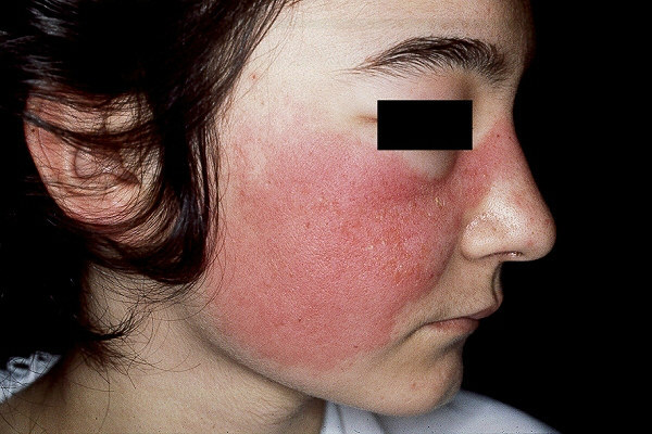
- Localised ACLE - a malar ‘butterfly' rash with redness and swelling over both cheeks, sparing the nasolabial folds, lasting hours to days
- Generalised ACLE - a diffuse or papular erythema of the face, upper limbs (sparing the knuckles), and trunk resembling a measles-like rash
- Toxic epidermal necrolysis-like ACLE - associated with lupus nephritis or cerebritis
- Other mucocutaneous features associated with SLE
- 30% subacute lupus erythematosus
- 11% non-scarring alopecia, 7% scarring alopecia
- 9% discoid lupus erythematosus
- 6% vasculopathy
- 5% Raynaud's
- 4% panniculitis
- 2% chilblains
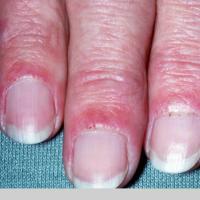
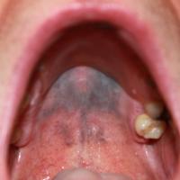
- Other features can include mouth ulcers, urticaria, hypocomplementaemic urticarial vasculitis, non-palpable livedo reticularis, bullous eruptions, oedema of the face and hands, and chelitis
-
Systemic features of SLE
-
Non-erosive arthritis
-
Episcleritis
-
Most patients have renal involvement, and such changes are very important in assessing the prognosis. Lupus-related renal complications account for 3% of all cases of end-stage renal failure
-
Cardiac eg hypertension and pericarditis
-
Pulmonary eg pleurisy and pneumonitis
-
Nervous system eg migraine and epilepsy
- American Rheumatism Association criteria of SLE
- Rash - malar
- Rash - discoid
- Photosensitivity
- Oral ulcers
- Non-erosive arthritis
- Serositis - pleurisy or pericarditis
- Renal involvement
- Neurological involvement - seizures or psychosis
- Haematological involvement - haemolytic anaemia, leukopenia, lymphopenia, thrombocytopenia
- Immunological disorder - anti-DNA antibody, anti-Sm antibody, antinuclear antibodies
- The presence of four or more of these manifestations is 96% sensitive and specific for SLE
-
Drug-induced SLE
-
Certain medications may rarely precipitate lupus in predisposed individuals. Symptoms generally take several months to develop, and drug-induced SLE does not usually involve the skin. Anti-DNA antibodies are absent. The most frequent drugs implicated are hydralazine, carbamazepine, lithium, phenytoin, the sulphonamides, and minocycline
-
Lupus in pregnancy and the neonate
-
In general, pregnancy does not cause flares of SLE. When flares do develop they often occur during the first or second trimester or during the first few months after delivery
-
SLE increases the risk of spontaneous abortion, intrauterine foetal death, pre-eclampsia, intrauterine growth retardation, and premature delivery. The risks of foetal loss are increased significantly if the SLE is associated with the Antiphospholipid syndrome
-
Neonatal lupus erythematosus (NLE) is thought to be caused by the transplacental passage of maternal autoantibodies, however, only 1-2% of infants with positive maternal autoantibodies develop neonatal lupus erythematosus. The most common clinical manifestations are dermatological, cardiac eg complete heart block, and haematological eg thrombocytopenia
-
Prognosis for both mother and child are best when SLE is quiescent for at least six months before the pregnancy and when the mother’s underlying renal function is stable and normal or near normal
-
The Antiphospholipid syndrome
-
The Antiphospholipid syndrome consists of persistently positive APL antibodies (two tests carried out 12 weeks apart) associated with arterial / venous thrombosis and / or adverse outcomes in pregnancy (mother or foetus)
-
The Antiphospholipid syndrome occurs in isolation (primary APL syndrome) in more than 50% of patients, but it can be associated with autoimmune disease and other conditions (secondary APL syndrome). 20-35% of patients who have SLE will develop a secondary APL syndrome
-
All patients with SLE require testing for the Antiphospholipid syndrome
-
Other associations
-
Include rheumatoid arthritis, morphoea, systemic sclerosis (predominantly with limited cutaneous systemic sclerosis ie hand involvement only), Sjögren's syndrome, thyroiditis, myaesthenia gravis, haemolytic anaemia, and thrombocytopenic purpura
Less common types of lupus erythematosus
Chilblain lupus
- Lesions persist beyond colder months
- Coexist with SLE and cutaneous LE
- Often ANA, anti-Ro positive. Negative for cryoglobulins, cold agglutinins
- Histology can be helpful if the diagnosis is in doubt
Lupus erythematosus tumidus
-
Lupus erythematosus tumidus is a dermal form of lupus
-
Papules, plaques, and nodules arise on the cheeks, upper chest, upper arms or back - the rash is photosensitive. Lesions are sometimes annular in shape
-
It tends to clear during the winter months and does not scar
-
The main differential diagnoses are Jessner's lymphocytic infiltrate, B-cell lymphoma, sarcoid, and granuloma faciale
Lupus profundus (syn. lupus panniculitis)
-
Can affect any age
-
The face is the most common site
-
Inflammation occurs in the adipose tissue, which presents as firm deep nodules
-
The end result is unsightly indented scars (lipodystrophy)
Clinical Images
Please refer to notes on image rights at bottom of the page with regards to individual image
ownership.
1
2
3
4
5
6
7
8
9
10
11
12
13
14
15
16
17
18
19
20
21
22
23
24
25
26
27
28
29
30
31
32
33
34
35
36
37
Investigations
Autoantibodies and other tests for lupus erythematosus
-
For patients with local cutaneous DLE further investigations are seldom needed. For all other patients refer to the section on Investigations
-
It is worth checking with your local pathology department as the tests performed may vary, and the spectrum of autoantibodies is continually evolving
Other bloods tests in SLE
- FBC and ESR are often abnormal (CRP usually normal) - anaemia is present in 75% of cases. Leucopenia, especially a lymphopenia, is said to be characteristic. Thrombocytopenia affects 20% of patients
- Rheumatoid factor is present in 40% of cases
-
The lupus anticoagulant is one of a number of antiphospholipid antibodies that may be found in up to 50% of patients
-
False-positive serology tests for syphilis are found in 25% of patients
-
Complement levels may be reduced
-
Cryoglobulins (and other cryoproteins) are found in up to 15% of cases, indeed cryoglobulinaemia may precede SLE by many years
Histopathology and immunohistology in lupus (reference: Rook's Textbook of Dermatology)
-
DLE - the characteristic histopathological changes observed include liquefaction degeneration of the basal cell layer, degeneration changes of the connective tissue such as hyalinisation and fibrinoid change and an inflammatory cell infiltrate (usually lymphocytic) especially around the skin appendages. Other features may include thickening of the basement membrane, follicular plugging, hyperkeratosis and incontinence of pigment
-
SCLE - features are similar to DLE but with increased epidermal atrophy, and less inflammatory infiltrate and follicular plugging
-
SLE - there is no single diagnostic pathological feature. A combination of features, including some of the above, may aid diagnosis
- Direct immunofluorescence (DIF) - may aid in diagnosis. Deposition of immunoglobulin, predominantly IgG (sometimes IgM), and / or complement at the dermoepidermal junction is a characteristic feature of lupus erythematosus. Skin biopsies for DIF are best taken from skin lesions; however, in acute cutaneous lupus erythematosus where the rash has faded, DIF will still be positive in 70-80% of cases if the biopsy is taken from sun-exposed skin
Management
General measures
DLE (chronic cutaneous) and SCLE
-
Are generally managed by specialists - effective treatment is required to reduce the scarring caused by DLE
-
First-line treatment - topical
-
A potent topical steroid (eg betnovate ®), or, occasionally a superpotent topical steroid (eg dermovate ®) should be applied thinly and accurately to the plaques. Once control is gained reduce to twice weekly maintenance, increasing for flares
- An alternative are the topical calcineurin inhibitors - pimecrolimus cream appears more effective than tacrolimus ointment
-
Intralesional corticosteroids are used very occasionally for resistant cases, although there is a greater risk of atrophy
- Second-line treatment - antimalarial therapy (used in combination with the above)
- Hydroxychloroquine (HCQ) - 200–400 mg daily. The daily maintenance dose of HCQ should not exceed 5 mg/kg/day. HCQ can safely be used in pregnancy
- Mepacrine 50-100 mg OD is indicated as follows:
- To use as an alternative if additional risk factors for retinal toxicity exist, such as concomitant tamoxifen therapy or impaired renal function (estimated glomerular filtration rate < 60 mL min–1 1.73 m–2)
- If HCQ not tolerated
- As an add in to HCQ if response suboptimal
- Consider mepacrine up to 200 mg daily in people resistant to standard dosing, when combination antimalarials or other therapies are contraindicated
- Chloroquine (CQ) < 2.5 mg/kg/day - third-line antimalarial option. Take care when prescribing CQ as the dosing depends on the salt used and is generally expressed in reference to chloroquine base, for example chloroquine phosphate 250 mg is approximately equivalent to chloroquine sulfate 200 mg and to chloroquine base 155 mg
- Retinal screening for antimalarials (not needed for mepacrine)
- Annual screening is recommended in all patients who have taken HCQ for > 5 years. This should include spectral domain optical coherence tomography and fundus autofluorescence imaging photography
- Annual screening may be commenced after 1 year of treatment if additional risk factors for retinal toxicity exist, such as concomitant tamoxifen therapy or impaired renal function (estimated glomerular filtration rate < 60 mL min–1 1.73 m–2), or if the daily dose of HCQ is > 5 mg kg–1
- Annual retinal assessment in all people with CQ after 1 year of therapy
- Consider intermittent use of antimalarial therapy in people with seasonal lupus (eg summer flares in photosensitive lupus erythematosus and winter flares in chilblain lupus erythematosus)
-
Other treatments
- Systemic steroids - concomitant, short-term, and tapering courses can be used for flares and severe/disseminated disease
- Dapsone - consider dapsone (typically commenced at 50 mg daily and escalated to 150 mg daily, depending on response and tolerability) as a first-line systemic treatment option in people with SCLE and bullous SLE
- Suboptimal response to antimalarial therapy - several other treatments have been used with variable success including acitretin (25-50 mg OD), methotrexate (up to 25 mg once a week), mycophenolate mofetil (typically 0.5 g BD, up to 1.5 g BD), clofazime, biologic therapies, IVIG, and thalidomide. The latter can be very effective, including in cases of chilblain lupus, although there are significant risks such as teratogenicity and polyneuropathy
-
Associated Raynaud's phenomenon - calcium-channel blockers can be helpful
Systemic lupus erythematosus
-
SLE is a multisystem condition that requires a multidisciplinary team approach. Patients with suspected SLE should be referred urgently to Secondary Care (if renal involvement refer to nephrology, otherwise rheumatology or dermatology)
-
Systemic steroids are required, sometimes at a high-dose, to manage flares, and most patients require a low-dose as maintenance
-
Antimalarials may have a role in those with marked photosensitivity
-
Immunosuppressive drugs may be required as steroid sparing agents
-
Cyclophosphamide can be useful for renal disease
-
Prognosis - the course of SLE is highly variable. Approximately three-quarters of patients will survive 15 years
Other resources
Disclaimer - the author PCDS cannot accept responsibility for any misleading or incorrect statements, and the management of individual patients remains the direct responsibility of the individual doctor. We do however hope that visitors to this site can contact us regarding comments that are considered misleading or incorrect so that we can continue to improve the site.
Image Rights - The PCDS would like to thank Dermatoweb, DermQuest (Galderma), and others who have contributed images. All named individuals and organisations maintain copyright for the relevant images.