Porphyria
LAST UPDATED: Nov 18, 2021
Introduction
The porphyria's result from altered metabolism in the haem biosynthesis pathway, leading to an accumulation of porphyrins. There are three main types - porphyria cutanea tarda, variegate porphyria and erythropoietic protoporphyria. This chapter also considers pseudoporphyria.
This chapter is set out as follows:
Clinical findings
Porphyria cutanea tarda (PCT)
- The the commonest of all the porphyrias
- Results from deficiency of the liver enzyme uroporphyrinogen decarboxylase (UROD), causing an accumulation of uroporphyrin and other highly carboxylated porphyrins
- The majority of case are acquired (type I), and associated with liver disease. Twenty‐five per cent have type II (familial) disease where the enzyme deficiency is hereditary
- Symptoms result from a reaction between the porphyrins in the skin and UV radiation, which goes on to cause cell damage. Although it is often worse in summer months the patient may not be able to clearly associate symptoms with UV exposure
-
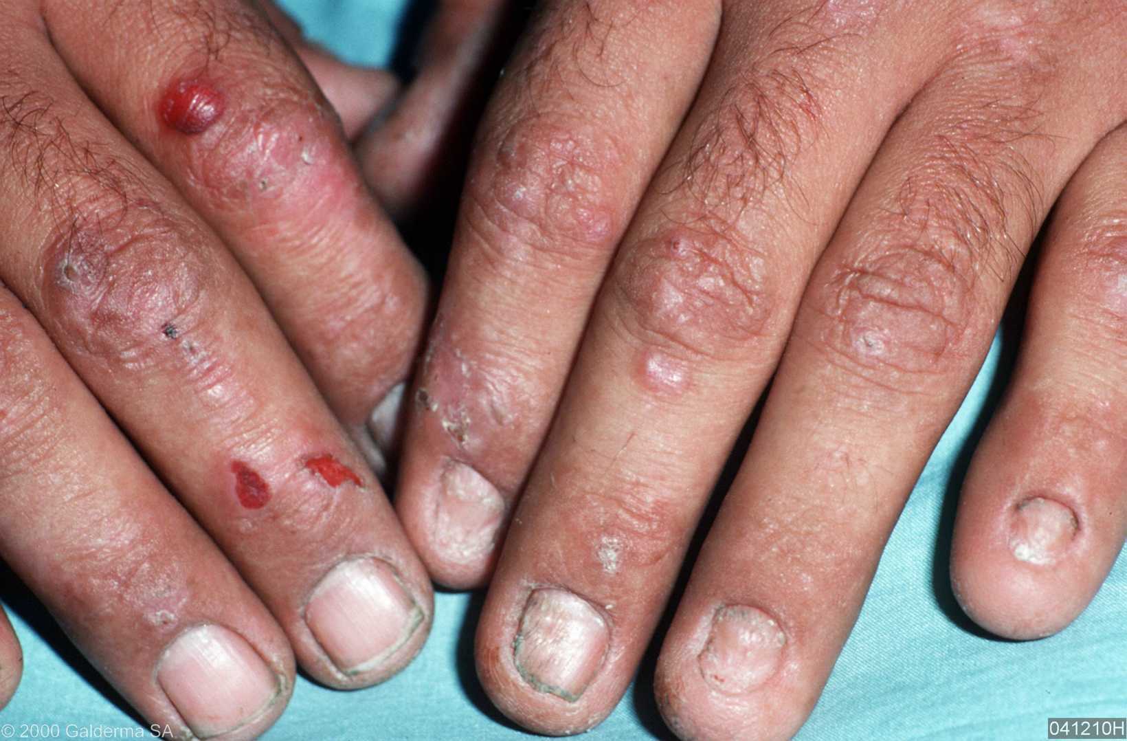
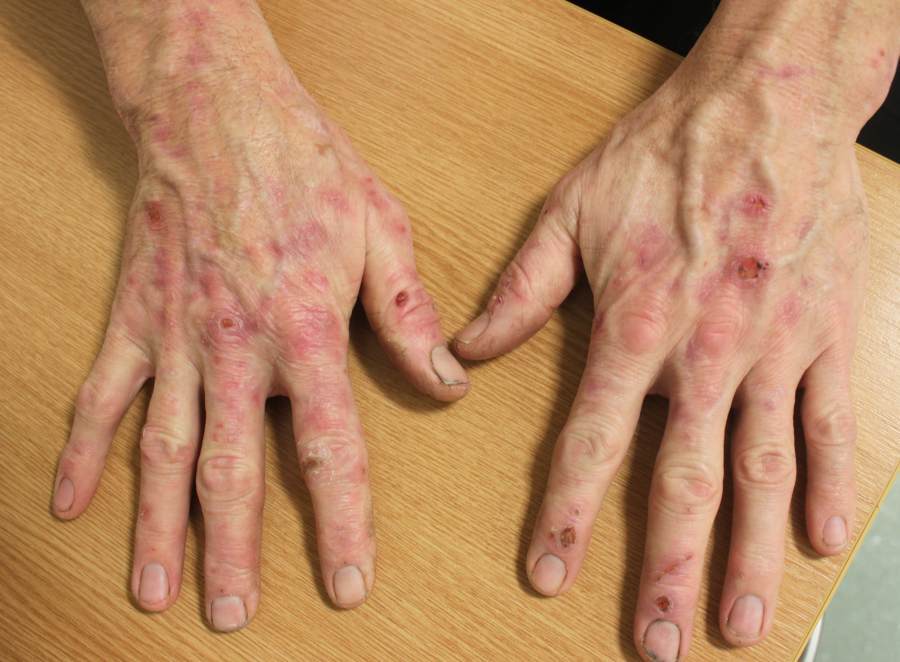
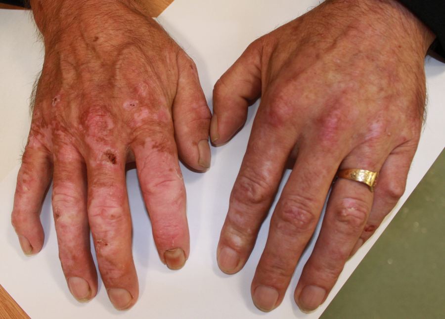
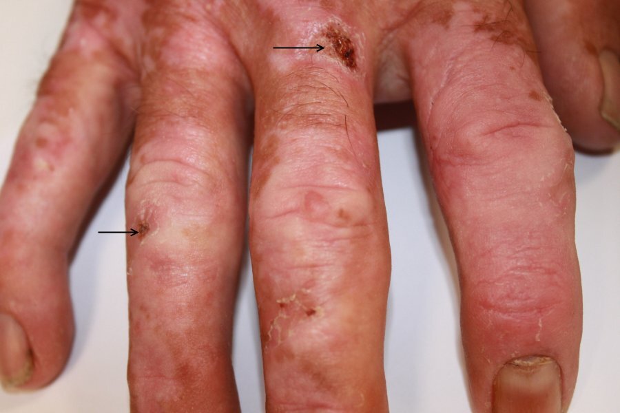
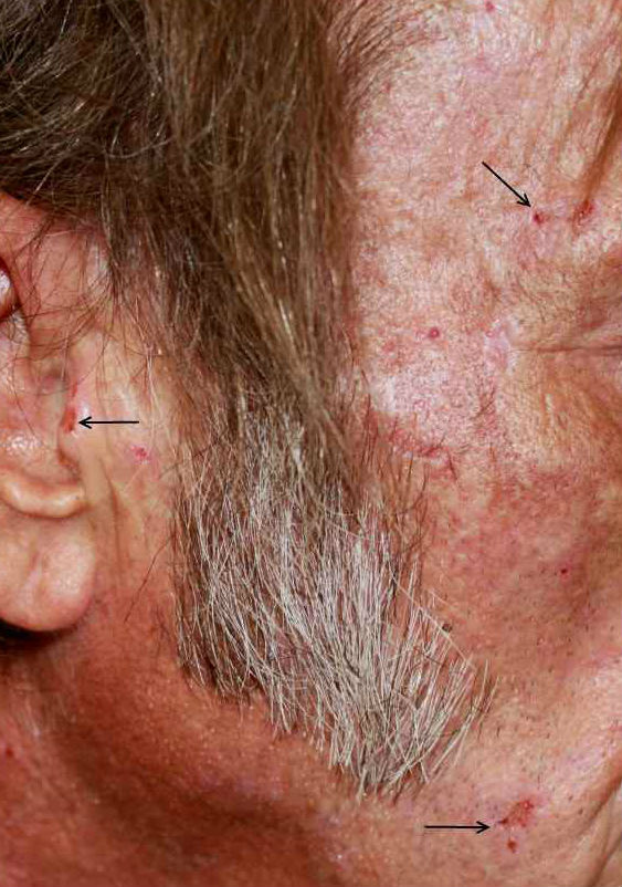
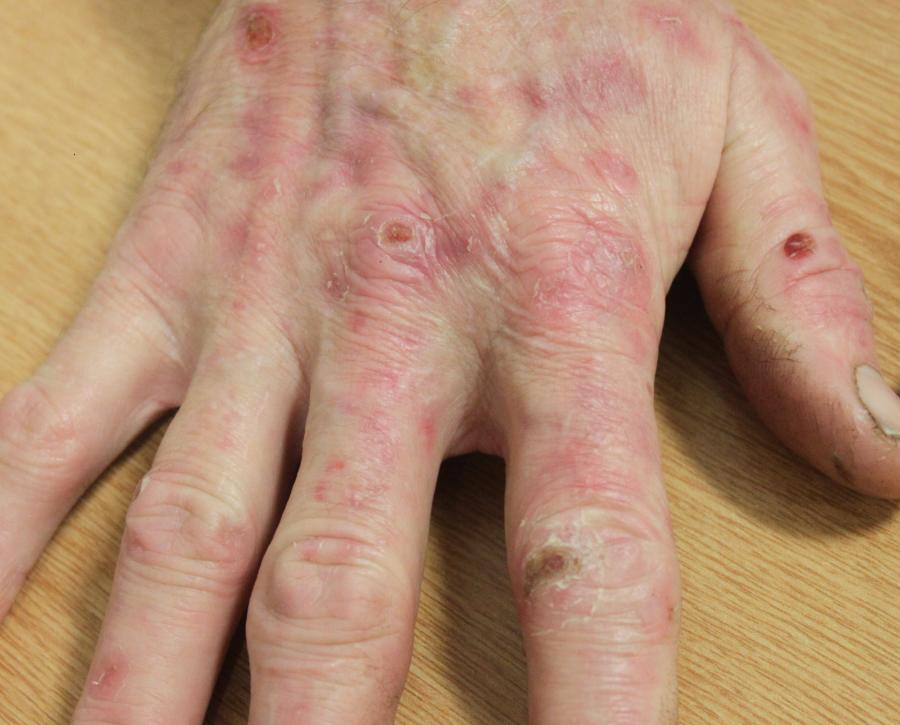
Clinical features - skin fragility and blisters on the backs of hands and sometimes bald areas of the scalp. Lesions heal slowly and often leave scars. Milia and areas of hyperpigmentation may develop. Mild cases may present with only shedding of the skin over the backs of the hands without blisters
Pseudoporphyria
-
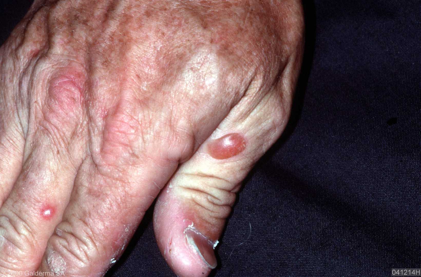
Clinical features similar to PCT but lack milia and scarring
-
This is not a true porphyria, but instead secondary to drugs, or occasionally dialysis for chronic renal failure, or sunbeds
-
The most common cause of pseudoporphyria are non‐steroidal anti‐inflammatory drugs (NSAIDs), especially naproxen and nabumetone. Oxaprozin, ketoprofen, mefenamic acid and diflunisal are also reported causes. Other drugs reported to induce pseudoporphyria include nalidixic acid, tetracyclines including minocycline, bumetanide, furosemide, isotretinoin and dapsone
-
In drug‐induced and sunbed‐related pseudoporphyria, the key to management is to remove the provoking factor by stopping the relevant drug or sunbed usage. However, symptoms may continue for several months after the discontinuation of a causative drug, and any scarring persists. Dialysis‐related pseudoporphyria generally persists until renal transplantation removes the need for dialysis
Variegate porphyria
-
A rare autosomal dominant condition with the highest incidence in South Africans of European extraction
-
Patients have skin signs of PCT along with acute systemic attacks comprising abdominal pain and Guillain–Barré like symptoms such as confusion, peripheral neuropathy and other neurological features that can be life-threatening
-
The acute attacks are usually trigged by drugs
Erythropoietic protoporphyria (EPP)
-
An autosomal dominant condition
-
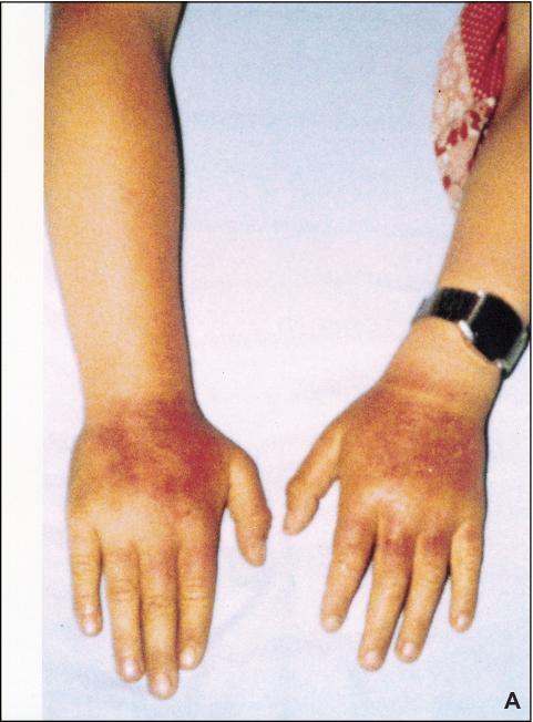
Presents in childhood with episodes of severe burning, redness and swelling of exposed areas
-
Symptoms arise within minutes of UV exposure and persist for 2-3 days
-
The main differential is Gunther's disease, also known as congenital erythropoietic porphyria (CEP). This is a mutilating type of porphyria associated with fragile skin, along with pain, bullae and scarring on exposed sites
Clinical Images
Please refer to notes on image rights at bottom of the page with regards to individual image
ownership.
Investigations
-
General screening - in a photosensitive patient with a low suspicion for porphyria only a plasma sample for porphyrins is required
- PCT and variegate porphyria - send a urine and plasma sample for porphyrins, morning samples are not required
- EPP - a plasma sample only is required to look for porphyrins in red blood cells
- Other important notes:
- All samples sent for porphyria must be kept in the dark, eg wrapped in tin foil, and the labs notified
- Which lab has done the tests? If the tests are negative but clinical suspicion remains it is advisable to repeat the samples and ask the labs to forward them to one that specialises in porphyria
Management
General measures - UV-protection
Provide a patient information leaflet on UV protection, which includes the following advice:
- That normal sunscreens are not effective as the reaction results from visible violet light (wavelengths 400-420nm), instead opaque sunscreens such as the Dundee formula are required (refer to the management section on Photodermatoses - an overview)
- Window films may also be required
PCT
-
Provide a patient information leaflet
-
Investigate the cause - a complete liver screen is required (20% haemochromatosis, 15% hepatitis C, 15% related to alcohol). Oestrogens (most commonly HRT) can also be causal
-
Refer for specialist management - first line treatment is usually low dose hydroxychloroquine (100 mg twice a week) or chloroquine (125 mg twice a week), higher doses cause liver damage. In patients with haemochromatosis, and in other cases where medical treatment is not effective, then manage with venesection
-
Long-term follow up is required as patients with poorly controlled PCT are more likely to get cirrhosis and liver carcinoma
Variegate porphyria
-
Patients are best managed by a department that specialises in porphyria
-
Family members need screening
Erythropoietic protoporphyria
-
Refer patient to a specialist
-
The most common treatment is oral beta-carotene
-
The α-melanocyte stimulating hormone Afamelanotide is the most effective treatment but is not currently available in the UK on the NHS
Other resources
Disclaimer - the author PCDS cannot accept responsibility for any misleading or incorrect statements, and the management of individual patients remains the direct responsibility of the individual doctor. We do however hope that visitors to this site can contact us regarding comments that are considered misleading or incorrect so that we can continue to improve the site.
Image Rights - The PCDS would like to thank Dermatoweb, DermQuest (Galderma), and others who have contributed images. All named individuals and organisations maintain copyright for the relevant images.