Pemphigus foliaceus (and its variants)
LAST UPDATED: Jul 07, 2024
Introduction
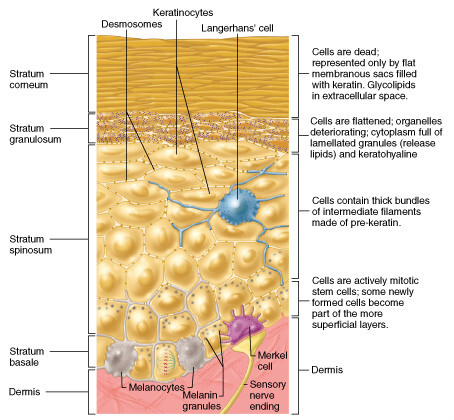
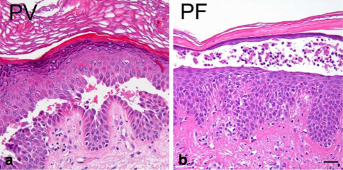
The pemphigus family is a group of rare immunobullous conditions affecting skin and/or mucous membranes. Pemphigus foliaceus is less common than its more serious relative pemphigus vulgaris, and is characterised by blistering lesions high in the epidermis, either in the granular layer (stratum granulosum) or just beneath the stratum corneum.
This chapter is set out as follows:
Aetiology
-
Pemphigus foliaceus (PF) is an immunobullous condition
-
Keratinocytes in the skin are cemented together at junctions known as desmosomes. In PF, autoantibodies bind to the protein desmoglein-1, which is found in desmosomes near the top of the epidermis. The result is the surface keratinocytes separate from each other, and are replaced by fluid, which forms a blister. Because the blister is very close to the surface of the skin the blisters rupture easily. In most cases the autoantibodies are IgG, but occasionally they are IgA
-
PF is sometimes provoked by UV exposure
-
Drugs may induce or exacerbate pemphigus - penicillamine, captopril and other ACEI as well as angiotensin II receptor blockers, nifedipine, NSAID and anti-malarials have all been associated with drug-induced PF. If the drug is stopped there is a 50% chance that PF will clear up
-
Endemic PF occurs quite commonly in rural parts of South America, where it is commonly known as fogo selvagem (wild fire). It appears to be set off by a virus transmitted by an insect bite
History
-
Pemphigus foliaceus affects people of all races, age and sex
-
It can occur at any age but most commonly presents between the ages of 50-60 years
Clinical findings
-
PF is usually confined to the skin with little or no mucosal involvement, which is in contrast to pemphigus vulgaris
-
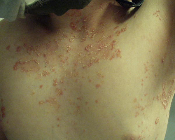
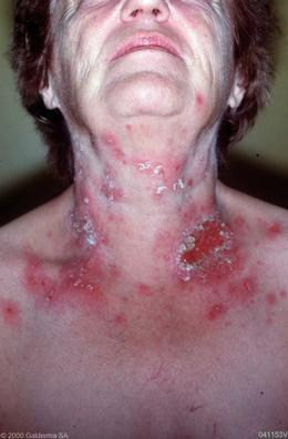
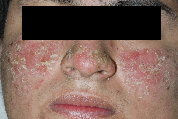
The onset is usually insidious with scattered, scaly lesions in a 'seborrhoeic' distribution, ie the scalp, face and upper trunk
-
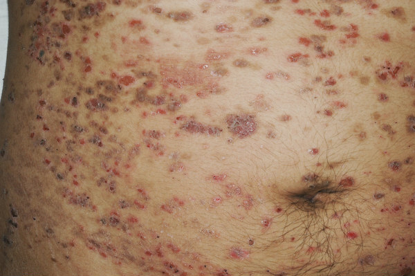
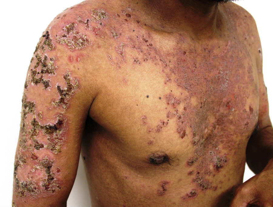
Erosions, crusting and erythema are common. Occasionally patients can become erythrodermic. Blisters may not be obvious as they easily rupture
-
The Nikolsky sign is positive - this is where firm sliding pressure with a finger will cause an erosion
-
The patient is usually otherwise well
Clinical variants
-
Pemphigus erythematosus affects the face and overlaps with lupus erythematosus, and may be exacerbated by UV exposure
-
Pemphigus herpetiformis resembles dermatitis herpetiformis in the early phase
Clinical Images
Please refer to notes on image rights at bottom of the page with regards to individual image
ownership.
Investigations
Bloods tests - indirect immunofluorescence
-
A request for skin antibodies covers both pemphigoid (skin basement membrane) and pemphigus (skin desmosome) antibodies. In PF the results are frequently positive for circulating desmoglein-1 antibodies
Skin biopsies - histology and direct immunofluorescence (reference: Rook's Textbook of Dermatology)
-
Two skin biopsies are required:
-
An intact blister should be excised and sent for histology
-
A second biopsy, of peri-lesional skin (within 2 cm of the blister), is also required for direct immunofluorescence (DIF). The sample must be put on top of a piece of plain gauze, which has been soaked in a small amount of normal saline, and placed into a dry pot, the specimen must be examined the same day. If the sample cannot be examined the same day it must be placed in a suitable transport media eg Michel's solution, in order to preserve the sample. The result of DIF can sometimes be false negative, and if needed a further biopsy sample can be taken from unaffected skin of the buttocks or thighs
-
Histology shows rounded-up separated keratinocytes (acantholytic cells) within blisters in the upper layers of the epidermis. Dyskeratotic cells in the granular layer of older lesions also help distinguish PF from pemphigus vulgaris
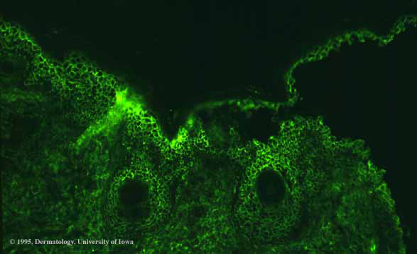
- DIF shows linear deposition of IgG and C3 predominantly in the upper layers of the epidermis
Management
Referral
-
Patients need referring urgently to dermatology for diagnosis and management
Topical steroids
-
Potent / superpotent topical steroids can be used for less severe cases
Systemic treatment (the majority)
-
Prednisolone 20-40 mg/day initially, then reduced down once control has been achieved
- If the condition continues to flare as the dose of steroid is reduced, a number of medications have been used as adjuncts including hydroxychloroquine, daspone, azathioprine and mycophenolate mofetil
Prognosis
-
Spontaneous remission may occur in some patients whilst in others the condition may persist for several years
Other resources
Disclaimer - the author PCDS cannot accept responsibility for any misleading or incorrect statements, and the management of individual patients remains the direct responsibility of the individual doctor. We do however hope that visitors to this site can contact us regarding comments that are considered misleading or incorrect so that we can continue to improve the site.
Image Rights - The PCDS would like to thank Dermatoweb, DermQuest (Galderma), and others who have contributed images. All named individuals and organisations maintain copyright for the relevant images.