Mucous membrane pemphigoid (syn. cicatricial pemphigoid)

LAST UPDATED: Aug 09, 2021
Introduction

Mucous membrane pemphigoid (MMP) is a chronic, immunobullous condition of the mucosa that may involve the skin, and usually results in permanent scarring of the affected area, particularly the conjunctiva. This entity includes patients formerly diagnosed as oral pemphigoid and some cases of linear IgA disease and epidermolysis bullosa acquisita.

Considerable variability exists in the clinical presentation of MMP, affected sites include the oral cavity, conjunctiva, nasopharynx, larynx, oesophagus, genitourinary tract, and anus. The skin is involved in approximately one-quarter of patients, often limited to the head, neck, and upper torso regions. Pathologically MMP is characterised by the presence of subepidermal blisters, with various basement membrane zone components as targets of the autoantibodies.

This chapter is set out as follows:


Aetiology

  • MMP is an immunobullous condition
  • The immunobullous conditions are characterised by pathogenic autoantibodies directed at target antigens whose function is either cell-to-cell adhesion within the epidermis or adhesion of stratified squamous epithelium to the dermis or mesencyme. The target antigens involved are components of desmosomes or the functional unit of the basement membrane zone known as the adhesion complex
  • In MMP, IgG and less commonly IgA autoantibodies are directed against varies basement membrane zone antigens including bullous pemphigoid antigens BP180 and BP230, along with laminin 5, laminin 6 and the β4-integrin subunit

History

  • MMP is predominantly a disease of late-middle to old age with a peak incidence at around 70 years. However, childhood cases have been reported
  • It is twice as common in women than men

Clinical findings

​Oral mucosa

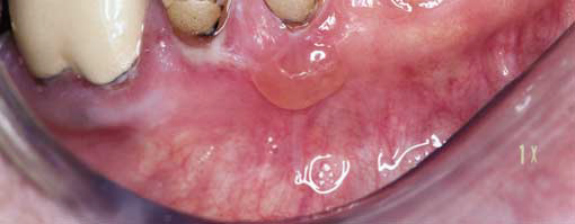
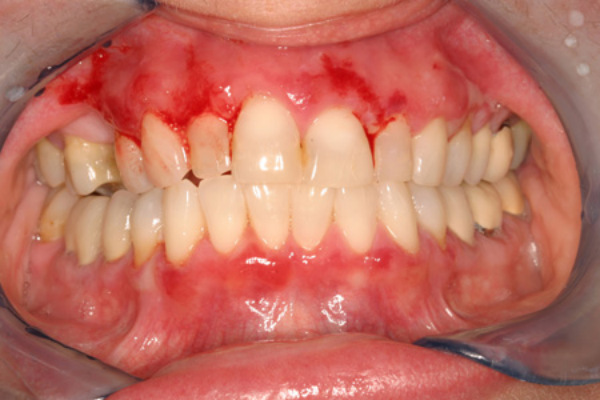
  • ​Oral lesions occur in the majority of patients
  • Gum involvement is common, and as with lichen planus the gums are bright red
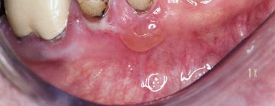
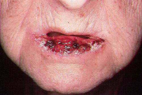
  • Compared to other conditions affecting the mouth, vesicles or small bullae may remain intact for some time. When erosions form they are slow to heal
  • Lesions can be persistent and extensive in the buccal mucosa, especially the hard palate
  • Progression of the condition in to the oesophagus, trachea and larynx, leading to stricture formation, can become life-threatening  

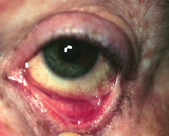
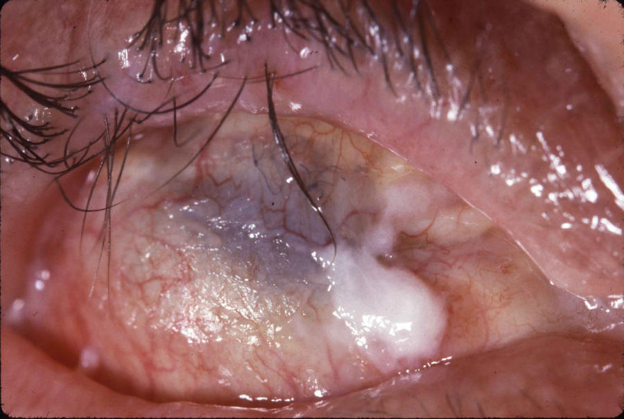
Eyes

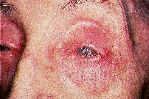
  • Lesions may start in one eye but later involve both
  • In some cases patients present with conjunctivitis and complain of grittiness or pain. This may come and go over a few years, before then progressing
  • On occasions painful erosions and blisters are the presenting feature, although the blisters may be difficult to visualise
  • Once established, lesions erode and heal to leave scar tissue, which impairs vision, or even causes blindness. Sometimes the scarring can occur without any preceding symptoms 

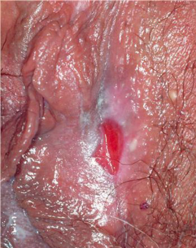
Genitalia 

  • ​The genitals are involved in half of female patients. Painful blisters and erosions on the clitoris and labia can lead to architectural changes similar to those seen in lichen sclerosis 
  • Penile involvement can lead to adhesions between the prepuce and glans penis 

Nose

  • Erosions and blisters can cause discomfort, crusting and nose bleeds

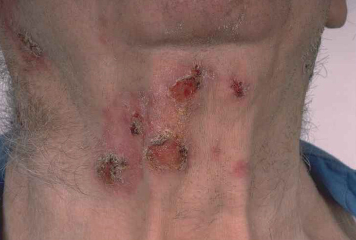
Skin 

  • The skin can be affected in two ways:
    • Localised areas of scarring blisters, predominantly of the scalp and of the skin close to affected mucosal surfaces
    • Less commonly a generalised bullous eruption similar to bullous pemphigoid 

Clinical Images

Please refer to notes on image rights at bottom of the page with regards to individual image ownership.
Investigations

Blood tests - indirect immunofluorescence (reference: Rook's Textbook of Dermatology)
  • A request for skin antibodies covers both pemphigoid (skin basement membrane) and pemphigus (skin desmosome) antibodies. In MMP, the result is often negative, however, a minority will be positive for circulating pemphigoid antibodies 
Skin biopsies (reference: Rook's Textbook of Dermatology)
  • In order to take the biopsies correctly refer to the section on investigations in the chapter Bullous pemphigoid - of course depending on the site of blisters, other specialist departments may need to be involved. Two samples are needed:
    • Histology of a blister reveals a subepidermal blister with a variable inflammatory cell infiltrate 
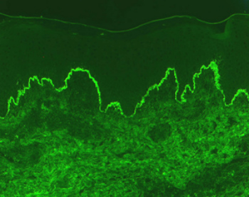
    • Direct immunofluorescence (DIF) of peri-lesional mucosa or skin is essential for diagnosis, and reveals linear deposition of IgG, C3, or less commonly IgA along the basement membrane zone
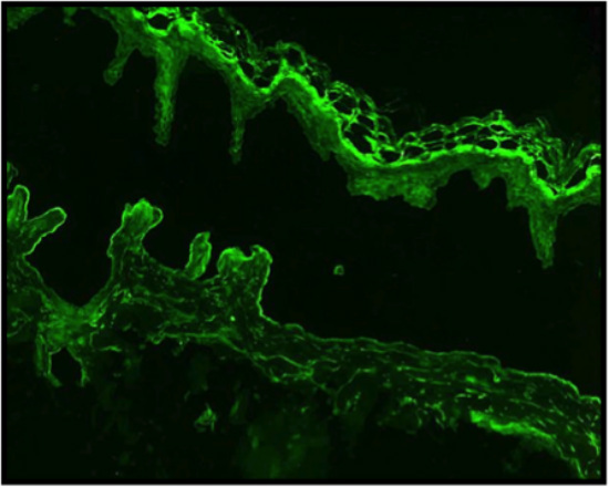
  • In cases of clinical diagnostic uncertainty the use of salt-split skin immunofluorescence studies can be useful in differentiating between MMP and bullous pemphigoid - the skin biopsy sample is placed in 1 mol/L salt prior to performing the immunofluorescence studies, which causes cleavage through the lamina lucida. Examination of the salt-split skin reveals IgG on the blister floor (dermal side of split skin) in MMF, while, in bullous pemphigoid the IgG can be found in blister roof (epidermal side of split skin) 

Management

  • Patients suspected of having MMP should be referred urgently to an appropriate specialist in Secondary Care 
  • MMP is a chronic, progressive disease that often responds poorly to treatment. Spontaneous resolution is rare. Some patients may experience long remissions with intermittent exacerbations
  • A multidisciplinary approach is essential in the management of MMP. Early recognition and treatment may decrease disease-related complications. In a patient who presents with involvement of one site, a thorough review of symptoms highlighting other potential areas of involvement should be obtained. The choice of agents for treatment of MMP is based upon the sites of involvement, clinical severity, and disease progression

Disclaimer - the author PCDS cannot accept responsibility for any misleading or incorrect statements, and the management of individual patients remains the direct responsibility of the individual doctor. We do however hope that visitors to this site can contact us regarding comments that are considered misleading or incorrect so that we can continue to improve the site.

Image Rights - The PCDS would like to thank Dermatoweb, DermQuest (Galderma), and others who have contributed images. All named individuals and organisations maintain copyright for the relevant images.

Quick Links

The following pharmaceutical companies have had no involvement in the content of this website or in our conference programmes


Almirall
Galderma
Glenmark
Johnson & Johnson
La Roche-Posay
LEO Pharma
Pierre Fabre
Schuco