Subcorneal pustular dermatosis (syn. Sneddon-Wilkinson disease)
LAST UPDATED: Aug 01, 2021
Introduction
Subcorneal pustular dermatosis is a rare, chronic, relapsing, bullous condition, characterised by blisters that become filled with pus. It was first described in 1956 by Drs Ian Sneddon and Darrell Wilkinson.
This chapter is set out as follows:
Aetiology
-
The cause is unknown
-
It is associated with a number of conditions, including:
-
IgA monoclonal gammopathy and myeloma
-
Pyoderma gangrenosum, inflammatory bowel disease and rheumatoid arthritis
History
-
It affects women more often than men (at a ratio of 4:1), usually starting after the age of 40
Clinical findings
Distribution
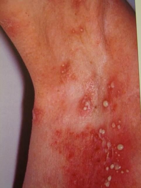
- Although any area of skin can be involved, it is less common for the forearms and lower legs to be affected. Flexural sites under the breasts, the axilla, and the groin are common sites of involvement
- The face and mucous membranes are almost never affected
Morphology
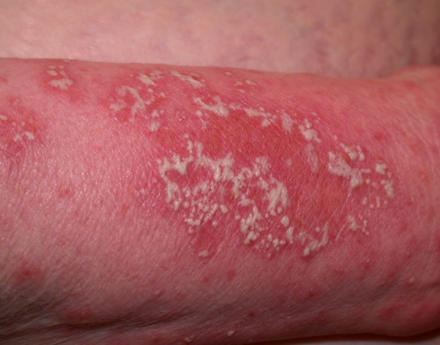
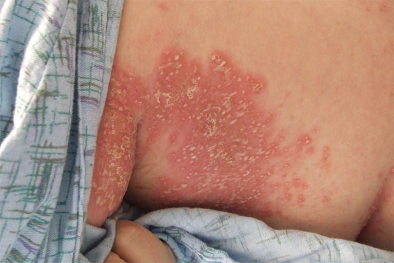
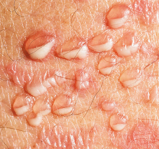
- Lesions arise as numerous small, sterile pustules. Characteristically the pus accumulates in the lower half of the pustule
- Lesions easily rupture and tend to coalesce, forming annular or serpiginous patterns with a scaly edge
- Eruptions fade. The interval before the next eruption can be days to a few months
Clinical Images
Please refer to notes on image rights at bottom of the page with regards to individual image
ownership.
Investigations
-
Blood tests
-
Should include a FBC, routine biochemistry, and immunoglobulins with protein electrophoresis to look for a gammopathy
-
Histology (reference: Rook's Textbook of Dermatology)
-
The classic histological finding in subcorneal pustular dermatosis is subcorneal pustules composed primarily of neutrophils and occasional eosinophils. However, this finding is not specific for subcorneal pustular dermatosis and can be found in other conditions such as pustular psoriasis, acute generalised exanthematous pustulosis, pemphigus foliaceus, bacterial impetigo, and dermatophytosis. In subcorneal pustular dermatosis, unlike in pustular psoriasis, the epidermis usually has minimal spongiosis. The dermis in subcorneal pustular dermatitis shows a perivascular infiltrate of neutrophils and occasional monocytes and eosinophils. Acantholysis is not prominent; however, it has been reported in older lesions. Direct and indirect immunofluorescence studies are typically negative
Management
-
Patients require referral to Secondary Care
- Dapsone 50-150 mg daily is the treatment of choice. This is often successful, with the lesions resolving over a month. Ongoing maintenance with a lower dose is sometimes needed
-
Other treatment options include sulfapyridine or sulfamethoxypyridazine, acitretin, colchicine, phototherapy and biologic therapies. Systemic steroids are generally ineffective and can precipitate a flare-up
-
Prognosis - the average duration is 5.8 years
Disclaimer - the author PCDS cannot accept responsibility for any misleading or incorrect statements, and the management of individual patients remains the direct responsibility of the individual doctor. We do however hope that visitors to this site can contact us regarding comments that are considered misleading or incorrect so that we can continue to improve the site.
Image Rights - The PCDS would like to thank Dermatoweb, DermQuest (Galderma), and others who have contributed images. All named individuals and organisations maintain copyright for the relevant images.