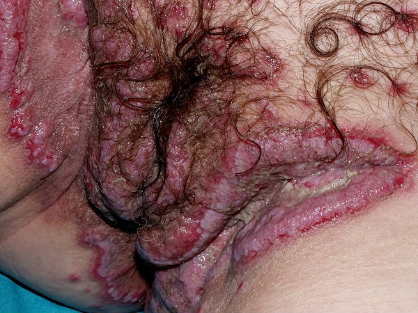
Pemphigus vulgaris and pemphigus vegetans
LAST UPDATED: Jul 07, 2024
Introduction
Pemphigus is derived from the Greek pemphix meaning blister or bubble. Pemphigus vulgaris is a rare immunobullous condition that is characterised by blisters and erosions on the skin and mucous membranes, most commonly the mouth.
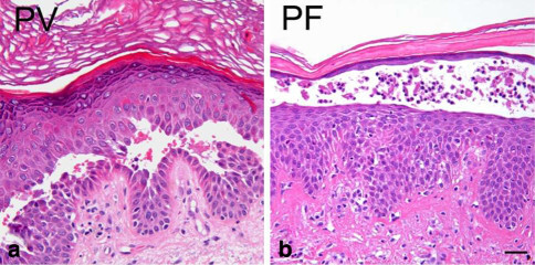
The blisters in both pemphigus vulgaris, and its even less common vegetating form, pemphigus vegetans, are suprabasal. Pemphigus vulgaris is the most common subtype of pemphigus, accounting for 70% of all pemphigus cases worldwide.
This chapter is set out as follows:
Aetiology
-
Pemphigus vulgaris is an immunobullous condition
-
The immunobullous conditions are characterised by pathogenic autoantibodies directed at target antigens whose function is either cell-to-cell adhesion within the epidermis or adhesion of stratified squamous epithelium to the dermis or mesencyme. The target antigens involved are components of desmosomes or the functional unit of the basement membrane zone known as the adhesion complex
-
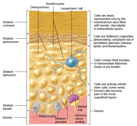
In pemphigus vulgaris IgG autoantibodies are directed to the pemphigus vulgaris antigen, also known as desmoglein-3, which is found in desmosomes in the keratinocytes near the bottom of the epidermis. The result is the keratinocytes separate from each other, and are replaced by fluid, the blister
History
-
Pemphigus vulgaris affects all races and both sexes
-
It most commonly presents between the ages of 50-60 years, but can affect any age, although rarely children
-
It is more common in the East compared to the West, and in those of Jewish ancestry, especially Ashkenazi Jewish heritage
-
Pemphigus is more common in patients with other autoimmune conditions eg myasthenia gravis and lupus
Clinical findings
Pemphigus vulgaris
-
Mucosal lesions
-
The vast majority of patients have mucosal lesions, and the mouth is the most common site of presentation
-
Mucosal lesions may precede cutaneous lesions by months, or may be the only manifestation of the condition
-
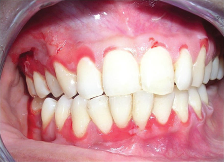
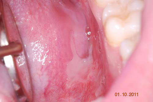
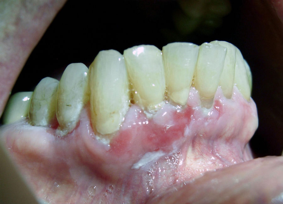
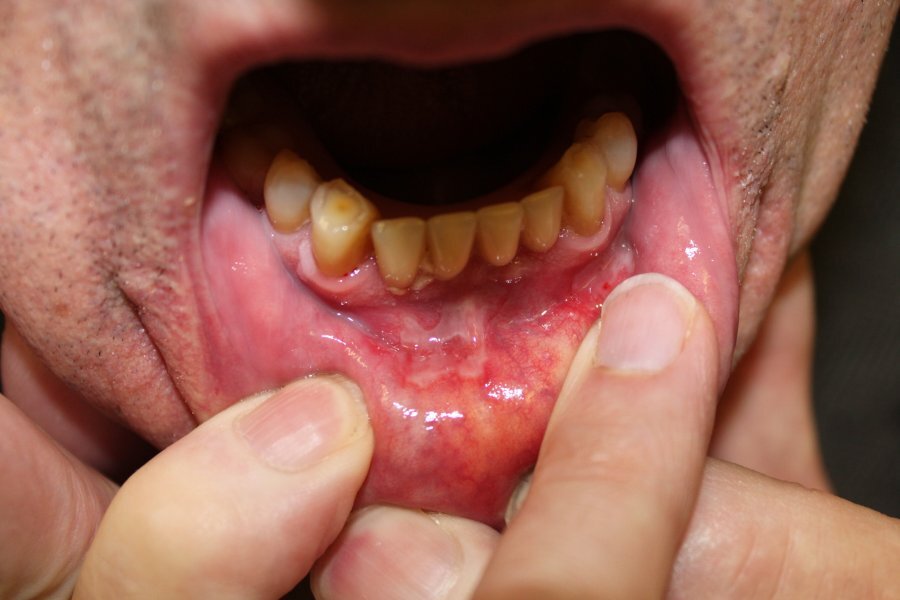
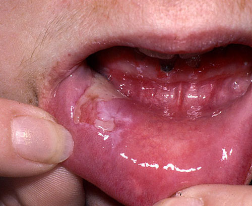
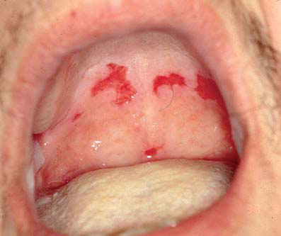
Bullae are rarely seen, instead, ill-defined, painful erosions mainly involving the lips, buccal mucosa and palate, which are slow to heal. Lesions on the border of the soft and hard palate are almost pathognomonic. There is significant whitening of the mucosa. Erosions can be seen on the gingival tips
-
The mucosa returns to normal when the condition is in remission
-
Other mucosal sites affected include the conjunctiva, oesophagus, nose, pharynx, larynx, and genitalia
-
Cutaneous lesions
-
Affect most patients
-
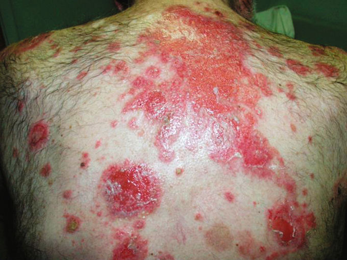
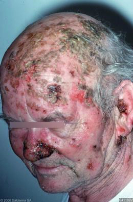
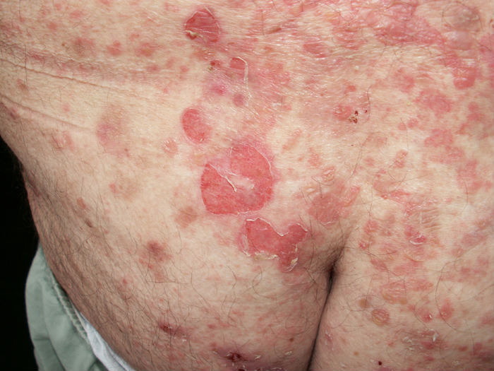
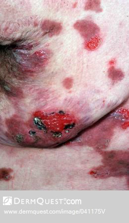
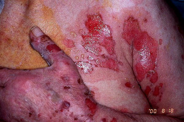
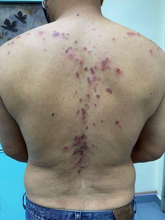
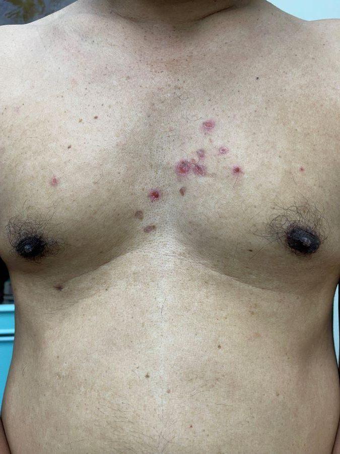
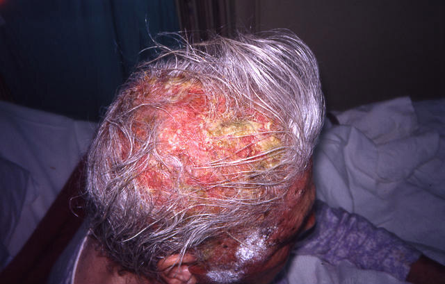
The most commonly affected sites include the scalp, face, axillae, groins and pressure points. The trunk can also be involved
-
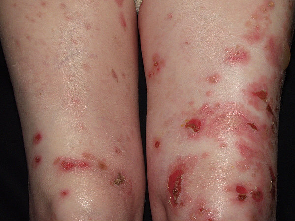
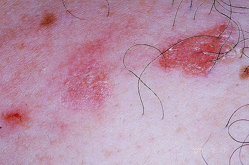
The primary lesion of pemphigus vulgaris is a flaccid blister filled with clear fluid that arises on healthy skin or on an erythematous base. Blisters are fragile and may rupture, producing painful erosions (the most common skin presentation). Lesions heal without scarring
-
The Nikolsky sign is positive - this is where firm sliding pressure with a finger will cause an erosion
-
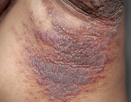
Flexural involvement readily forms vegetating granulomas
-
Nails
-
Acute or chronic paronychia, subungual haematomas, and nail dystrophies affecting one or several fingers or toes have been reported
Pemphigus vegetans
-
A rare variant of pemphigus vulgaris
-
The principle finding are vegetating lesions in the flexures, although any site can be affected
-
Nearly all patients have oral involvement
Clinical Images
Please refer to notes on image rights at bottom of the page with regards to individual image
ownership.

1

2

3

4

5

6

7

8

9

10

11

12

13

14

15

16

17

18

19

20

21
Investigations
Blood test - indirect immunofluorescence
-
A request for skin antibodies covers both pemphigoid and pemphigus antibodies. In pemphigus vulgaris, circulating pemphigus antibodies are detected in over 80% of cases
Skin biopsies (reference: Rook's Textbook of Dermatology)
-
Two skin biopsies are required
-
An intact blister should be excised and sent for histology
-
A second biopsy, of peri-lesional skin (within 2 cm of the blister), is also required for direct immunofluorescence (DIF). The sample must be put on top of a piece of plain gauze, which has been soaked in a small amount of normal saline, and placed into a dry pot, the specimen must be examined the same day. If the sample cannot be examined the same day it must be placed in a suitable transport media eg Michel's solution, in order to preserve the sample. The result of DIF can sometimes be false negative, and if needed a further biopsy sample can be taken from unaffected skin of the buttocks or thighs
-
Histology shows suprabasal clefts and blisters, containing some acantholytic cells
-
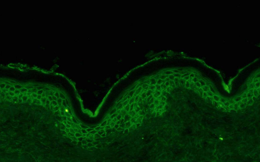
DIF shows IgG deposited on the surface of keratinocytes throughout the epidermis
Management
General measures
-
Patients will need referring urgently to dermatology
- Supportive measures include wound care and reducing the risk of secondary infection by using antiseptic regimes, eg Dermol 500 lotion ® as a wash and / or topical emollient
-
Patients should minimise activities that may traumatise the skin and mucous membranes during active phases
Topical steroids
-
Potent / super-potent topical steroids may be used in milder cases, especially earlier in the natural history of the condition
Systemic corticosteroids
-
Are the preferred treatment option, but should be used in conjunction with topical treatments
-
Prednisolone 1-1.5 mg/kg/day is usually sufficient to control the condition, the dose should be reduced as the condition improves
Adjunctive treatments
- If the skin continues to flare as the dose of oral steroids is reduced, a number of other medications have been used as adjuncts including azathioprine, mycophenolate mofetil, dapsone and cyclophosphamide
Prognosis
-
Pemphigus typically has a chronic course, although there is great variability in disease duration and time taken to disease remission between individuals
-
Disease activity generally decreases with time and most relapses occur in the first two years, although the prognosis is worse in Jews and older patients
-
Treatment should be stopped when the disease is inactive, but can be re-started for flares
Other resources
Disclaimer - the author PCDS cannot accept responsibility for any misleading or incorrect statements, and the management of individual patients remains the direct responsibility of the individual doctor. We do however hope that visitors to this site can contact us regarding comments that are considered misleading or incorrect so that we can continue to improve the site.
Image Rights - The PCDS would like to thank Dermatoweb, DermQuest (Galderma), and others who have contributed images. All named individuals and organisations maintain copyright for the relevant images.