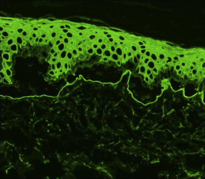
Epidermolysis bullosa acquisita
LAST UPDATED: Nov 11, 2025
Introduction
Epidermolysis bullosa acquisita (EBA) is a rare immunobullous condition in which patients may have chronic acquired, trauma-induced, subepidermal blistering, or a clinical picture similar to bullous pemphigoid. EBA has a distinctive immunopathology. It is one of the rarest subepidermal bullous conditions in Western Europe with an incidence of about 0.25 per million, but may be more common in the middle and far East.
Epidermolysis bullosa (EB), a group of genetically determined skin fragility conditions, is discussed in a related chapter.
This chapter is set out as follows:
Aetiology
-
EBA is an immunobullous condition. In several affected patients, autoantibodies have found to be directed to type VII collagen, which is the major structural component of anchoring fibrils attaching the basement membrane to the underlying dermis
-
There is an association with inflammatory bowel disease and lymphoproliferative disorders
-
Drug-induced EBA has been described
History
-
EBA can occur at any age, although it most commonly arises in the fourth and fifth decade of life
-
Males and females of all races can be affected
Clinical findings
There are two main presentations:
EBA affecting trauma-prone extensor skin surfaces
-
This is the most common form of EBA
-
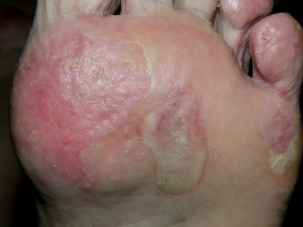
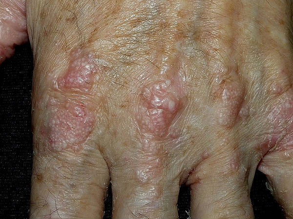
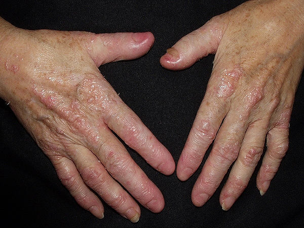
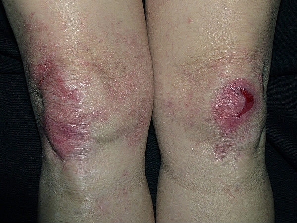
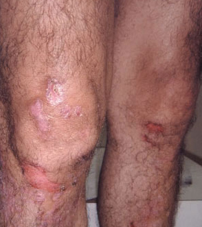
Tense vesicles and bullae (serous or haemorrhagic) arise primarily on areas of trauma such as the extensor surfaces of hands, elbows, knees, ankles and feet
-
Lesions heal with significant scarring, milia and hyperpigmentation
-
Mucosal involvement is variable, but can be severe
-
It resembles the inherited form of dystrophic epidermolysis bullosa
Generalised inflammatory EBA
-
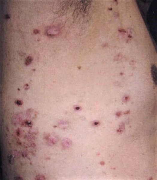
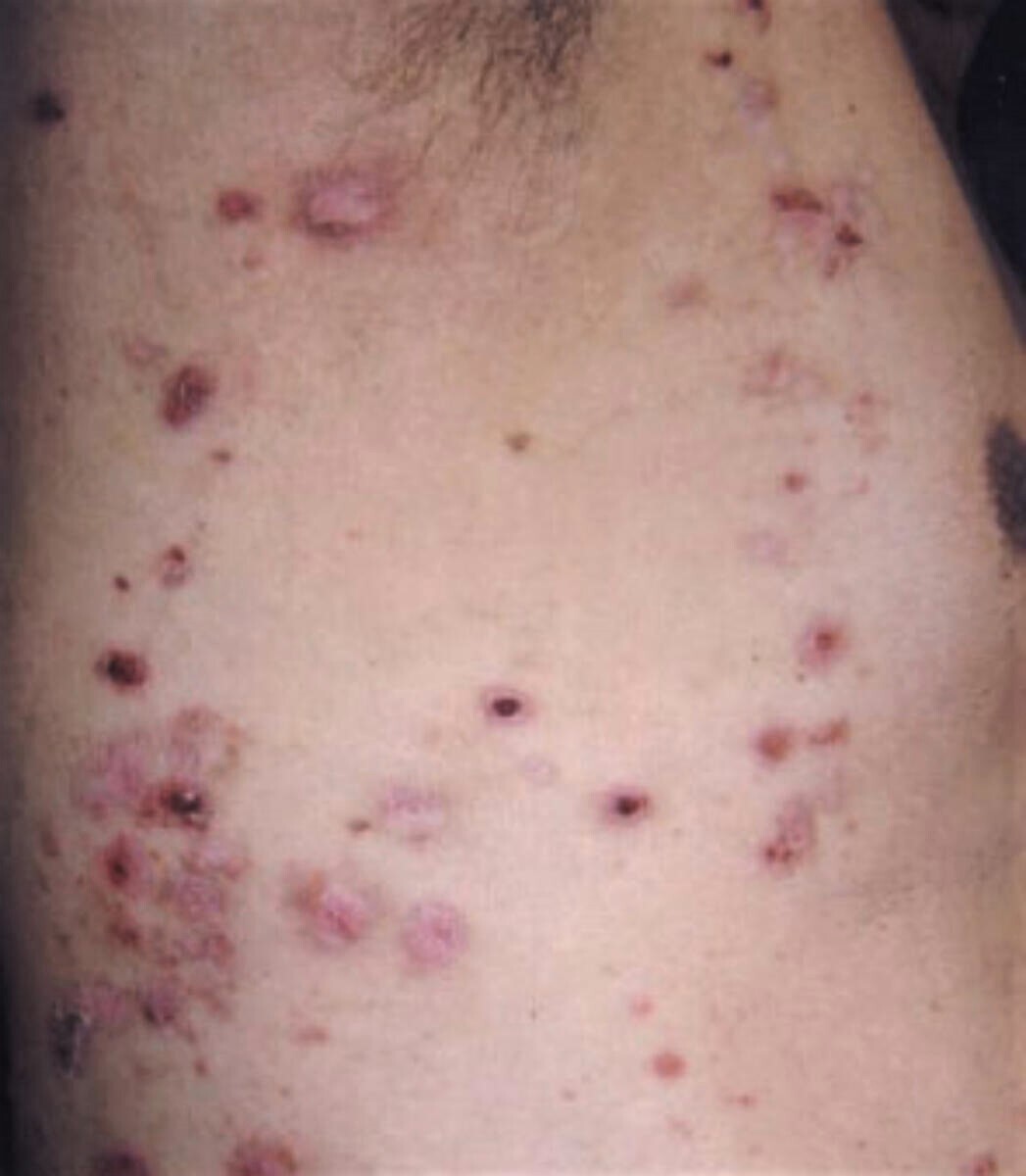
Some presentations are indistinguishable from bullous pemphigoid
-
Lesions heal with less scarring
-
Mucosal involvement is variable, but can be severe
-
Some evolve in to a more typical mechanobullous picture
Those patients in whom mucosal symptoms predominate, are now regarded as mucous membrane pemphigoid.
Clinical Images
Please refer to notes on image rights at bottom of the page with regards to individual image
ownership.
Investigations
Blood tests - indirect immunofluorescence
-
A request for skin antibodies covers both pemphigoid and pemphigus antibodies. In EBA, circulating IgG autoantibodies that target the skin basement membrane (pemphigoid antibodies) are present in approximately half the patients
Skin biopsies
-
Two skin biopsies are required
-
An intact blister should be excised and sent for histology
-
A second biopsy, of peri-lesional skin (within 2 cm of the blister), is also required for direct immunofluorescence (DIF). The sample must be put on top of a piece of plain gauze, which has been soaked in a small amount of normal saline, and placed into a dry pot, the specimen must be examined the same day. If the sample cannot be examined the same day it must be placed in a suitable transport media eg Michel's solution, in order to preserve the sample
-
Findings in EBA (reference: Rook's Textbook of Dermatology)
-
Histology shows a subepidermal blister. In the inflammatory phase there is a heavy neutrophil infiltrate. In the mechanobullous, non-inflammatory, state, there is usually an absent or sparse infiltrate
-
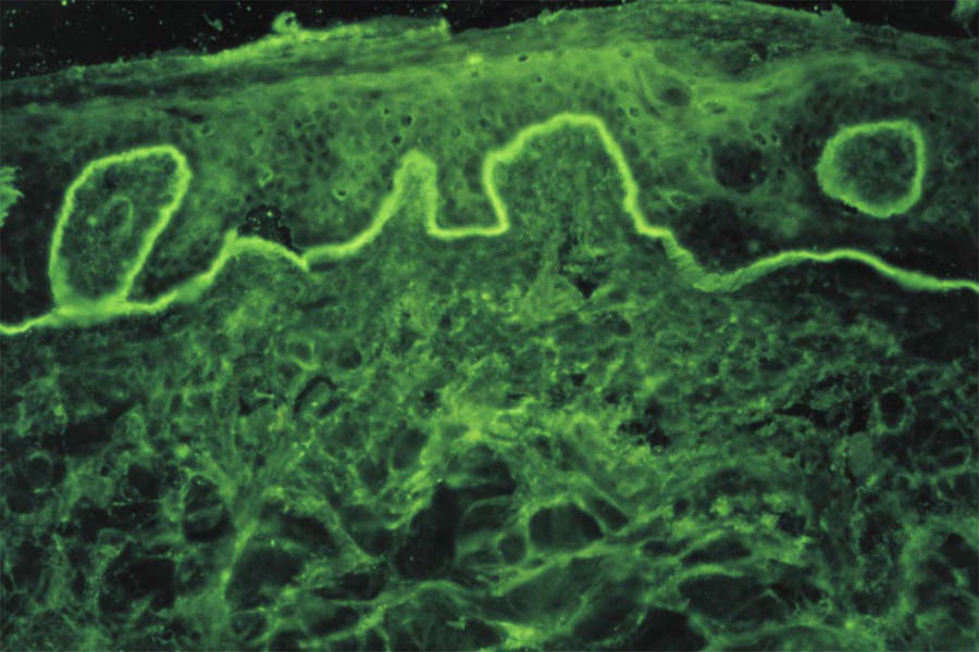
DIF show IgG deposited in a line along the dermoepidermal junction. Linear IgA and IgM may also be seen
-
The use of salt-split skin immunofluorescence studies can be useful in differentiating between epidermolysis bullosa acquisita and bullous pemphigoid - the skin biopsy sample is placed in 1 mol/L salt prior to performing the DIF technique, which causes cleavage through the lamina lucida. Immunofluorescence on salt-split skin in epidermolysis bullosa acquisita reveals IgG in the blister floor (dermal side of split skin), while, in patients with bullous pemphigoid the IgG localises in the blister roof (epidermal side of split skin)
Management
Patients will often be referred to Secondary Care to obtain a diagnosis, and also with respect to management
- The primary aim of treatment is to protect the skin and reduce stop blister formation, promote healing and prevent secondary infection, scarring and deformities
- Given the rarity of this condition, there is little data that can conclude an evidence-based management strategy for the inflammatory component of EBA. Many treatment have been tried including corticosteroids, dapsone, azathioprine, ciclosporin, colchicine, mycophenolate mofetil, intravenous immunoglobulin and the biologic Rituximab
- The mechanobullous variant often proves very resistant to treatment
- The prognosis is variable - patients with a bullous pemphigoid-like picture may go into remission. However those with mechanobullous disease tend to be chronic
Other resources
- DEBRA is the patient support organisation for people in the UK directly affected by any form of inherited EB or acquired EB, known as epidermolysis bullosa acquisita (EBA). Family members, carers, healthcare professionals and researchers working in the field of EB, can also join DEBRA as a member for free. Benefits of DEBRA membership include nationwide access to their EB Community Support Team, financial support and benefits advice, EB information and resources, respite breaks, and opportunities to connect with the EB community. For more information please use the following link Become a DEBRA member
- Notable resources within the DEBRA website include:
Disclaimer - the author PCDS cannot accept responsibility for any misleading or incorrect statements, and the management of individual patients remains the direct responsibility of the individual doctor. We do however hope that visitors to this site can contact us regarding comments that are considered misleading or incorrect so that we can continue to improve the site.
Image Rights - The PCDS would like to thank Dermatoweb, DermQuest (Galderma), and others who have contributed images. All named individuals and organisations maintain copyright for the relevant images.