Acne keloidalis nuchae (syn. folliculitis keloidalis nuchae)
LAST UPDATED: Nov 21, 2021
Introduction
Acne keloidalis nuchae is a chronic inflammatory process involving the hair follicles of the neck, leading to hypertrophic scarring in papules and plaques.
The term acne keloidalis nuchae is somewhat of a misnomer because the lesions do not occur as a result of acne vulgaris, but rather a folliculitis. Moreover, histologically lesions are not keloidal.
This chapter is set out as follows:
Aetiology
Possible aetiologies include:
-
Close shaving of the neck - this often exacerbates the condition as the sharp, curved hairs re-enter the skin and invoke an acute inflammatory response
-
Constant irritation from shirt collars
-
Possibly, chronic low-grade bacterial infections
History
-
Acne keloidalis nuchae is most prevalent in African Americans; however, it has occasionally been reported in Hispanics and Asians, and, rarely, in whites
-
It is very uncommon in females
-
The condition most commonly arises between the ages of 14 and 25 years. Lesions manifesting prior to puberty or in patients older than 50 years are unusual
-
Some patients have a history of significant acne vulgaris
Clinical findings
-
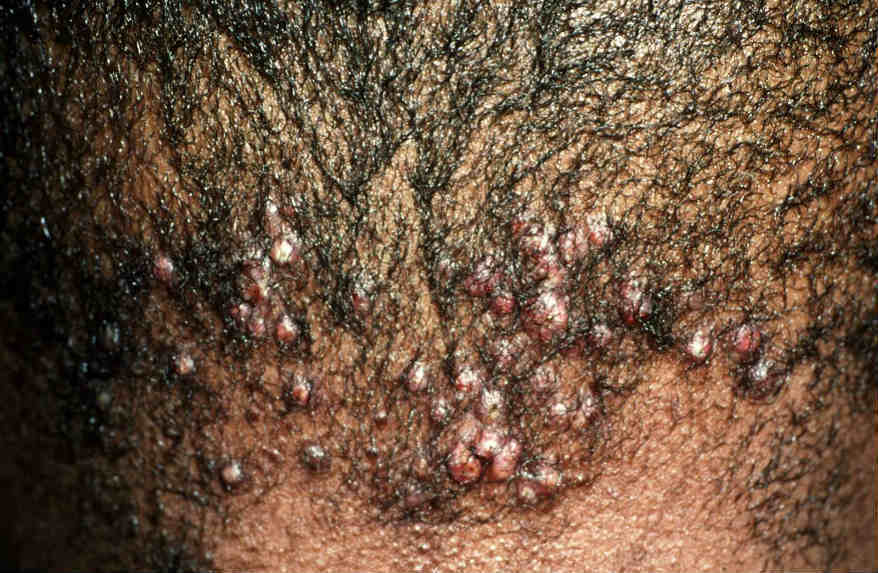
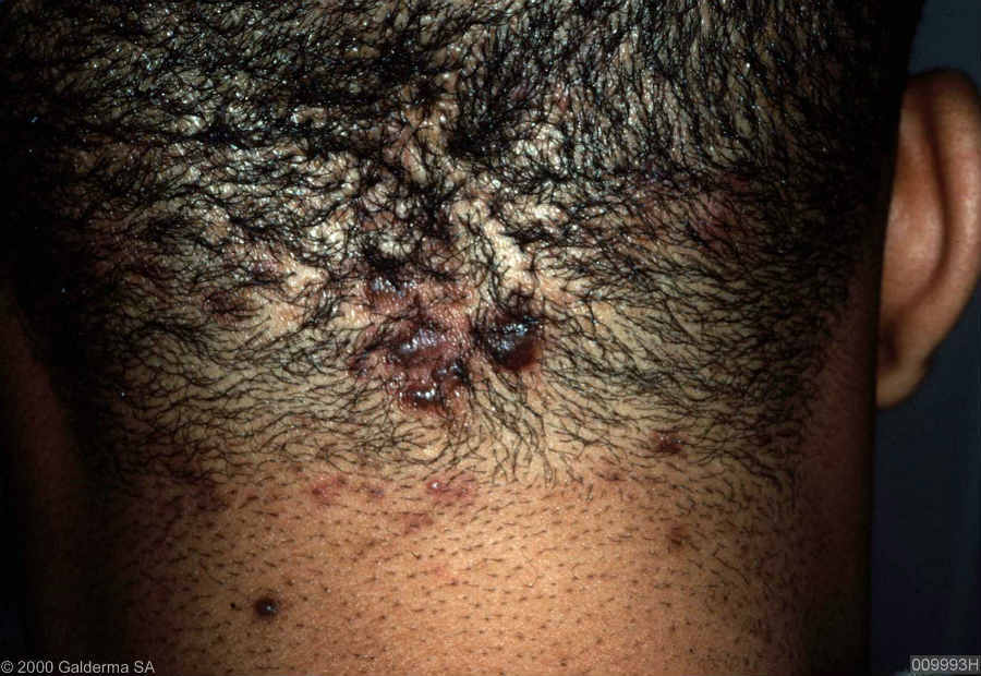
Early lesions manifest as follicular papules or pustules, developing on the nape of the neck just below the hairline. They may extend into the adjacent scalp
-
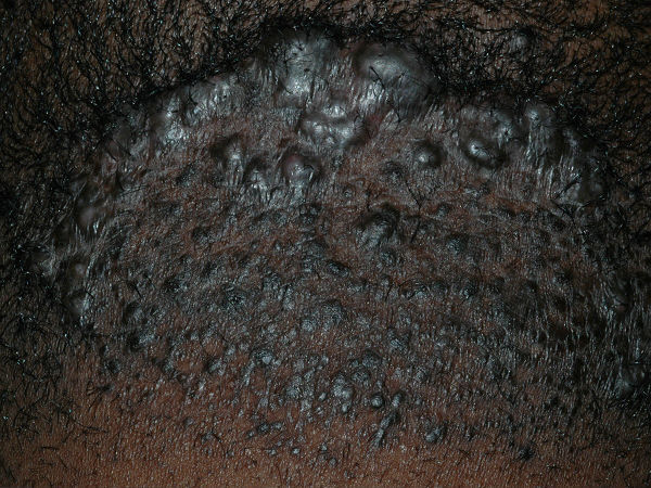
The papules may remain discrete or may fuse into horizontal bands or irregular plaques
-
Inflammation can be very subtle in some, while persistent and troublesome in others with discharging sinuses
-
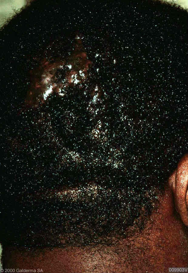
Scarring alopecia eventually ensues
Clinical Images
Please refer to notes on image rights at bottom of the page with regards to individual image
ownership.
Investigations
- Diagnosis is usually clinical. If a diagnostic biopsy is required histology shows a dense superficial and deep inflammatory process with dermal scarring and follicular disruption
Management
Treatment of acne keloidalis nuchae (AKN) is difficult, and numerous modalities have been used with varying degrees of success:
Step 1: general measures
-
Making sure clothing and equipment, such as high collars and helmets, do not rub the back of the neck
-
Avoid a short haircut, and shaving of the hair on the affected scalp
-
Patients should discontinue hair greases or pomades as these are thought to aggravate the condition
-
In order to reduce secondary infection wash the affected area daily using an antimicrobial cleanser, eg Dermol ® lotion or Hibiscrub ®
Step 2: steroids
-
Either super-potent topical steroids eg Dermovate ® cream BD, or intralesional steroids such as triamcinolone acetonide given every 2-3 weeks, may help gradually improve symptoms
Step 3: antibiotics
-
When pus or serous drainage is present a culture should be taken. Antibiotic use, which may include oral antibiotics, should be tailored according to bacterial sensitivities. For more severe or persistent infection, consider a 3-6 month course of clindamycin 300 mg BD and rifampicin 300 mg BD. Before commencing treatment the BNF advises checking U&E and LFT, if these are normal then further checks are usually unnecessary as liver damage is rare, and if it does occur it is usually within the first six weeks of treatment. Patients should be advised to stop treatment and seek urgent medical attention should they develop signs of hepatotoxicity (fever, malaise, vomiting, jaundice)
-
If the AKN is not improving significantly, and there is no significant infection, consider a three month trial of an oral tetracycline (eg lymecycline 408 mg OD or doxycycline 100 mg OD) acting as a skin anti-inflammatory. If this helps, treatment can be continued for longer periods if required
Step 4: other treatments for consideration in a specialist's setting
-
Oral isotretinoin
-
Short courses of systemic steroids if inflammation is very marked
-
Physical treatments:
-
Cryosurgery - the area is frozen for 20 seconds, allowed to thaw, and is then frozen again one minute later. The process may be painful for patients, and the treated site often becomes hypopigmented because of destruction of the melanocytes, and may remain so for up to 12-18 months
-
Surgical excision of the most troublesome areas, with or without intralesional steroids
-
Laser ablation
Step 5: maintenance therapy
-
Once the condition has settled it is important to continue with the general measures referred to in step 1
-
Intermittent courses of topical steroids may also be required
Disclaimer - the author PCDS cannot accept responsibility for any misleading or incorrect statements, and the management of individual patients remains the direct responsibility of the individual doctor. We do however hope that visitors to this site can contact us regarding comments that are considered misleading or incorrect so that we can continue to improve the site.
Image Rights - The PCDS would like to thank Dermatoweb, DermQuest (Galderma), and others who have contributed images. All named individuals and organisations maintain copyright for the relevant images.