Anetoderma

LAST UPDATED: Nov 25, 2021
Introduction

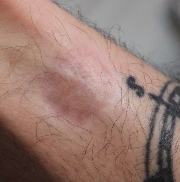
The term anetoderma (anetos: slack) refers to a circumscribed area of slack skin associated with a loss of dermal substance on palpation and a loss of elastic tissue on histological examination. Primary anetoderma implies there is no associated localised underlying cutaneous disease, whereas secondary anetoderma can be attributed to some associated condition. 

This chapter is set out as follows:


Aetiology

Primary anetoderma
  • Is strongly associated with antiphospholipid antibodies, with or without a prothrombotic state. It is probable that these antibodies underlie the association historically noted with syphilis, and more recently with borreliosis (Lyme disease) and systemic lupus erythematosus. In a few cases there appears to be an underlying structural defect of connective tissue. Familial cases are reported 
Secondary anetoderma
  • Is seen in association with another identifiable condition including the following:
    • Skin infections eg chickenpox, syphilis
    • Connective tissue conditions, especially lupus erythematosus
    • Inflammatory dermatoses such as acne and granuloma annulare
    • Skin lesions eg pilomatrixoma, juvenile xanthogranuloma

History

  • Women are affected more than men
  • Most cases arise between the ages of 20-40 years, although any age group can be affected 
  • In secondary anetoderma there may be a history of preceding inflammation
  • Lesions are asymptomatic 

Clinical findings

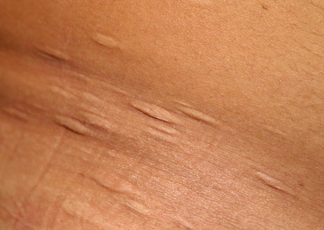
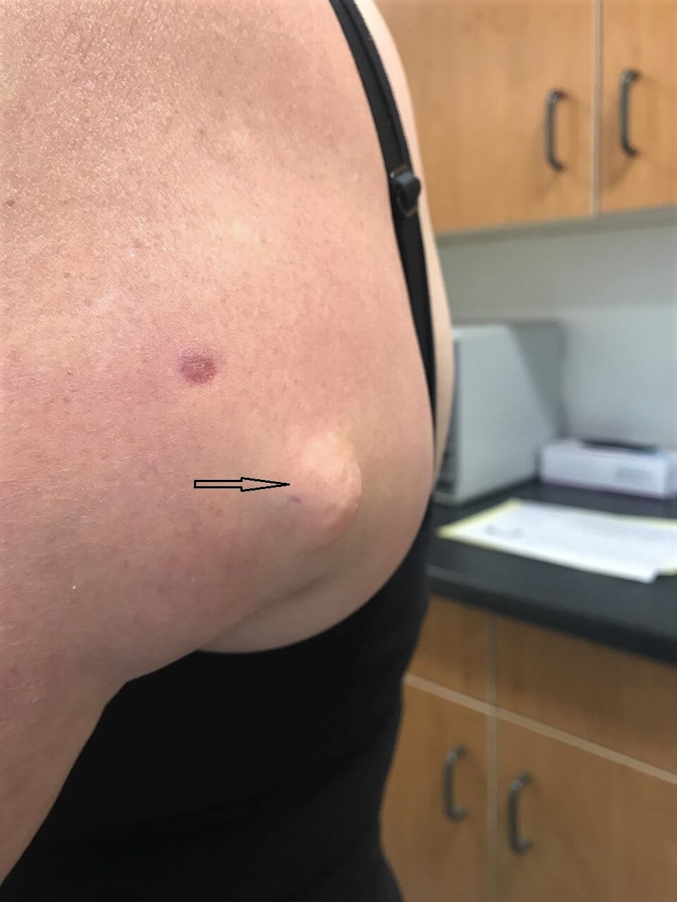
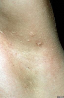
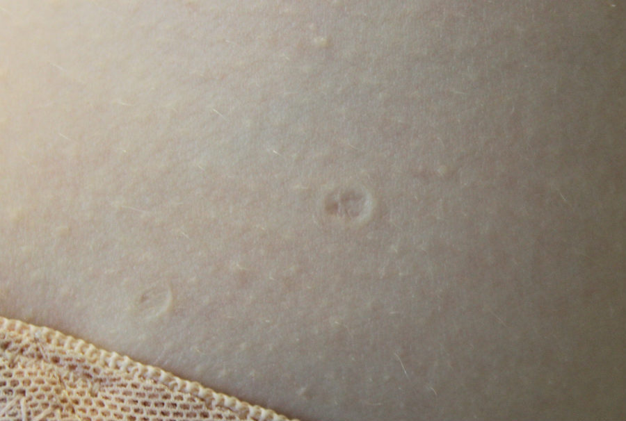
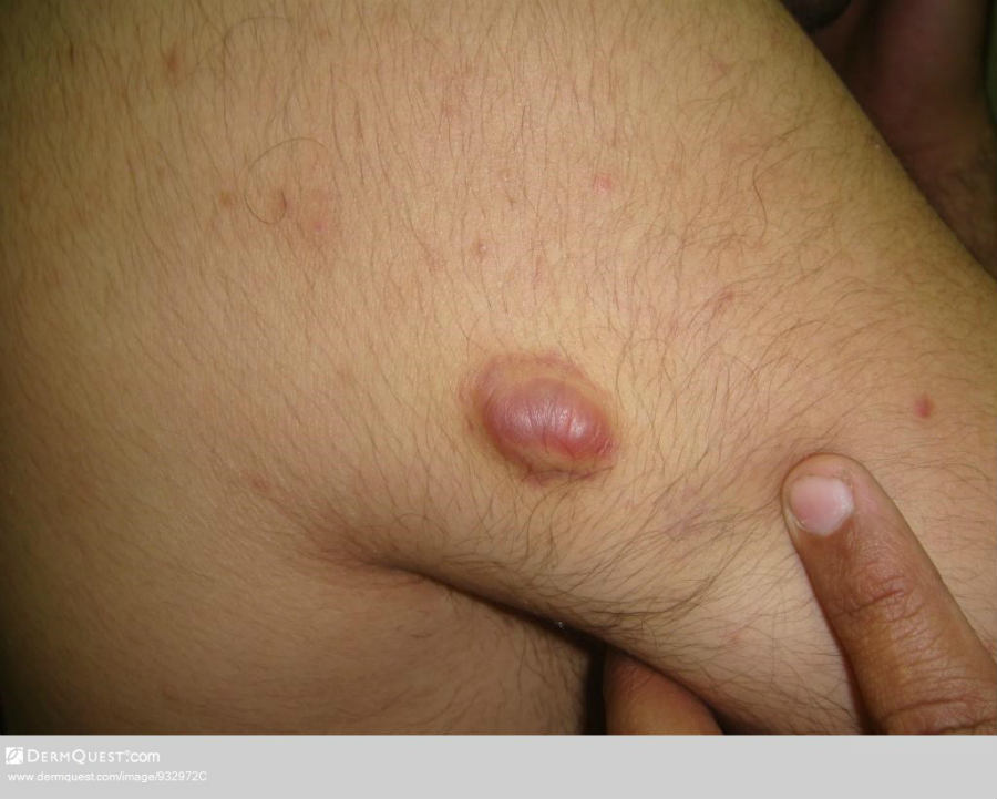
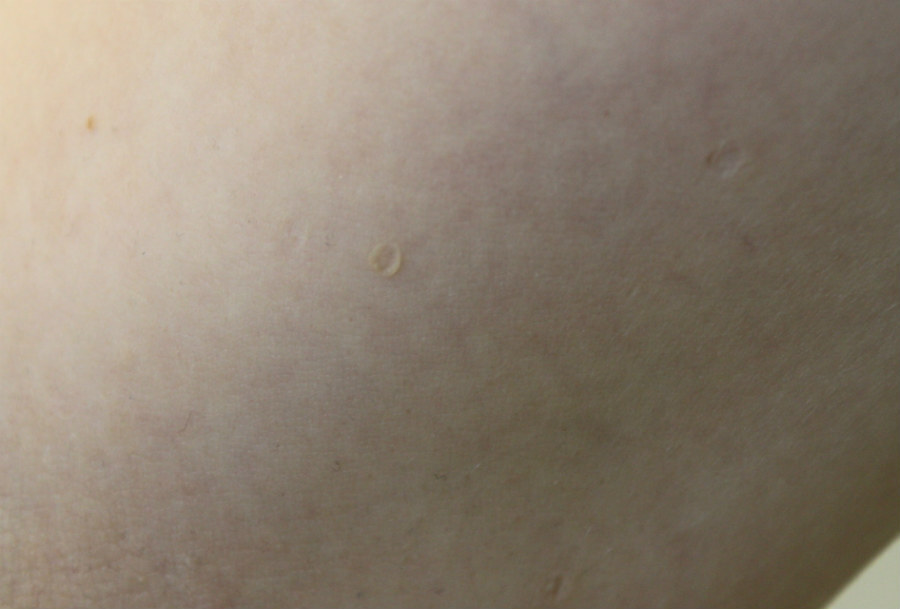
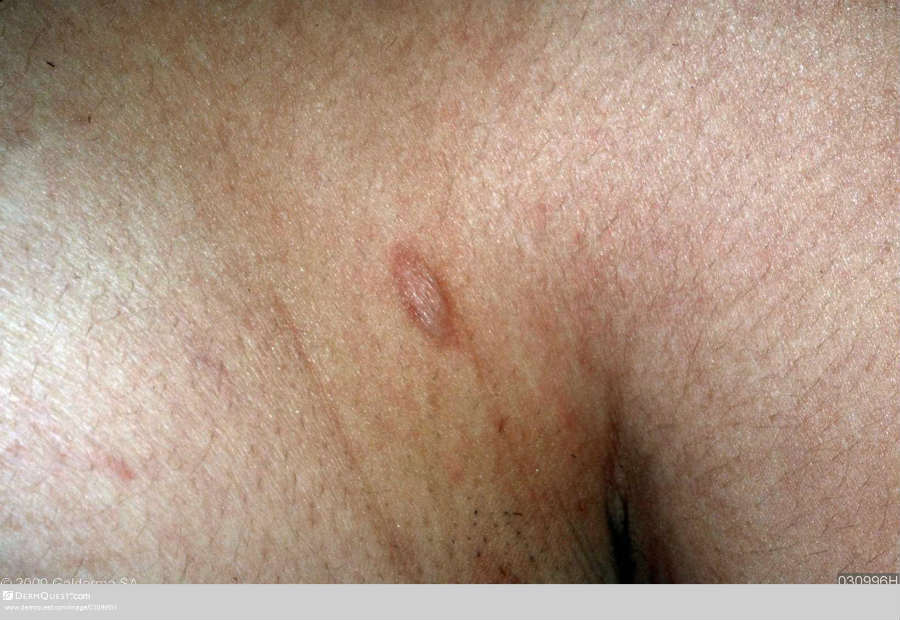
  • The typical lesion of anetoderma is well-defined, 1-2 cm in diameter, with wrinkly skin overlying a palpable depression, the so-called button-hole sign. Sometimes the lesion may have a buldging, herniated appearance
     
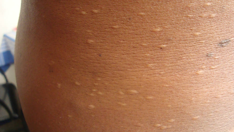
  • In primary anetoderma crops of round-oval pink macules 0.5–1.0 cm in diameter develop predominantly on the trunk, thighs and upper arms. Each macule extends for a week or two to reach a size of 2–3 cm. Slowly, each lesion fades and leaves a macule of wrinkled atrophic skin, which yields on pressure. The number of lesions varies widely, and new lesions often continue to develop for many years
     
  • Differential diagnosis - anetoderma is one of several elastolytic disorders that include cutis laxa, mid-dermal elastolysis, and granulomatous slack skin:
    • Acquired cutis laxa differs from anetoderma by its generally widespread sagging or lax skin 
    • Mid-dermal elastolysis is characterised by selective loss of elastic fibers in the mid dermis, which results in widespread fine wrinkling 
    • Granulomatous slack skin is a rare variant of cutaneous T-cell lymphoma manifesting as erythematous pendulous folds of lax skin

Clinical Images

Please refer to notes on image rights at bottom of the page with regards to individual image ownership.
Investigations

  • If a skin biopsy is taken the pathologist will need to use special stains in order to demonstrate the loss of elastic fibres in the upper to mid-dermis 

Management

  • There is no specific treatment
  • In the case of secondary anetoderma, treatment should be directed against the underlying cause 

Disclaimer - the author PCDS cannot accept responsibility for any misleading or incorrect statements, and the management of individual patients remains the direct responsibility of the individual doctor. We do however hope that visitors to this site can contact us regarding comments that are considered misleading or incorrect so that we can continue to improve the site.

Image Rights - The PCDS would like to thank Dermatoweb, DermQuest (Galderma), and others who have contributed images. All named individuals and organisations maintain copyright for the relevant images.

Quick Links

The following pharmaceutical companies have had no involvement in the content of this website or in our conference programmes


Almirall
Galderma
Glenmark
Johnson & Johnson
La Roche-Posay
LEO Pharma
Pierre Fabre
Schuco