Skin adnexal tumours (syn. appendageal tumours)
LAST UPDATED: Jun 12, 2023
Introduction
Skin adnexal tumours (SATs) comprise a diverse range of neoplasms that arise from skin appendage stem cells, required for the maintenance of structures such as the hair follicle, sebaceous and eccrine glands. Morphologically, they are classified according to the adnexal component of normal skin that they show differentiation towards, namely the hair follicle, sebaceous gland, and sweat duct (which is further divided into eccrine and apocrine subtypes). Histological analysis is required to reach a diagnosis, as their clinical appearance is often nonspecific. SATs are predominantly benign, but malignancy is well recognised, and the presentation of multiple SATs may be a cutaneous manifestation of an underlying genetic syndrome.
This chapter is set out as below, please read the section on history as this sets out a logical approach to this complex area of dermatology:
History
Overview
A retrospective health record audit of SATs received at the Department of Cellular Pathology, Royal Victoria Infirmary, Newcastle upon Tyne Hospitals NHS Foundation Trust, during the period November 2012 to October 2017 found a total of 1615 cases of SATs from 1359 patients:
- The average age at presentation was 55 years (range 11 months to 97 years) and the male to female ratio was 0.77
- 1570 (97.2%) were benign and 45 (2.8%) were malignant
- The majority of SATs (58%, 915 of 1570) were of sweat duct differentiation, with eccrine being more common than apocrine. The next most frequent tumour differentiation pattern in the benign tumours was hair follicle differentiation (37%, 580 of 1570), followed by sebaceous differentiation (4.5%, 71 of 1570). In contrast to adults, hair follicle SATs were the most common SAT type among children
- In terms of specific diagnoses, the most frequently diagnosed SAT in adults was hidrocystoma (492 of 1570). Pilomatrixomas (306 of 1570) were the next most frequently encountered, followed by trichilemmomas, cylindromas and spiradenomas. In children, pilomatrixoma occurred most often
- The majority (60%) of malignant tumours were categorized as ‘malignant tumours with apocrine and eccrine differentiation’. In this WHO category, eccrine porocarcinomas (eight of 45) and microcystic adnexal carcinomas (eight of 45) were reported as being the most frequent. Malignant sebaceous differentiation type tumours (35.6%) were all sebaceous carcinomas (16 of 45). Malignant hair follicle differentiation type tumours accounted for only 4% (two of 45) of malignant cases
- Prebiopsy diagnosis was correct in 28% of cases, which was not unexpected given that the clinical appearance of SATs is generally non-specific
Most SATs are solitary, sporadic, and of no concern being unrelated to an underlying genetic condition. However; several cancer predisposition syndromes related to SATs can now be confirmed with genetic testing, which has implications for the patient and the family.
A comprehensive list of SATs is found below in the sections on clinical findings and images, lesions are listed alphabetically. Additional notes on the most clinically important tumours and syndromes can be found immediately below.
The most clinically relevant skin adnexal tumours
- Skin adnexal carcinomas, eg, sebaceous carcinoma, porocarcinoma, microcystic adnexal carcinoma, Paget's disease of the nipple, and extra-mammary Paget's disease
-
Sebaceous adenoma / epithelioma / carcinoma - even a solitary lesion can be associated with the Muir-Torre syndrome (see below)
-
Multiple / large numbers of benign appendageal tumours
- Such patients are more likely to have an associated syndrome - see below
- Some conditions with multiple tumours, eg syringomas, have no significant associations
-
Naevus sebaceous
-
Usually presents at birth or in young children as a slightly raised yellow to tan plaque on the scalp or face
-
Rarely can transform into BCC - refer to clinical findings for more information
-
Syringocystadenoma papilliferum
-
Often occurs in association with the above, or occasionally presents separately as a pink plaque or linear arrangement of papules
-
Rarely can transform into BCC - refer to clinical findings for more information
Associated syndromes
-
Birt-Hogg-Dube syndrome
- Caused by mutations in the FLCN gene and inherited as an autosomal dominant trait.
-
Characterised by multiple fibrofolliculomas and trichodiscomas over the face, neck and upper trunk. Skin lesions tend to appear in the third and fourth decades. Large numbers of skin tags can be found. Other skin changes can include large lipomas, angiolipomas, and collagenomas
- Associations - an increased risk of renal and colon carcinoma, the risk for renal carcinoma is estimated to be 16-30%. A history of spontaneous pneumothorax and / or lung cysts supports the diagnosis
Cowden syndrome
An autosomal dominant condition with variable expression
Skin changes - patients tend to present as young adults with increasing numbers of hair follicle tumours (in particular trichilemmomas), multiple skin tags, acral keratoses (rough warty lesions on hands and feet), and lipoma
Oral changes - papillomata of the lips, tongue, buccal mucosa, and gingivae are seen in nearly all patients by the second decade, extensive papillomatosis may give a cobblestone appearance
- 80% of patients have macrocephaly
Associations - a very high incidence of carcinomas; namely breast, thyroid, genitourinary, colon, and melanoma
CYLD cutaneous syndrome (syn. Brooke-Spiegler syndrome)
An autosomal dominant disorder
Characterised by the development of trichoepitheliomas (skin-coloured papules over the central face), spiradenomas (painful nodules on head, neck, and trunk), and cylindromas (solitary or multiple tumours on the scalp) in early adulthood. The number of tumours increases over time, and in severe cases, tumours can cover most of the scalp. Rarely, one of these lesions can undergo malignant transformation
Associations - an increased risk of developing benign or malignant tumours of the salivary and parotid glands
-
Gardner syndrome
-
An autosomal dominant condition
- A form of familial adenomatous polyposis (FAP) that is characterised by multiple colorectal polyps and various types of tumours, both benign (noncancerous) and malignant (cancerous)
- Characterised by epidermoid cysts (50–65% of cases) that occur at an earlier age than normal (around puberty) and arise in less common locations such as the face, scalp, and extremities. Other features can include fibrous tissue tumours, osteomas, abnormalities of the skull / facial bones, and dental problems
-
Associations - colonic malignancy develops in over 40% of reported cases. There is also an increased risk of other carcinomas, such as those of the small bowel, stomach, pancreas, thyroid, central nervous system, liver, bile ducts, and/or adrenal glands
-
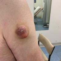
Muir-Torre syndrome (figure 43)
-
An autosomal dominant condition
- A rare variant of the Lynch syndrome (the most common inherited syndrome that predisposes to cancer)
- Diagnosed by a combination of any sebaceous adenoma (in 25-60% of cases), sebaceous epithelioma, sebaceous carcinoma or multiple keratoacanthomas PLUS one of the following visceral carcinomas - predominantly colorectal but also gastric, small bowel, endometrial, ovarian, urinary tract, and hepatobiliary
-
Patients found to have one or more of the above sebaceous tumours should have the histology sample tested for microsatellite instability (which may be done automatically, if not contact the histology department and request the test). Lesions showing high levels of instability can suggest an inherited DNA mismatch repair defect, making the diagnosis of the Muir-Torre syndrome more likely
Management of patients with suspected syndromes
-
All patients need referring to a genetics clinic for further investigations
-
For more information on rare syndromes refer to the following resources:
Clinical findings
Site affected
While appendageal tumours can arise on any part of the body, the list below sets out the most commonly affected sites for each individual tumour. Following on from this list is more detailed information on each tumour.
Scalp
- Trichilemmal cyst
- Naevus sebaceous (solitary, can also affect the face. Tan-coloured plaque)
- Syringocystadenoma papilliferum (often arises within a naevus sebaceous)
- Cylindroma (solitary or multiple, can also affect the face. Smooth, firm and rubbery, pink-red nodules)
Periorbital
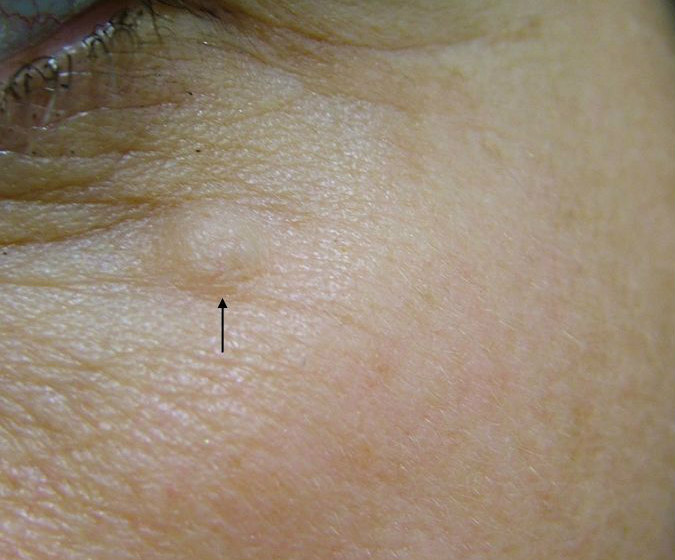
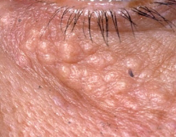
- Apocrine hidrocystoma (predilection for the lower eyelid, particularly the medial canthus. Dome-shaped translucent papule, often pigmented)
- Eccrine hidrocystoma (as opposed to the above they tend not to involve the eyelid margin but rather are distributed around the eyelid skin, and have a lighter colour)
- Syringoma (eyelids and cheeks. Multiple small, skin-coloured to yellow, rounded or flat-topped papules)
- Sebaceous carcinoma (eyelids, especially upper eyelid. Solid yellow-orange lesion, often with a translucent appearance)
Central face
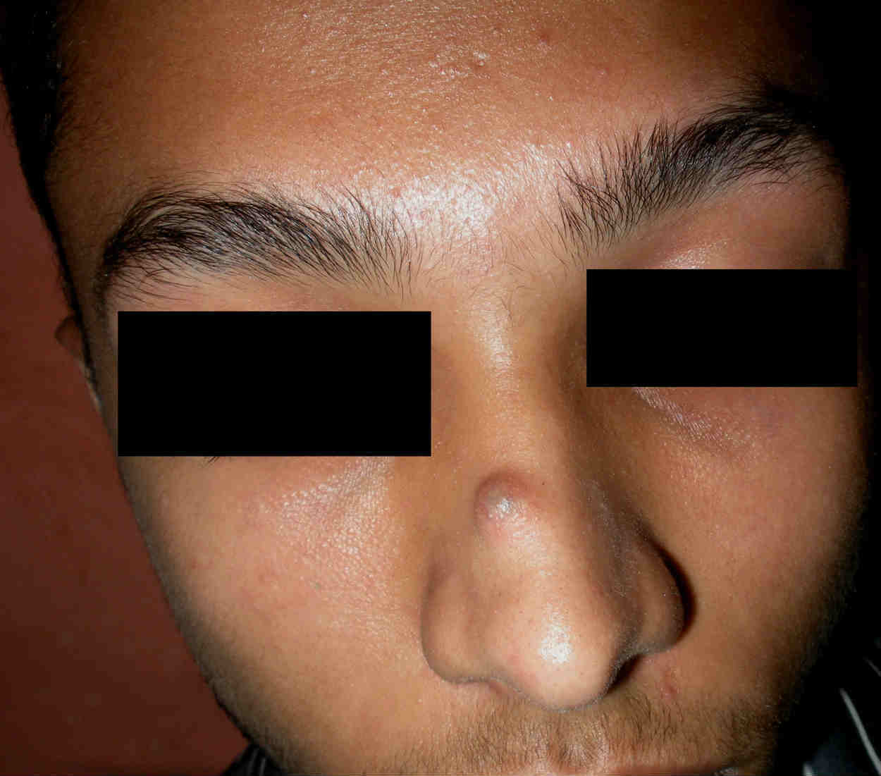
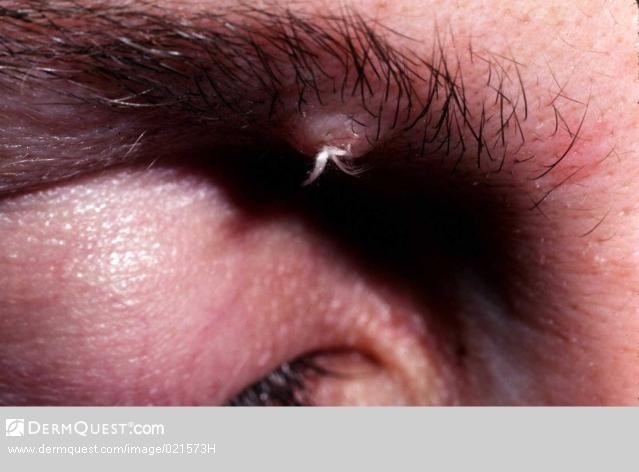
- Trichofolliculoma (a skin-coloured papule or small nodule with several hairs protruding together in a small tuft)
- Trichoepithelioma (a solitary lesion looks like a BCC. Multiple lesions are small pearly papules)
- Trichodiscoma (multiple, discrete flat-topped papules, 2-3 mm in diameter)
- Trichilemmoma (solitary or multiple. Small, well-defined, smooth, asymptomatic papules or verrucoid growths)
- Fibrofolliculoma (multiple small papules, centered around nose, anterior neck and chest. Lesions may be oval in shape but are not all uniform in size)
Lips
- Microcystic adnexal carcinoma (a subtle, elevated or depressed sclerotic papule or nodule)
Other predominantly facial sites
- Sebaceous gland hyperplasia (multiple 1-3 mm sized soft, white-yellow or skin coloured papules)
- Sebaceous adenoma (a yellow-tinged, facial nodule under 1 cm in diameter)
- Pilomatricoma (head and neck most common sites but can affect anywhere. A stony hard, deep-seated lesion, 3-30 mm in diameter)
- Inverted follicular keratosis (solitary papule, which can become inflamed, pruritic and reach a considerable size)
- Tumour of the follicular infundibulum (an irregular, often scaly, papule / nodule)
Mixed tumour of the skin (an asymptomatic, slow growing, intra-dermal or subcutaneous firm, white / yellow nodule)
Trunk
- Paget's disease of the nipple
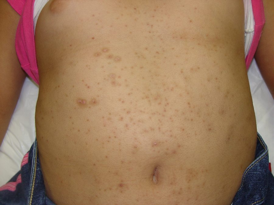
- Eruptive vellus cysts (multiple small, red or brown papules, mainly on the chest)
- Spiradenoma (trunk and proximal limbs most common sites. Solitary, firm, dome-shaped nodule. Can be painful)
Axillae
- Apocrine carcinoma (head and anogenital skin can also be affected. Presents as an expanding nodule, which can ulcerate)
Extremities
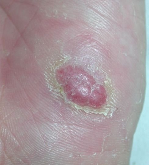
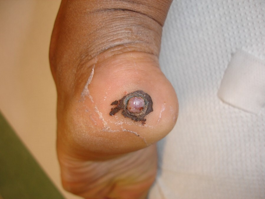
- Eccrine poroma (soles of the feet, palms less commonly. Solitary, soft, nodular, moist lesion with a sessile or broadly pedunculated base)
- Malignant eccrine poroma (lower limbs. The trunk and less commonly the head can also be affected. Lesions are ulcerated, red and dome-shaped nodules, with shiny surfaces)
Anogenital skin
- Extramammary Paget's disease (well-defined, pink-red patches)
- Hidradenoma papilliferum (the vulva more than perianal skin. A rounded, firm, slow growing nodule)
Site: non-specific
- Hidradenoma (an isolated red or bluish nodule)
- Hidroacanthoma simplex (a verrucous plaque, usually with a hyperkeratotic brown surface)
A-Z of appendageal tumours
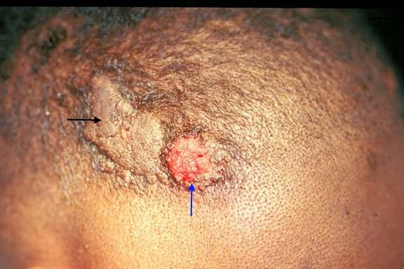
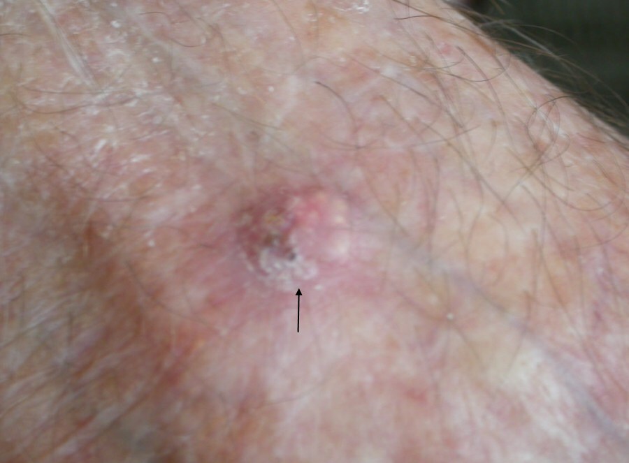
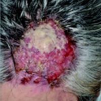
Apocrine carcinoma (figure 1)
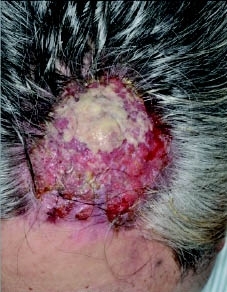
- A primary cutaneous apocrine carcinoma is a rare and highly aggressive cutaneous adenocarcinoma, metastatic spread occurs in upto 30% of cases
- Age - more common in middle-aged women
- Distribution - the axilla is the most common site, other affected sites include the head and anogenital skin
- Morphology - the lesion presents as an expanding nodule, which can ulcerate. As with many other appendageal tumours the diagnosis is not apparent until after histological analysis
- Management - in Secondary Care, primarily surgical
-
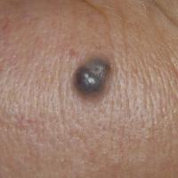
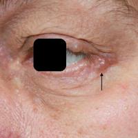
Apocrine hidrocystoma (figures 2-4)
-
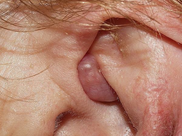
A relatively common, benign tumour of the apocrine secretory glands
-
Age - adults
-
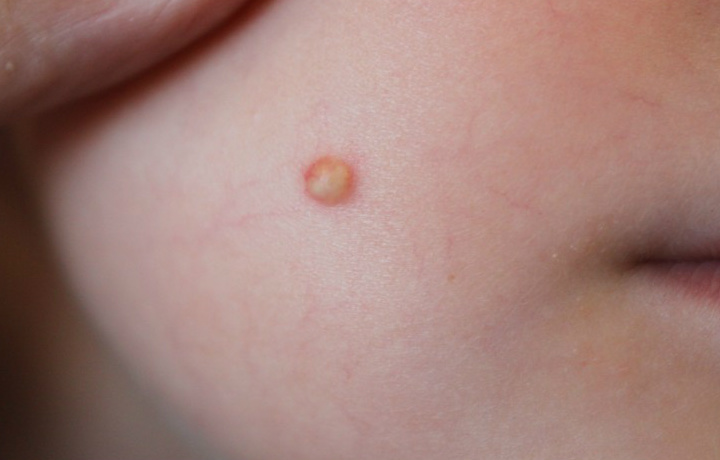
Distribution - the most common site is around the eye, with a predilection for the lower eyelid, particularly the medial canthus. Other body sites can be affected
-
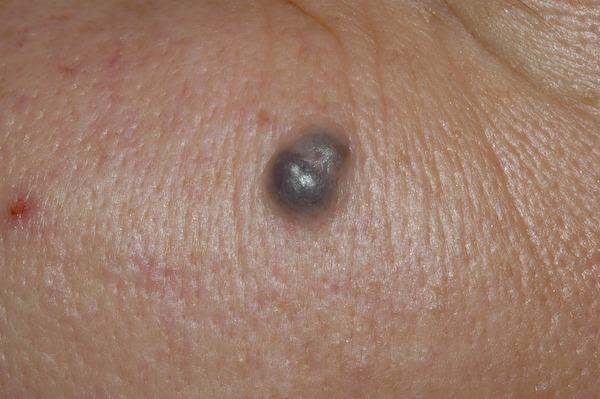
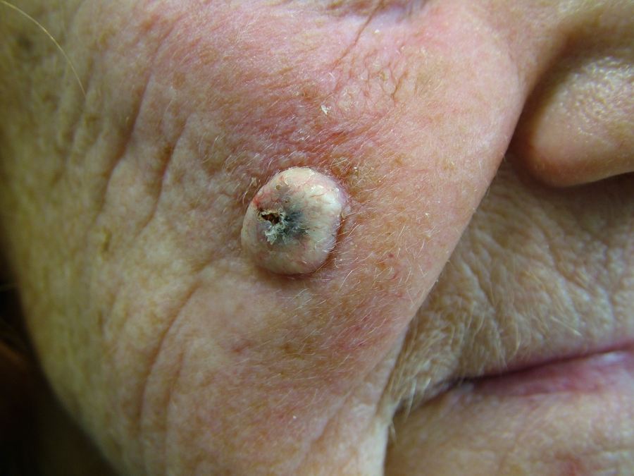
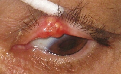
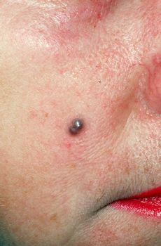
Morphology - usually a solitary, smooth, well-defined, dome-shaped translucent papule / nodule. Skin coloured to blue-black, pigmentation may affect all or part of the cyst. On palpation lesions are fluctuant and cystic
-
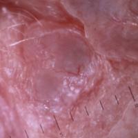
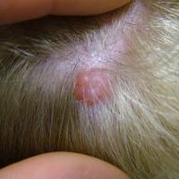
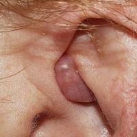
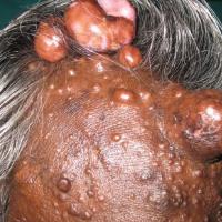
Cylindroma (figures 5-7)
-
A tumour of unknown origin affecting women more than men
-
Solitary cylindromas occur sporadically and typically are not inherited. Multiple tumours are observed in an autosomal dominantly inherited manner
-
Can be seen in conjunction with spiradenomas suggesting a similar derivation of both tumours
-
Age - usually early adult life but can be younger
-
Distribution - the scalp and less so the face
-
Morphology
-
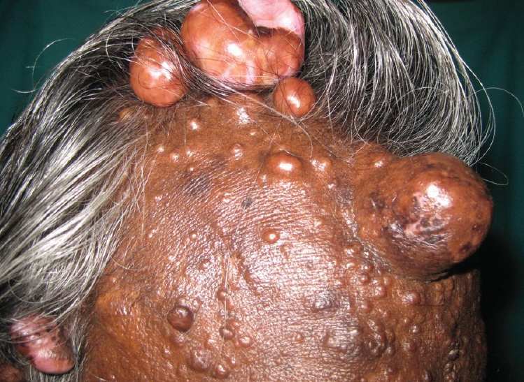
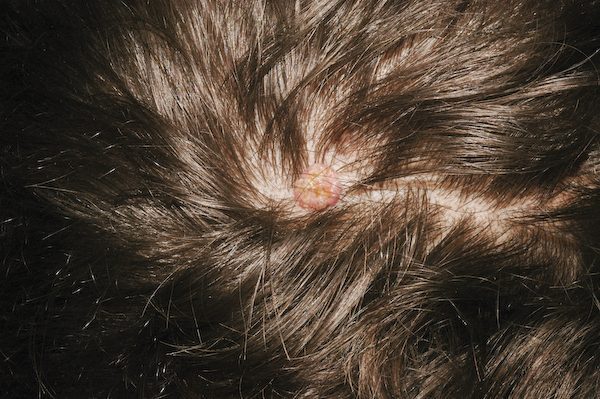
Smooth, firm and rubbery, pink-red nodules. Can be pedunculated
-
When nodules enlarge and coalesce on the scalp they form the distinctive turban tumour feature
- Associations - a combination of cylindromas, spiradenomas, and trichoepitheliomas are found in the CYLD cutaneous syndrome (refer above to the section on history)
-
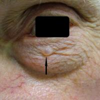
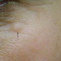
Eccrine hidrocystoma (figures 8-9)
-
A tumour composed of mature, deformed eccrine sweat units
-
Age - mainly middle-aged women
-
Distribution - cheeks and periorbital
-
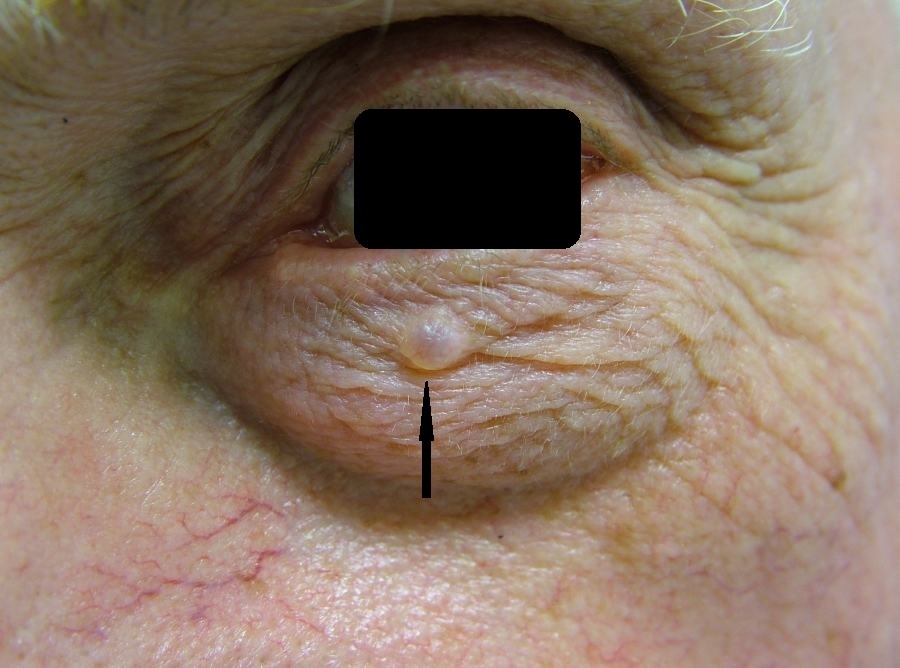
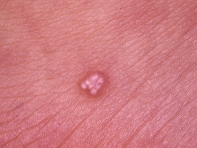
Morphology - small and tense thin-walled cysts ranging from 1 to 6 mm in diameter. They are typically dome-shaped and have an amber, brown, or bluish tint. Eccrine hidrocystomas usually do not involve the eyelid margin but rather are distributed around the eyelid skin; this, along with a lighter colour differentiates it from the apocrine hidrocystoma
-
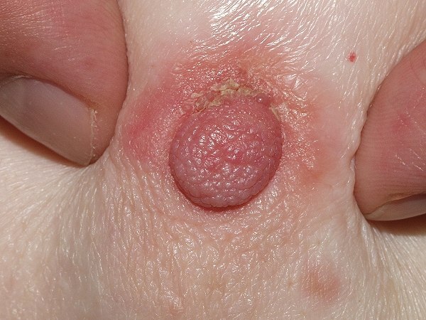
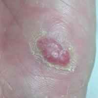
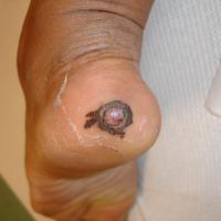
Eccrine poroma (figures 10-11)
-
A tumour arising from the eccrine duct epithelium
-
Age - middle-aged onwards
-
Distribution - the soles and sides of the feet are the most common sites, the palms can also be affected
-
Morphology - usually arise as a soft, non-tender, papular / nodular, moist lesion with a sessile or broadly pedunculated base, ranging in colour from tan to red / pink. Size can be up to 3 cm in diameter, but most are smaller
-
Lesions occasionally undergo malignant transformation into a malignant eccrine poroma (syn. porocarcinoma) - refer below for more information
-
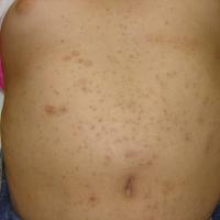
Eruptive vellus cyst (figure 12)
-
An occlusion and cystic dilatation of vellus hair follicles
-
Age - usually present in the second decade of life
-
Distribution - the chest
-
Morphology - usually multiple, small red or brown papules
-
If treatment is required topical retinoids may help. Surgical treatments such as curettage, or laser therapy are other options but scarring, especially on the chest can result
- For more information refer to the relevant chapter
-
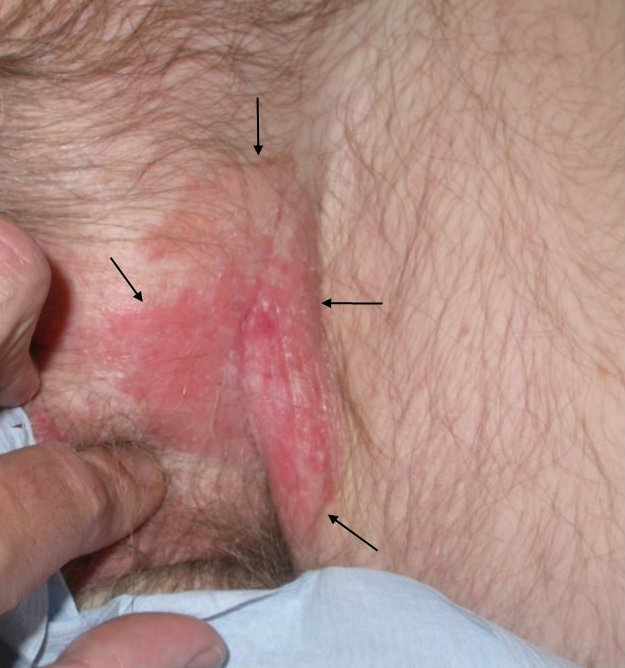
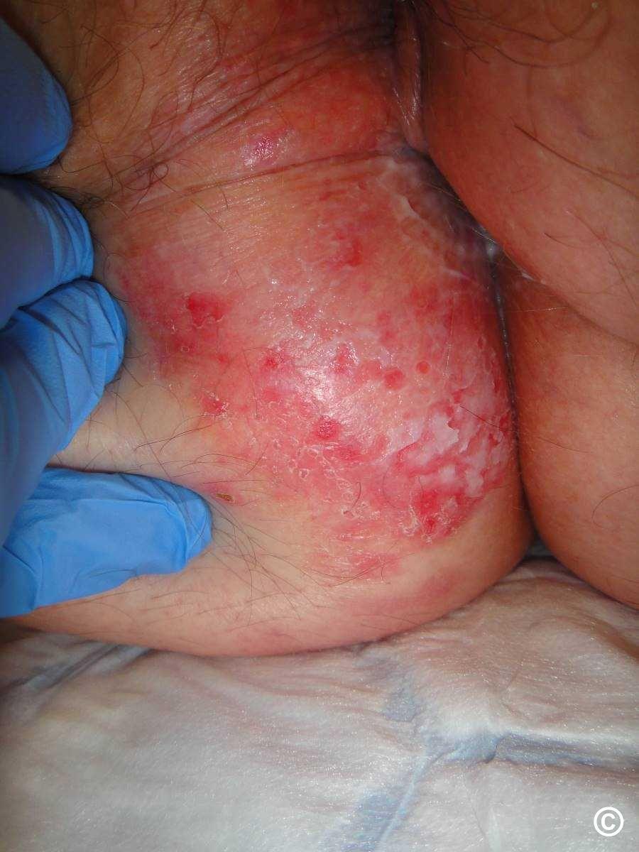
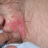
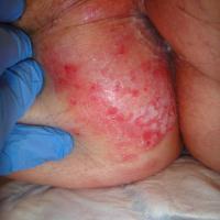
Extramammary Paget disease - EMPD (figures 13-14)
- Extramammary Paget disease (EMPD) is an uncommon intraepithelial adenocarcinoma composed of neoplastic cells showing glandular differentiation
- Clinical features can include:
- Mild-moderate itch
- Affects the ano-gential skin, and rarely the axillae
- Presents as an asymmetrical pink-red well-defined and slightly elevated plaque. The surface can have erosions, crusts, and scale
- For more information refer to the relevant chapter
-
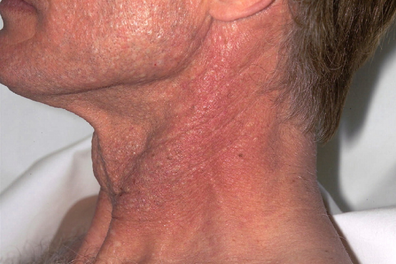
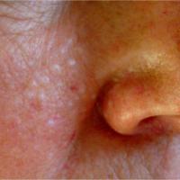
Fibrofolliculoma (figures 15-16)
-
A rare tumour of the perifollicular connective tissue
-
Age - usually appear in the third and fourth decades
-
Distribution - affect the upper trunk, neck, face (often centred around the nose) and sometimes the ears, the number and size of the fibrofolliculomas may increase with age
-
Morphology - slightly paler than the surrounding skin, small papules, which may be oval in shape but are not all uniform in size
- Associations - multiple lesions can suggest the Birt-Hogg-Dube syndrome (refer above to the section on history)
-
Hidradenoma (figure 17)
-
Can be apocrine or eccrine in origin
-
Age - mainly adults
-
Distribution - any body site
-
Morphology - an isolated red or bluish nodule
-
Hidradenoma papilliferum
-
An apocrine tumour of the female anogenital skin
-
Can be either asymptomatic or ulcerate and bleed
-
Age - middle-aged women
-
Distribution - the vulva is much more commonly affected than the perianal skin. Rarely other body sites can be affected
-
Morphology - a rounded, firm, slow growing nodule
-
Hidroacanthoma simplex (figure 18)
-
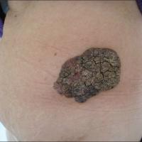
A rare intraepidermal benign tumour, derived from the eccrine duct epithelium
-
Distribution - has a predilection for the extremities, although the trunk can also be affected
-
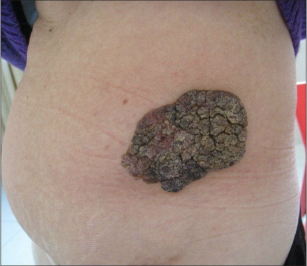
Morphology - a verrucous plaque, usually with a hyperkeratotic brown surface. It can mimic a seborrhoeic keratosis
-
Inverted follicular keratosis (figure 19)
-
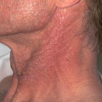
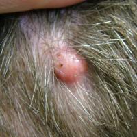
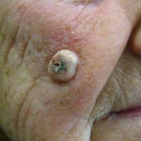
A hair follicle tumour
-
Age - middle-aged and older individuals
-
Distribution - the head and the neck are the most common sites
-
Morphology - most commonly presents as a solitary papule, which can become inflamed, pruritic and reach a considerable size. Can mimic malignant lesions, especially squamous cell carcinoma, both clinically and pathologically
-
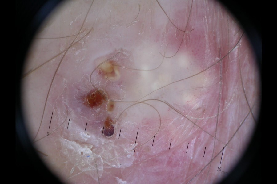
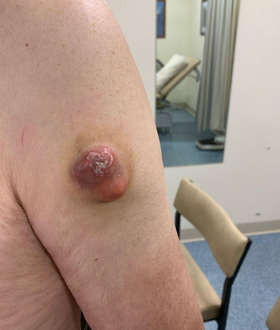
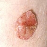
Malignant eccrine poroma - syn. porocarcinoma (figure 20)
-
The most common of the sweat gland carcinomas
-
Age - most cases arise in older patients. Women are affected more than men
-
Distribution - most commonly present on the lower limbs. The trunk and less commonly the head can also be affected
-
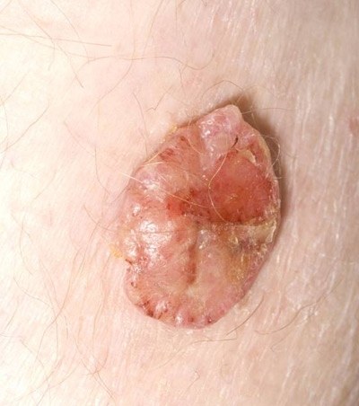
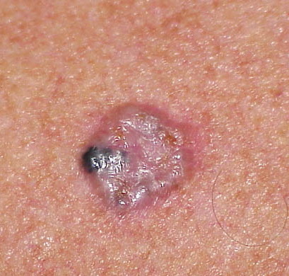
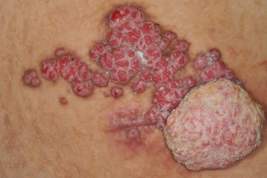
Morphology - the most common presentation is that of an ulcerated, red, dome-shaped nodule with a shiny surface. They can also present as a wart-like plaque. Tumours tend to grow slowly and can become very large
-
18% of lesions arise from a pre-existing benign eccrine poroma (refer above for more information) - malignant change should be considered in lesions that start to grow, become painful, ulcerate or bleed
-
Lesions have metastatic potential so wide excision in Secondary Care and follow up are required
-
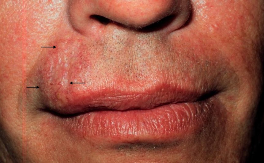
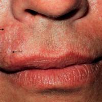
Microcystic adnexal carcinoma - syn. sclerosing/syringomatous sweat duct carcinoma, malignant syringoma (figure 21)
-
A relatively rare, low-grade eccrine gland carcinoma
-
Lesions can be painful if not treated promptly as perineural spread is possible
-
Age - any age although young to middle-aged patients are most frequently affected
-
Distribution - typically occurs on the head and neck, particularly the upper lip and other parts of the central face
-
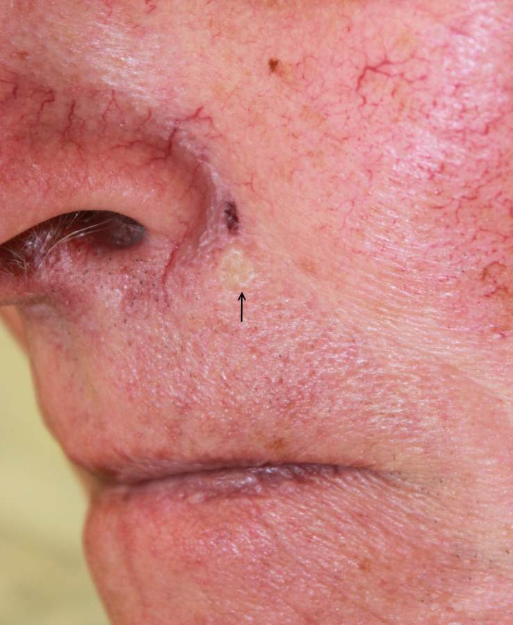
Morphology - often a rather subtle, elevated or depressed sclerotic plaque or nodule
- Management - in Secondary Care, primarily surgical
-
Metastatic spread is rare but local recurrence is common and can be extensive with neurological involvement. Mohs surgery should be considered as a first line treatment
-
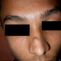
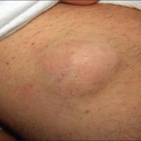
Mixed tumour of the skin - syn. chondroid syringoma (figures 22-23)
-
A rare tumour
-
Age - most commonly arise in middle-aged individuals
-
Distribution - 80% are located on the face, although any site can be affected
-
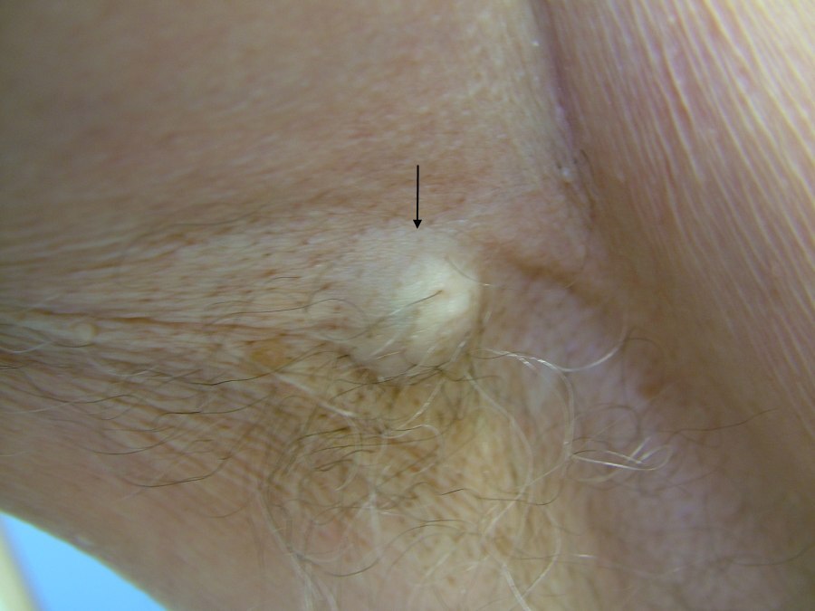
Morphology - the usual presentation is of an asymptomatic, slow growing, intradermal or subcutaneous firm, white / yellow nodule. Lesions can reach a large size
-
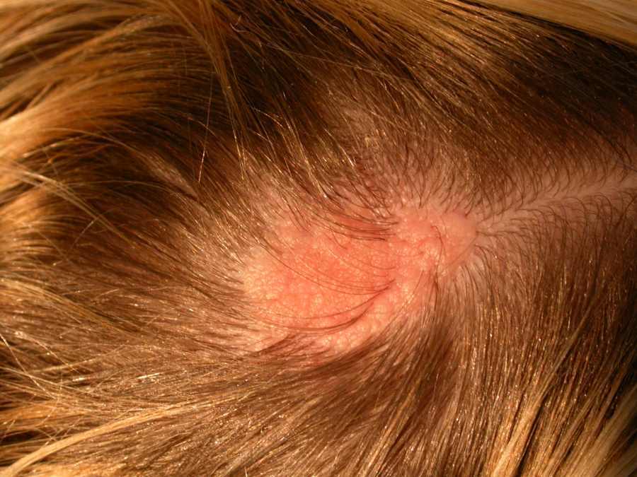
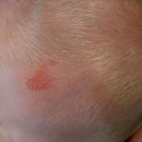
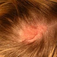
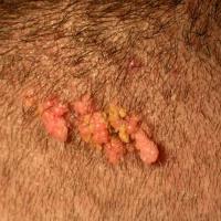
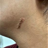
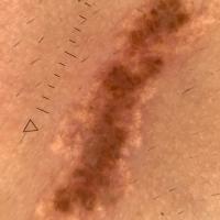
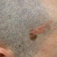
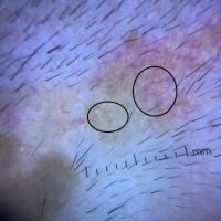
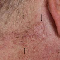
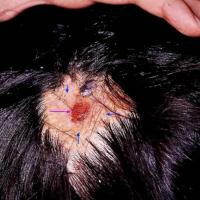
Naevus sebaceous of Jadassohn - syn. sebaceous naevus, organoid naevus (figures 24-33)
-
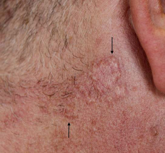
A naevus sebaceous is an organoid naevus comprised predominantly of sebaceous glands (an organoid naevus is characterised by hyperplasia of organoid epithelial structures such as the sebaceous glands, sweat glands and hair follicles)
-
They are seen in 0.3% of all neonates, with an equal incidence in males and females
-
Lesions are congenital and usually seen at birth, although they are occasionally first reported later in life
-
Distribution - the scalp, followed by the face are the most common sites
-
Morphology
-
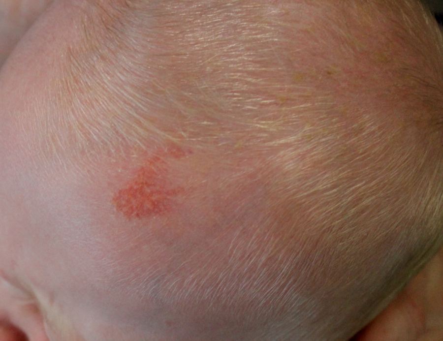
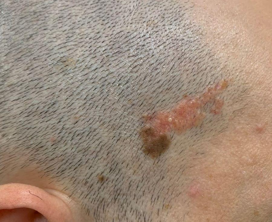
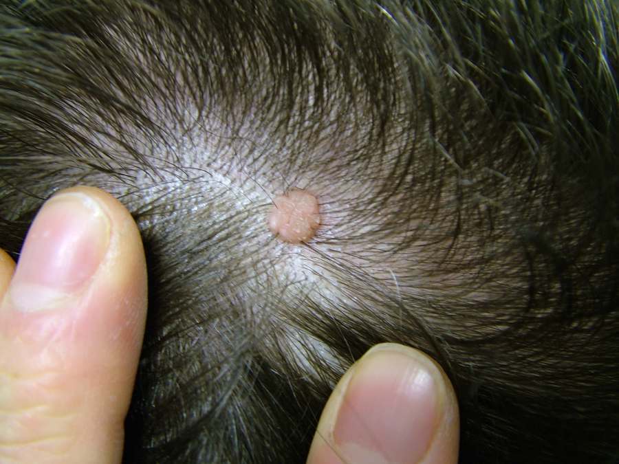
They present as slightly raised yellow to tan plaques
-
Smooth or velvety surface
-
If on the scalp the lesions are devoid of hair
-
They become papillomatous at puberty
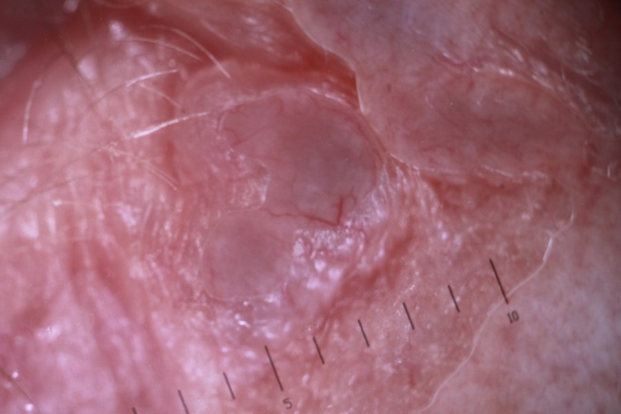
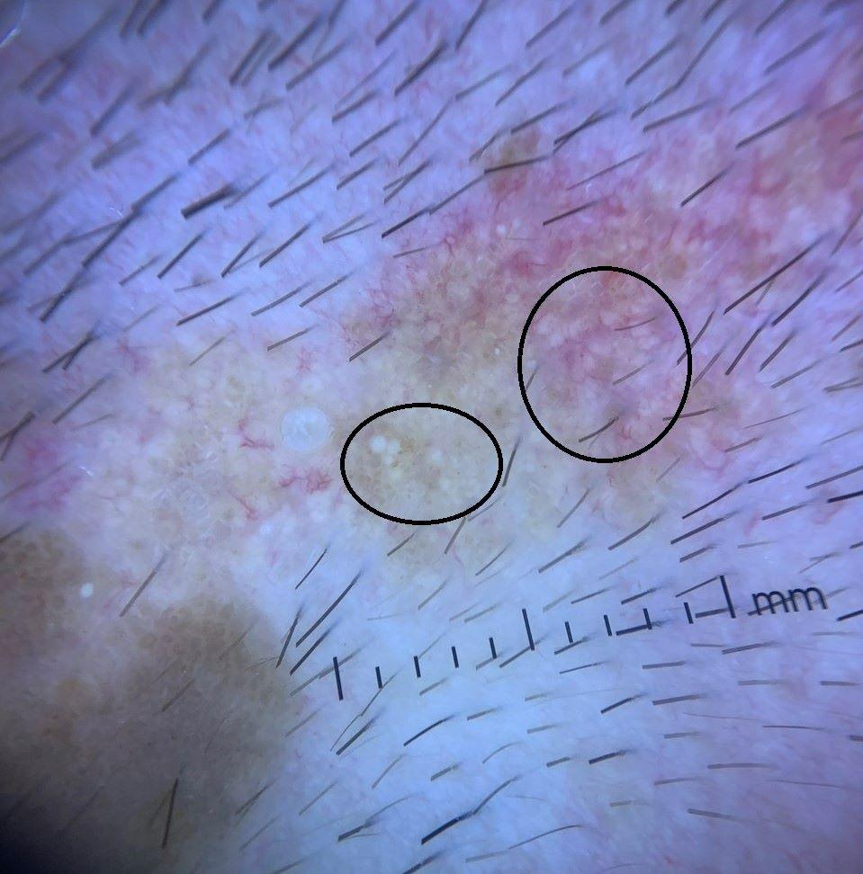
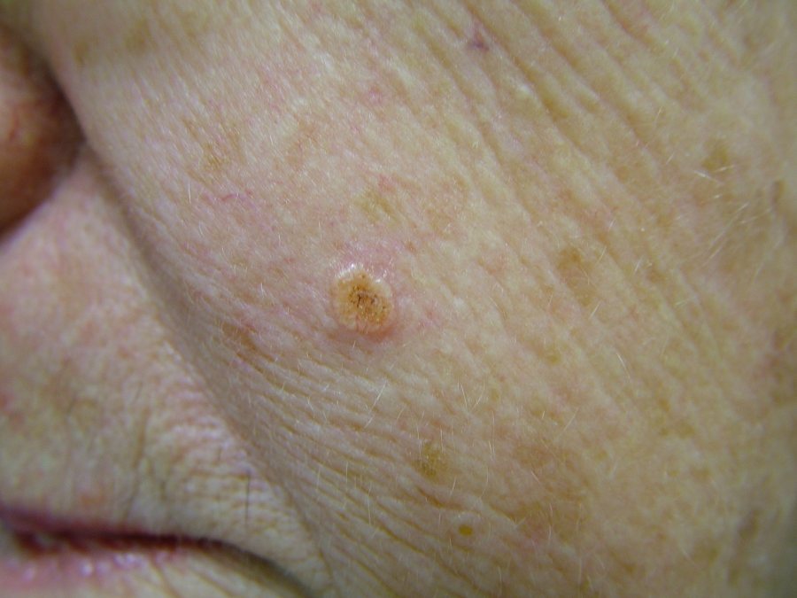
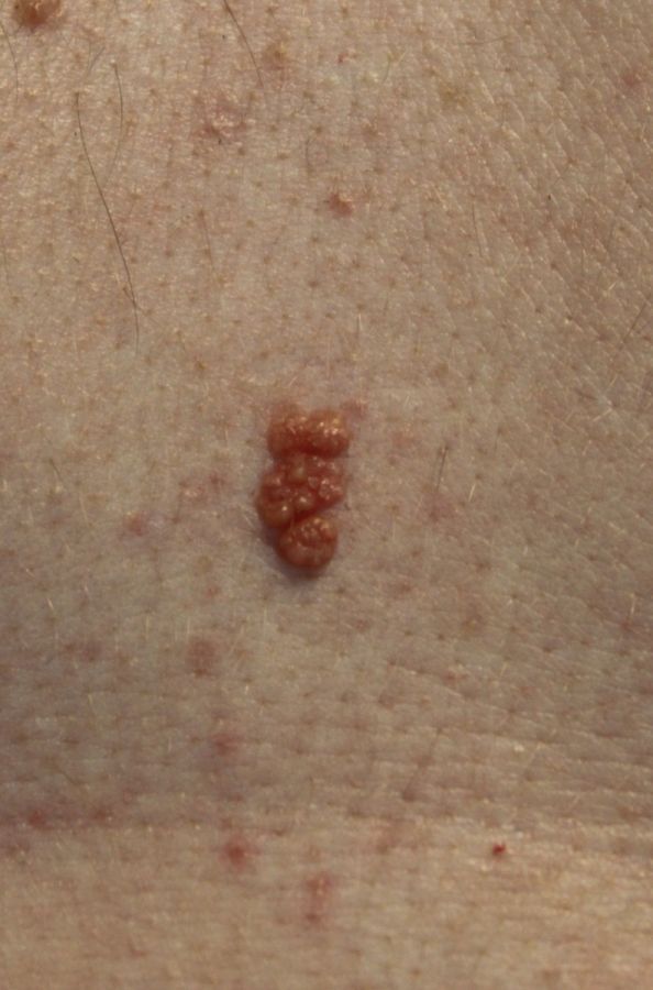
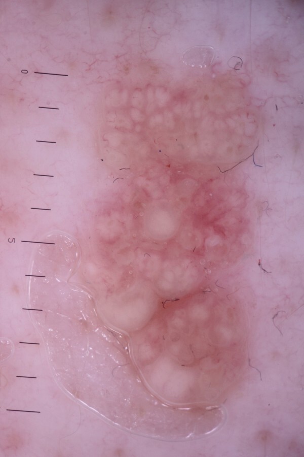
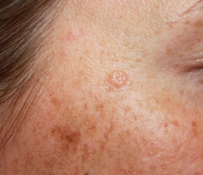
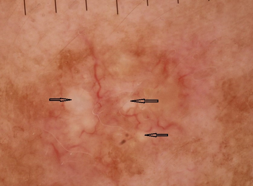
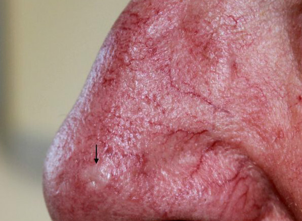
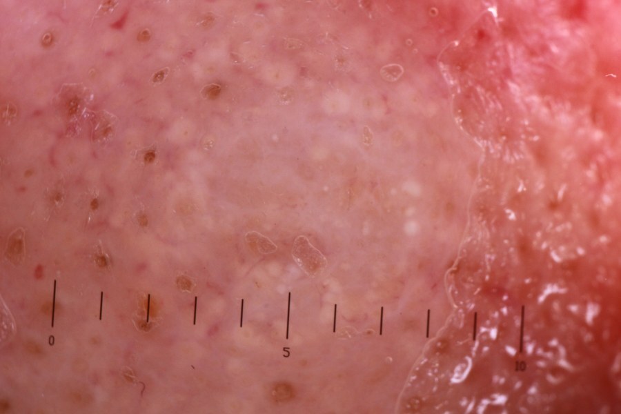
- Dermoscopic appearance - as with lesions of sebaceous gland hyperplasia, there are multiple, grouped, small, white-yellow globules within the lesion
- A naevus sebaceous can occasionally transform into a basal cell carcinoma (BCC) - in the past it was believed that this happened in approximately 4% of cases, however, it is now believed that many of the lesions previously diagnosed as BCC where instead trichoblastoma's, which are benign. As a result there is a move away from excising all naevus sebaceous lesions and instead offering watch and wait if the patient chooses to do so
- Other associations - large lesions are occasionally associated with a wide range of developmental abnormalities and the epidermal naevus syndrome
-
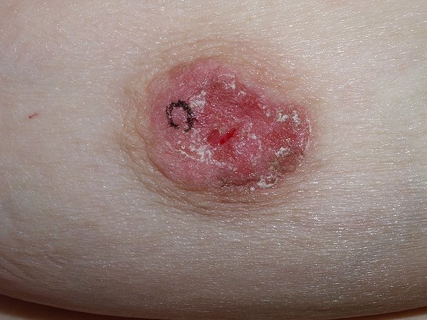
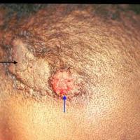
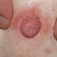
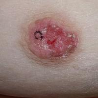
Paget’s disease of the nipple (figures 34-35)
-
Paget's disease and extramammary Paget's disease (EMPD) are uncommon intraepithelial adenocarcinomas composed of neoplastic cells showing glandular differentiation. Adenocarcinoma cells do not necessarily need to be part of a gland but may possess secretory properties (refer above for further information on EMPD)
-
The neoplastic cells in Paget's disease probably originate from the apocrine duct-derived epithelial cells of the breast, either as in situ adenocarcinoma of the breast or a more widespread infiltrating cancer, although this is sometimes difficult to detect on clinical examination or by mammogram
-
Paget's disease affects women and only rarely men
-
Age - it is rare before the fourth decade and most commonly arises in the fifth and sixth decades
-
Clinical features are as follows:
-
The condition is unilateral, rarely bilateral
-
Initial changes can be subtle with small, moist and intermittently crusted areas on the nipple
-
Over time surfaces changes persist and slowly evolve into a sharply marginated, crusty plaque with an irregular outline - areas affected include the nipple, areola and in later stages the skin of the breast
-
Occasionally there may be a blood stained discharge, nipple retraction or a palpable lump
-
Patients with suspected Paget's disease should referred urgently (two-week wait) to a breast clinic
-
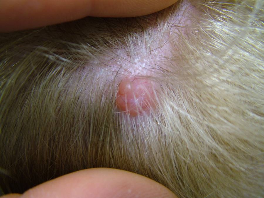
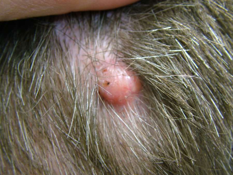
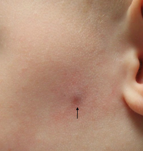
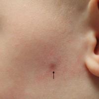
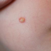
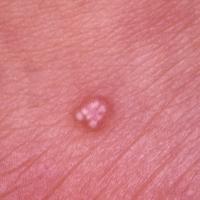
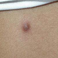
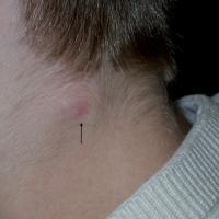
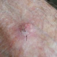
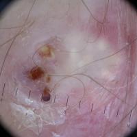
Pilomatricoma - syn. pilomatrixoma (figures 36-43)
-
A hamartoma of the hair matrix, and the most common of the hair follicle tumours
-
Age - the majority of patients are under 20 when they present, although can arise at any age
-
Distribution - mainly the head, neck and upper extremities
-
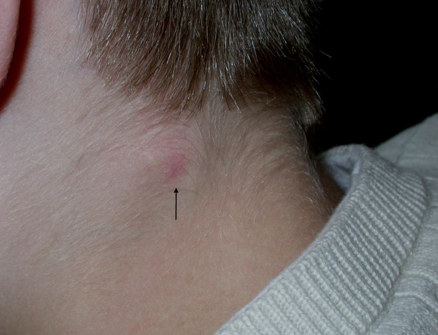
Palpation - a stony hard, deep-seated lesion, 3-30 mm in diameter, with normal overlying skin
-
Occasionally lesions arise in older patients when they often grow more rapidly and may occur at atypical sites
-
Malignant change is rare and most commonly occurs in large lesions
- For more information refer to the relevant chapter
-
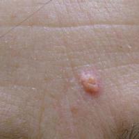
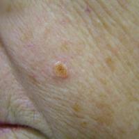
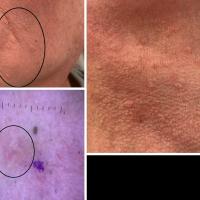
Sebaceous adenoma and sebaceomas (figures 44-46)
-
Benign tumours composed of incompletely differentiated sebaceous cells
-
Distribution - the face and scalp are the most common sites
-
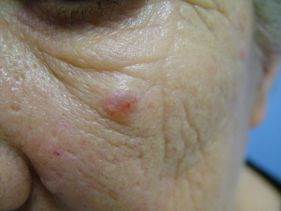
Morphology - A yellow-tinged facial nodule, usually under 1 cm in diameter
-
Associations - patients found to have one or more such lesions should be suspected as having the Muir-Torre syndrome (refer to section above on history)
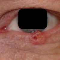
Sebaceous carcinoma (figures 47-48)
A malignant tumour composed of cells showing differentiation towards the sebaceous epithelium
Women are more commonly affected than men
Age - most lesions arise in middle-aged and older patients, the average age of presentation is approximately 70 years
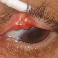
Distribution - sebaceous carcinoma most commonly affects the upper eyelid, probably as a result of a higher number of meibomian glands in this location
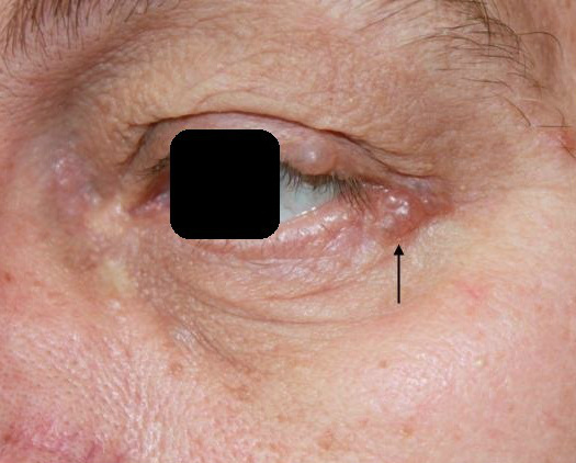
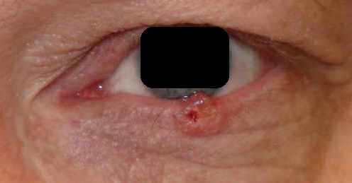
Morphology - sebaceous cell carcinoma is known for its ability to masquerade as other diseases, such as chalazion or blepharitis. The most common findings are that of a solid, yellow-orange lesion, often with a translucent appearance
Management - in Secondary Care, primarily surgical. Complete surgical excision is required as local recurrence rates range from 10% to 35%, metastases can occur
Associations - as with other sebaceous gland tumours (except sebaceous gland hyperplasia ) patients should be suspected as having the Muir-Torre syndrome (refer to section above on history)
-
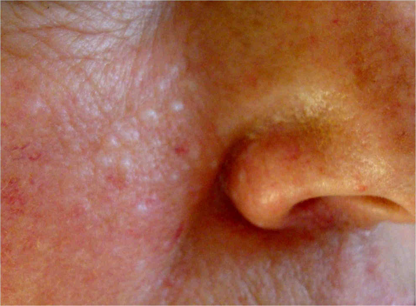
Sebaceous gland hyperplasia
-
Multiple 1-3 mm sized soft, white-yellow or skin coloured papules on the face
-
This conditions has no associations and is discussed elsewhere on the website - refer to the relevant chapter
-
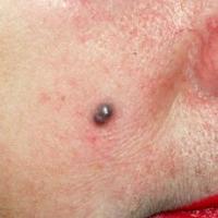
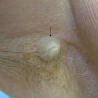
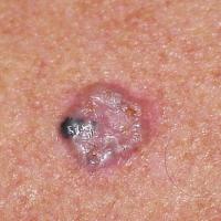
Spiradenoma - syn. eccrine spiradenoma (figures 49-51)
-
The histogenesis of spiradenomas remains in question, but many lesions demonstrate apocrine differentiation
-
Age - most commonly arises in people aged 15 to 35 years
-
Distribution - the front of the trunk and proximal limbs are the most common sites
-
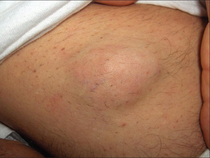
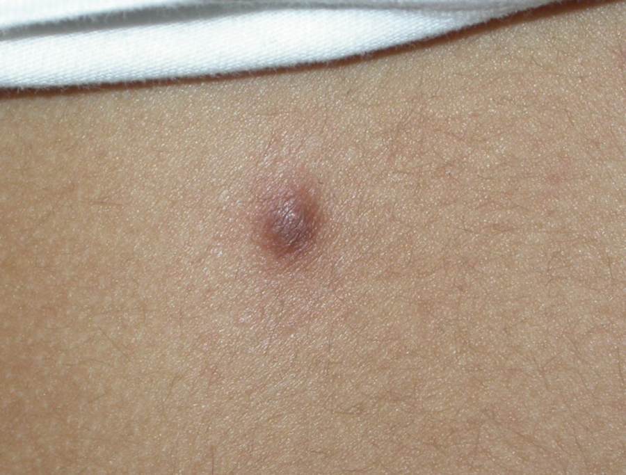
Morphology - usually presents as a solitary, 1 cm, firm, dome-shaped nodule. Some are painful and tender to touch. Their colour can vary but many have a bluish element
-
Associations:
-
Spiradenomas can arise in cylindromas, it has been suggested that the two may be part of the same spectrum
- Associations - a combination of cylindromas, spiradenomas, and trichoepitheliomas are found in the CYLD cutaneous syndrome (refer above to the section on history)
-
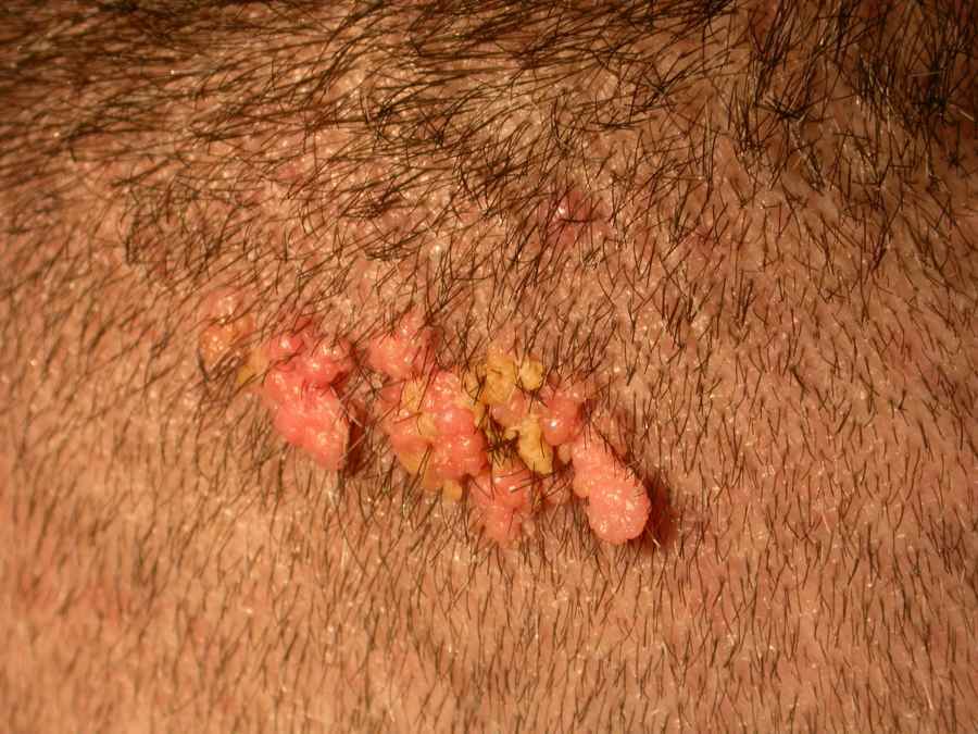
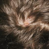
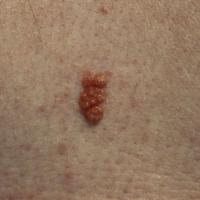
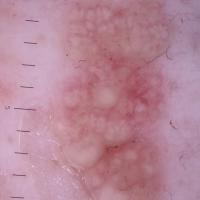
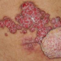
Syringocystadenoma papilliferum (figures 52-55)
-
Although classified as an organoid naevus the exact nature of this lesion remains controversial
-
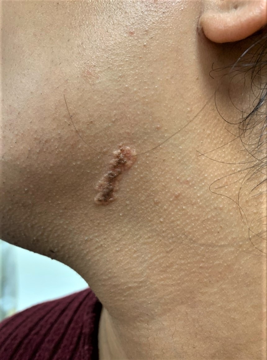
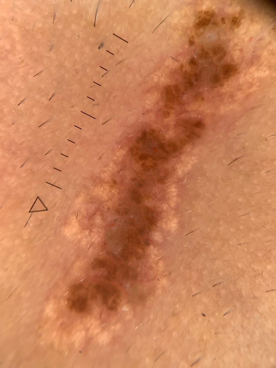
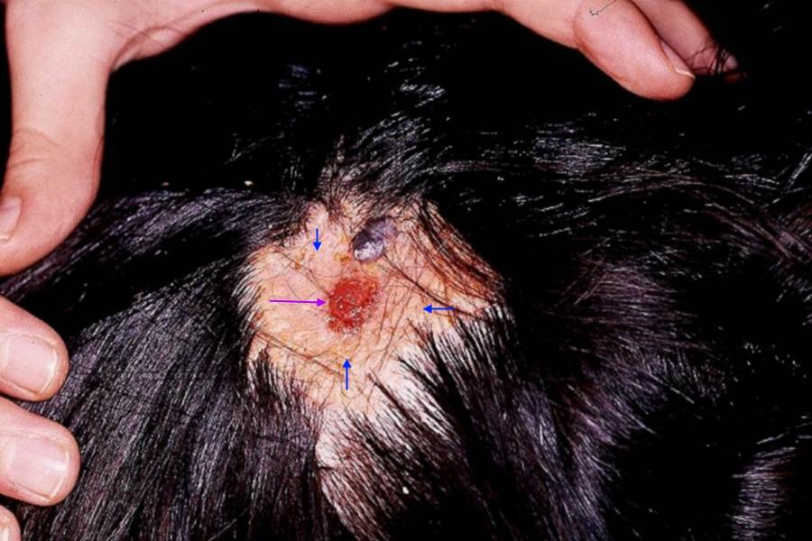
Many cases arise in sebaceous naevi at or after puberty. They can occasionally present as an isolated finding, mainly on the scalp / face at birth or early infancy. On the scalp they appear as a pink plaque or linear arrangement of papules. On other parts of the body they present as a linear lesion following the lines of Blaschko
-
A rare solitary form presents as a nodule or cutaneous horn
-
As with the naevus sebaceous lesions can occasionally transform into BCC
-
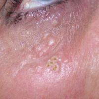
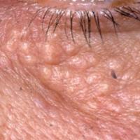
Syringoma - syn. syringocystadenoma, syringocystoma (figures 56-57)
-
A benign eccrine gland tumour more commonly found in women
-
Age - most commonly arises in adolescence
-
Distribution - the face, especially the cheeks and eyelids
-
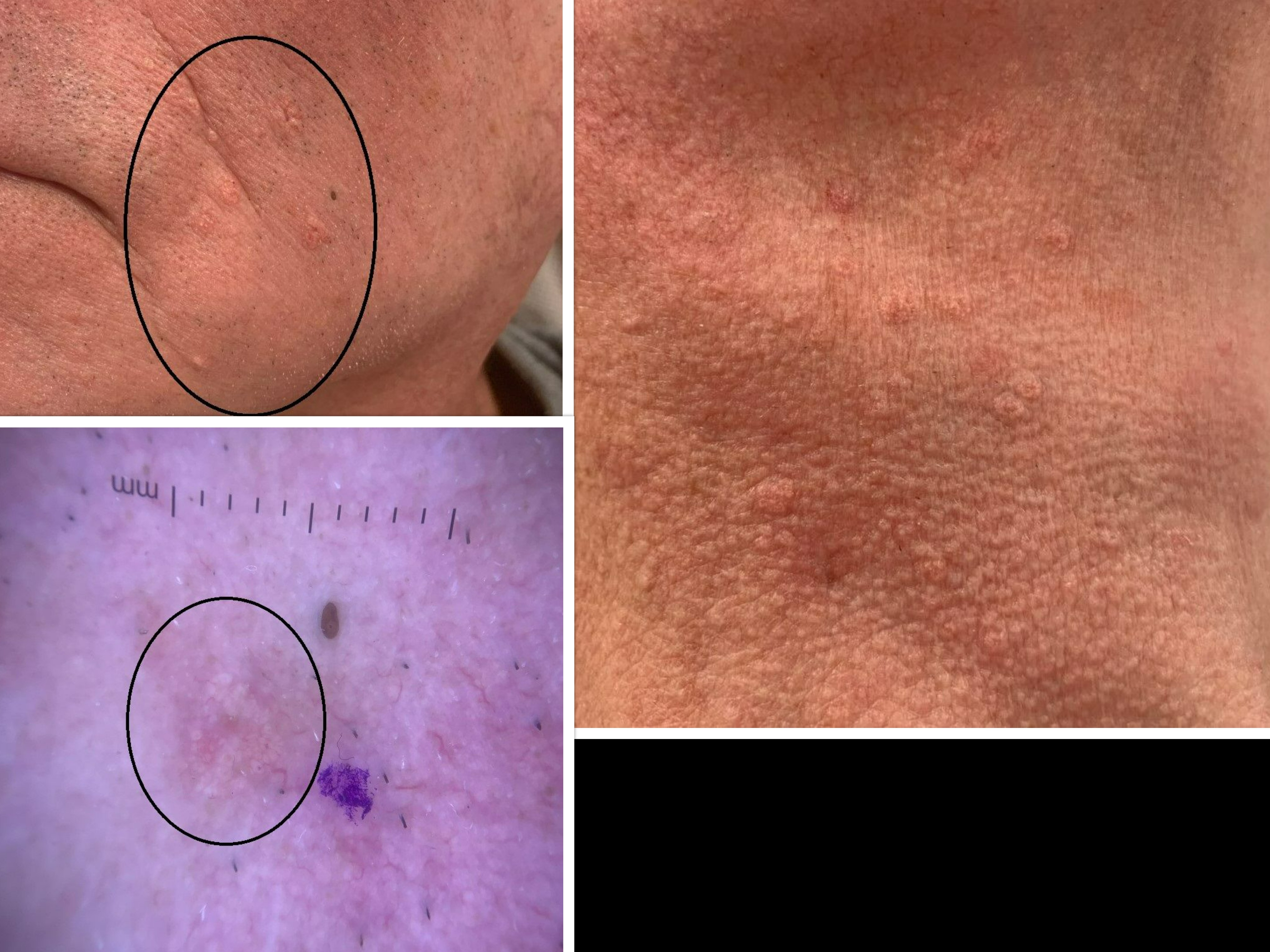
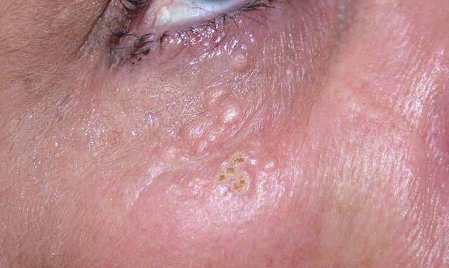
Morphology - multiple small, 1 to 5 mm, skin-coloured to yellow, rounded or flat-topped papules
-
Trichilemmal cyst
-
An external root-sheath tumour
-
Relatively common
-
Females more than males
-
Distribution - mainly seen on the scalp, rarely the extremities
-
Morphology - firm nodules
-
Trichilemmoma
-
A proliferation of the external root sheath of the hair follicle
-
Age - affect young and middle-aged adults
-
Distribution - the central face
-
Morphology - solitary or multiple. Small, well-defined, smooth, asymptomatic papules or verrucoid growths
-
Associations - if multiple can be part of the Cowden syndrome (refer above to the section on history)
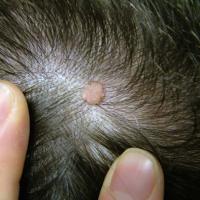
Trichoadenoma (figure 58)
- Lesions of the upper dermis. These cyst‐like structures have an appearance similar to the infundibular portion of the hair follicle but on high power microscopy no recognisable hair shafts are seen
- More common in adults. Congenital lesion or occurrence in childhood is rarely seen
- Non‐specific nodule, usually on the face, although there are some reports of lesions on the buttocks
- May occur within a naevus sebaceous or coexist with a dermal naevus
-
Trichoblastoma
-
A tumour of the hair gem cell composed of follicular germinative cells
-
Trichoblastomas and trichoepitheliomas appear to be part of the same spectrum. The main difference between trichoblastomas and trichoepitheliomas is the depth at which they arise in the dermis - trichoblastomas are found in the deep dermis and subcutaneous tissue, whereas trichoepitheliomas are more superficial
- Age - adults, most commonly arising in the fifth and sixth decades
- Distribution - head and neck
- Morphology - a dermal or subcutaneous skin-coloured papule / nodule, usually less than 1 cm in diameter
- Can occur within a naevus sebaceous
-
Trichodiscoma
-
A hamartoma of the hair follicle mesenchyme
-
Distribution - mainly the central area of the face
-
Morphology - multiple, discrete, flat-topped papules 2-3 mm in diameter
- Associations - multiple lesions can suggest the Birt-Hogg-Dube syndrome (refer above to the section on history)
-
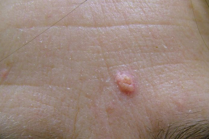
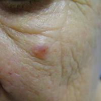
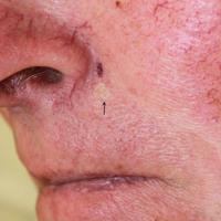
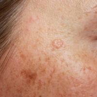
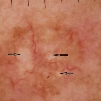
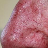
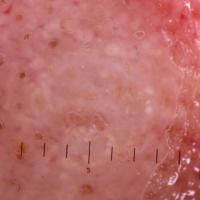
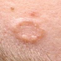
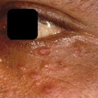
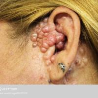
Trichoepithelioma (figures 59-67)
-
A hamartoma of the hair germ cell with focal, primitive follicular differentiation
-
Age - mainly young adults
-
Clinically, mainly presents on the face as:
-
A solitary nodule resembling a BCC - lesions need excising for diagnostic purposes, however, histologically they also can be difficult to distinguish from BCC
-
Multiple, small, pearly papules on the central face, inherited as autosomal dominant
-
A desmoplastic trichoepithelioma (syn. sclerosing epithelial hamartoma) can look even more like a BCC with a depressed centre and raised, rolled edges in many cases
- Associations - a combination of cylindromas, spiradenomas, and trichoepitheliomas are found in the CYLD cutaneous syndrome (refer above to the section on history)
-
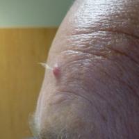
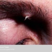
Trichofolliculoma (figures 68-69)
-
A hamartoma of the pilosebaceous follicle
-
Age - mainly young adults
-
Distribution - the face, with a predilection for the central face around the nose
-
Morphology - a skin-coloured papule or small nodule with several hairs protruding together in a small tuft
-
Tumour of the follicular infundibulum
-
A hair follicle tumour
-
Distribution - usually found on facial skin
-
Morphology - solitary follicular infundibulum tumours have no distinctive clinical features and usually present as an irregular, often scaly, papule / nodule up to 1.5 cm in diameter. Occasionally can be multiple
- Associations - occasionally with the Cowden syndrome (refer above to section on history)
Clinical Images
Please refer to notes on image rights at bottom of the page with regards to individual image
ownership.
1
2
3
4
5
6
7
8
9
10
11
12
13
14
15
16
17
18
19
20
21
22
23
24
25
26
27
28
29
30
31
32
33
34
35
36
37
38
39
40
41
42
43
44
45
46
47
48
49
50
51
52
53
54
55
56
57
58
59
60
61
62
63
64
65
66
67
68
69
Management
-
Patients with a suspected skin adnexal carcinoma (clinical or histological) need to be referred urgently (two-week wait) to dermatology or plastic surgery (refer to local guidelines), except for Paget's disease when patients need referring to a breast clinic
-
For lesions associated with the syndromes listed in the section on history, patients should be referred to an appropriate genetics clinic
-
Otherwise refer to the section on clinical findings for more information
Disclaimer - the author PCDS cannot accept responsibility for any misleading or incorrect statements, and the management of individual patients remains the direct responsibility of the individual doctor. We do however hope that visitors to this site can contact us regarding comments that are considered misleading or incorrect so that we can continue to improve the site.
Image Rights - The PCDS would like to thank Dermatoweb, DermQuest (Galderma), and others who have contributed images. All named individuals and organisations maintain copyright for the relevant images.