Atypical (dysplastic) melanocytic naevus
LAST UPDATED: Aug 12, 2025
Introduction
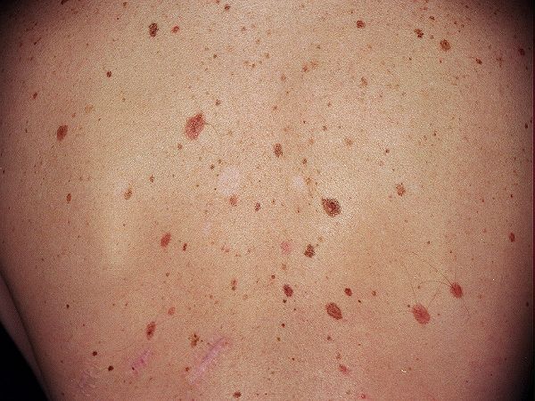
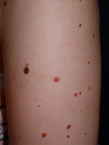
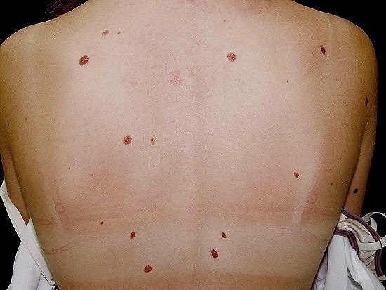
Atypical melanocytic naevi (AMN) are skin lesions whose clinical and histologic features sit somewhere on the proliferative continuum from a typical benign melanocytic naevus to a melanoma. The Atypical Mole syndrome (AMS) is defined by an individual with more than 50 naevi of which three or more are atypical. Although AMN infrequently progress to melanoma, the larger the number of atypical (and typical) melanocytic naevi, the greater the risk. The term dysplastic naevus is given to the histological appearance of an AMN.
This chapter is set out as follows:
Aetiology
-
AMN may be inherited as an autosomal dominant trait or occur sporadically - clinical and histological appearances of AMN occurring in a familial setting appear to overlap with those occurring sporadically
-
It is likely that both genetics and UV radiation have a role to play in their development
-
The prevalence of AMN in white populations has been reported to be between 2-5%. Celtic patients are much more prone to developing AMN, whereas AMN are rare in black, Asian or Middle Eastern populations
-
It is difficult to assess the risk of individual atypical naevi, however, the risk of solitary lesions transforming to melanoma is very low. As the number of atypical naevi increases so does the overall patient risk, the presence of five atypical moles represents a relative risk of 6.36 (range 3.8-10.33) for the development of melanoma
History
- AMN can develop throughout a person's lifetime, although the majority arise in childhood
-
Always ask about family history
-
Patients with sporadic atypical naevi typically have a relatively small number of lesions
-
Patients with a genetic cause may have large numbers of naevi, typical and atypical, and a family history of melanoma (refer to section on management)
Clinical findings
Distribution
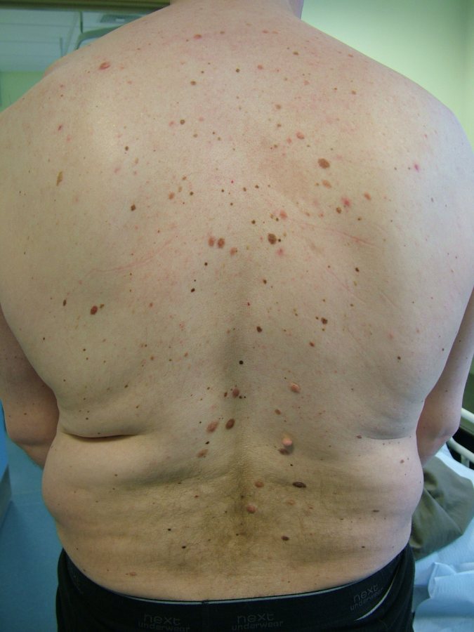
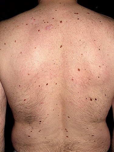
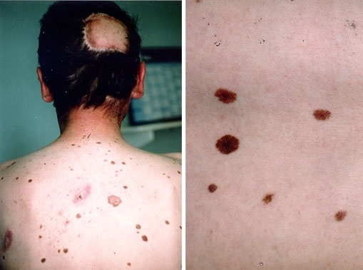
- Can arise on any body site although they are most commonly found on the trunk, upper limbs, scalp and buttocks
Morphology
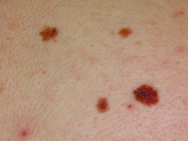
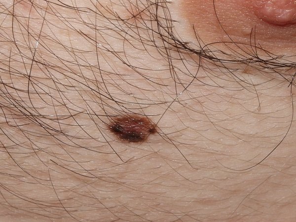
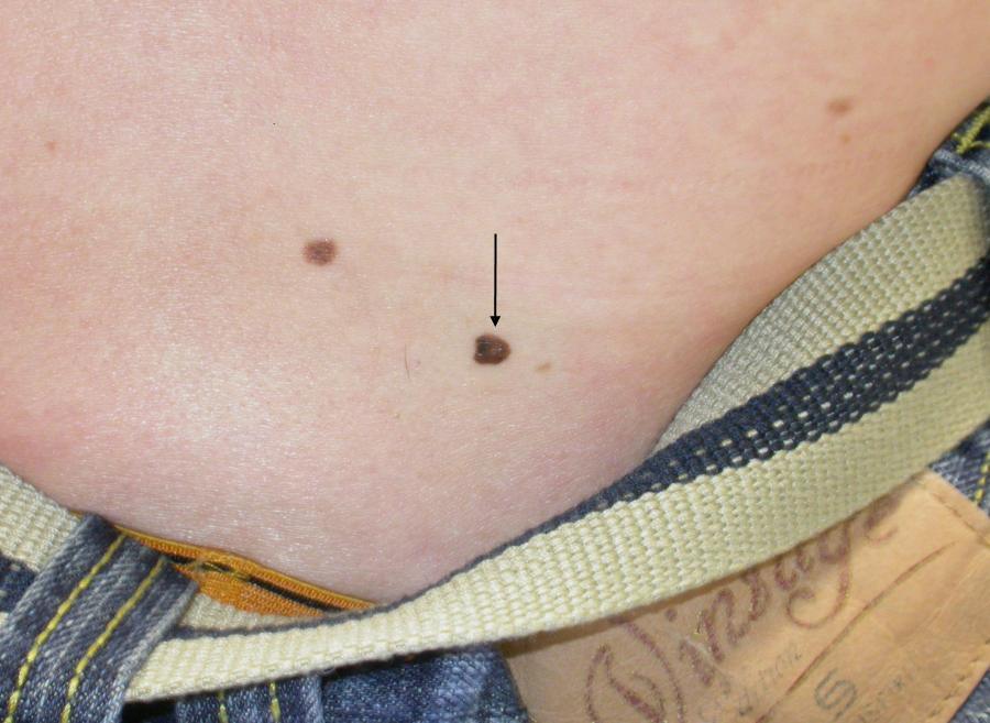
- Size - tend to be large, commonly more than 7 mm in diameter
- Shape and border - borders often irregular resulting in unusual shapes
- Colour - can vary from light-dark brown, pink and occasionally black. May be single colour or variegated. Can have an erythematous edge
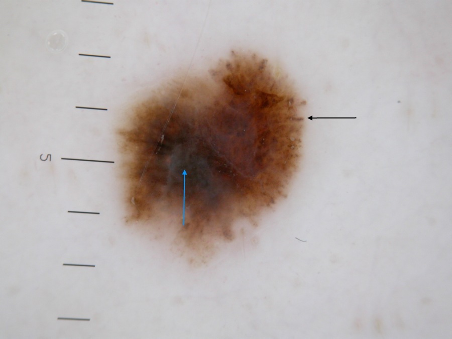
Dermoscopic features
- In patients with several lesions, they will often show a similar pattern
- Beware of a lesion looking different to the others
- As discussed in the chapter Melanoma - an overview the dermoscopic examination of melanocytic lesions requires considerable experience, accordingly any lesion found to have clinical OR dermoscopic features suspicious of melanoma should be referred urgently to Secondary Care
Clinical Images
Please refer to notes on image rights at bottom of the page with regards to individual image
ownership.
Management
General advice - for all
- In general, the larger the number of naevi, typical or atypical, the greater the risk of melanoma - in patients with >100 common naevi on their body, the relative risk of developing cutaneous melanoma is approximately sevenfold higher compared to those with only 15 or less naevi. The arm appears to correlate most closely to total body naevus counts - 11 or more naevi on an arm predicts an increased risk of having >100 total body naevi
- Patients need to be taught on appropriate UV protection and the self-examination of moles and informed to look for new lesions and change in existing lesions - approximately 20% of all melanomas arise from pre-existing naevi, with the remaining majority arising de novo
- Patients should take photographs of their moles and store the images on a home computer - the patient and partner / relative should perform regular skin examinations to look for change
Patients not needing referral
- Patients with only a few AMN that have not changed, and without a family history of melanoma can be managed as above
Routine referral
- Larger numbers of naevi with a mixture of typical and atypical melanocytic naevi. Patients require a thorough dermoscopic assessment and photography, from which the patient should receive a copy of their images to assist in self-monitoring
- The Familial Atypical Mole and Melanoma syndrome (FAMMM) - this is defined by patients with large numbers (>50) of typical and atypical melanocytic naevi AND a family history of melanoma. 20-40% of affected families have a mutation in the CDKN2A gene. Individuals with FAMMM are 25 times more likely to develop melanoma compared to unaffected individuals and so require long-term follow-up in Secondary Care. For more information refer to the related chapter on melanoma
Urgent referral
- Any 'atypical mole' which is changing in size, shape or colour, or causing other diagnostic uncertainty should be referred urgently to Secondary Care (2ww/USC pathway) in case of melanoma
- Incidental histological finding - any melanocytic lesion showing moderate-severe (high-grade) dysplasia will require reviewing at the local skin cancer MDT; as part of the histology review some such lesions may be upgraded to melanoma
Disclaimer - the author PCDS cannot accept responsibility for any misleading or incorrect statements, and the management of individual patients remains the direct responsibility of the individual doctor. We do however hope that visitors to this site can contact us regarding comments that are considered misleading or incorrect so that we can continue to improve the site.
Image Rights - The PCDS would like to thank Dermatoweb, DermQuest (Galderma), and others who have contributed images. All named individuals and organisations maintain copyright for the relevant images.