Chronic superficial scaly dermatitis syn. parapsoriasis; digitate dermatosis

LAST UPDATED: Nov 21, 2021
Introduction

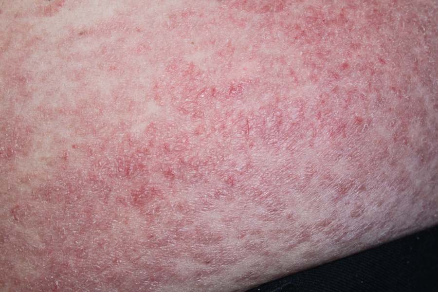
Chronic superficial scaly dermatitis is an uncommon skin condition characterised by round-oval and sometimes polymorphous, pink-red, slightly scaly patches on the trunk and limbs. Much controversy surrounds the best terminology for this condition ie chronic superficial scaly dermatitis or parapsoriasis, and the potential for progression of some cases into mycoses fungoides, which is a form of cutaneous t-cell lymphoma.

This chapter is set out as follows:


Aetiology

  • The cause is unknown

History

  • The onset is usually insidious
  • Symptoms are usually minimal, occasionally the skin can itch
  • Lesions are more prominent in winter than in summer. If they clear with treatment, lesions tend to recur in the same, or adjacent, areas when treatment is stopped

Clinical findings

Distribution

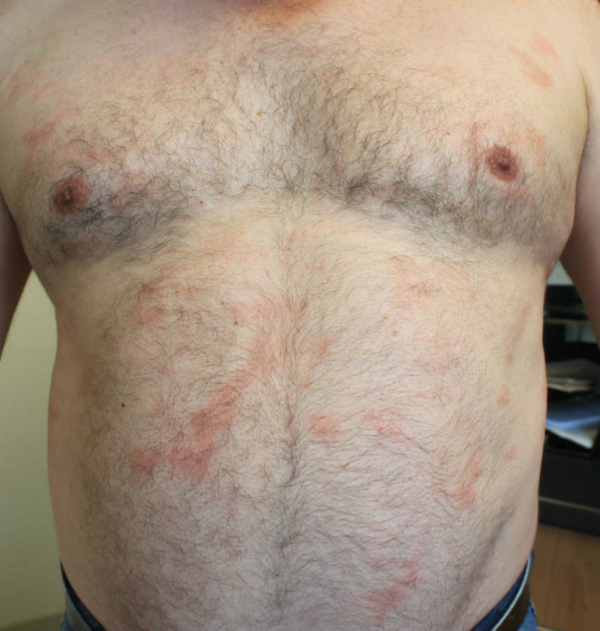
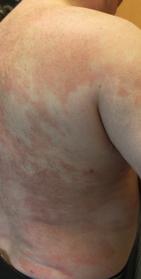
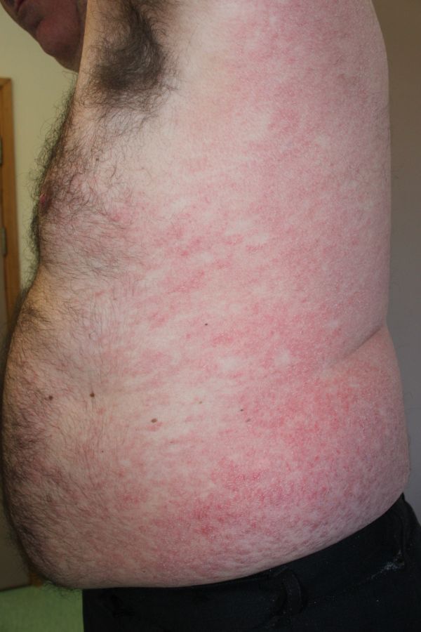
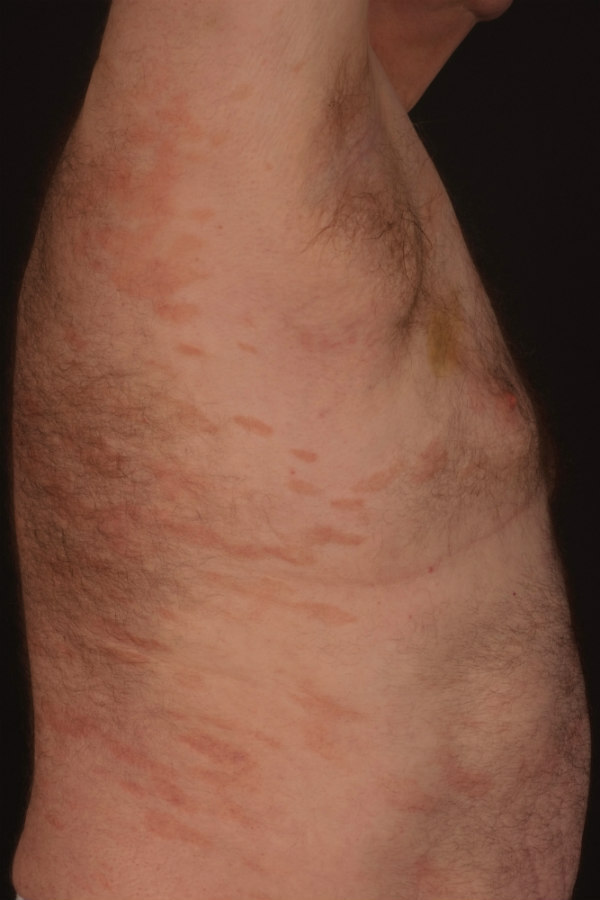
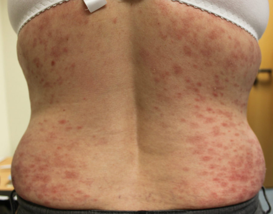
  • The commonest site is the trunk, followed by the limbs. It is seldom seen on the face, palms or soles

​Morphology  

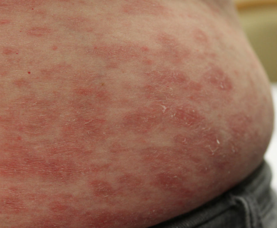
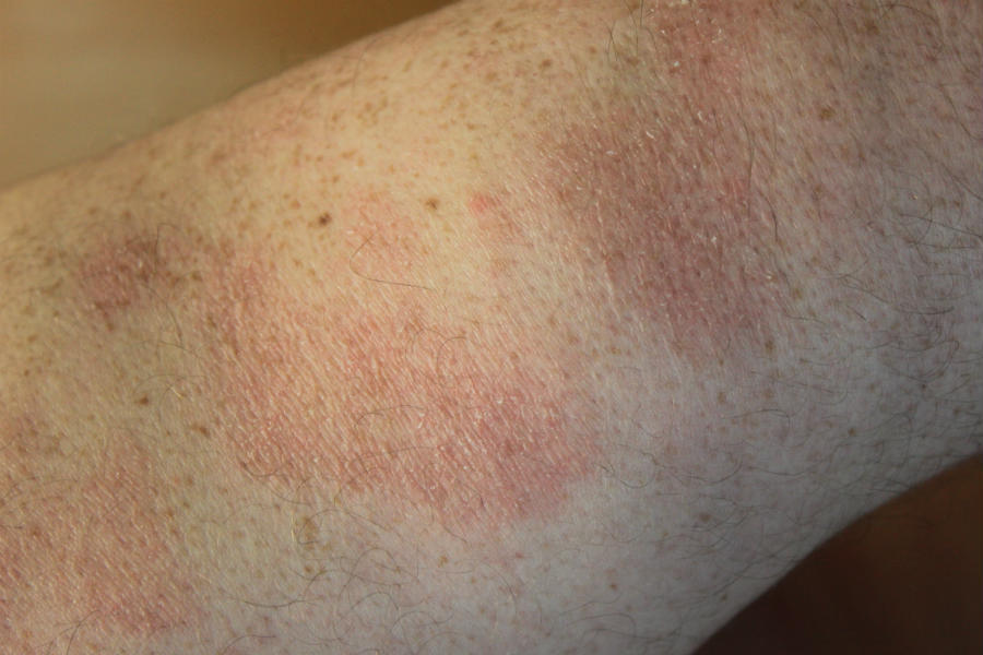
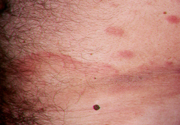
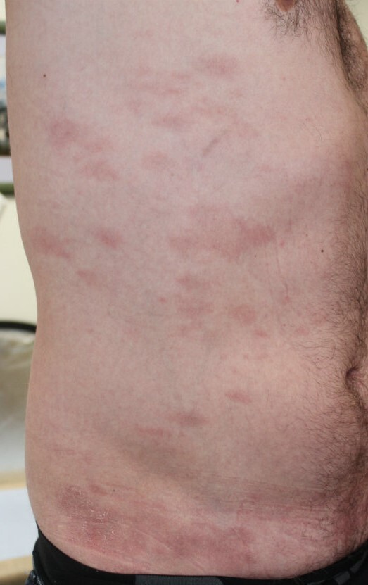
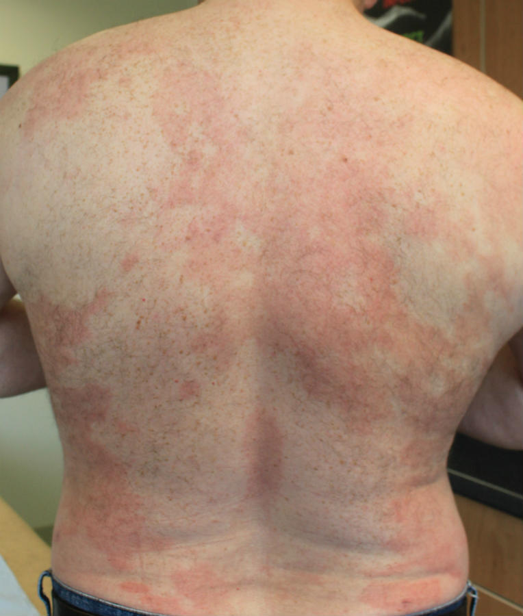
  • Patches are generally 2.5-5 cm in diameter, and round or oval, but finger-like processes are also common especially on the abdomen, and this accounts for the alternative name digitate or finger-like dermatosis
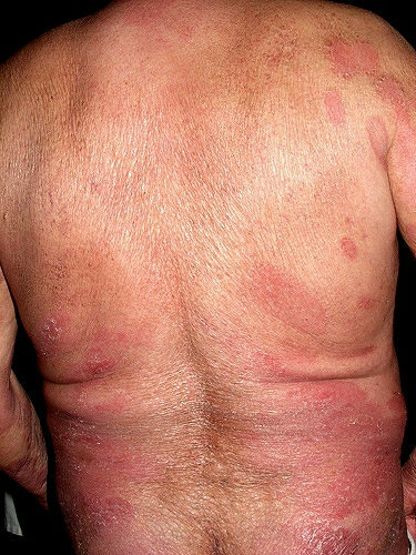
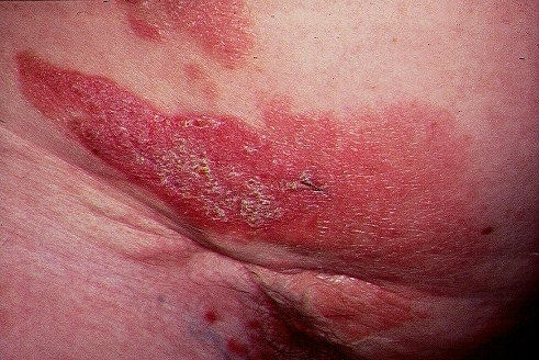
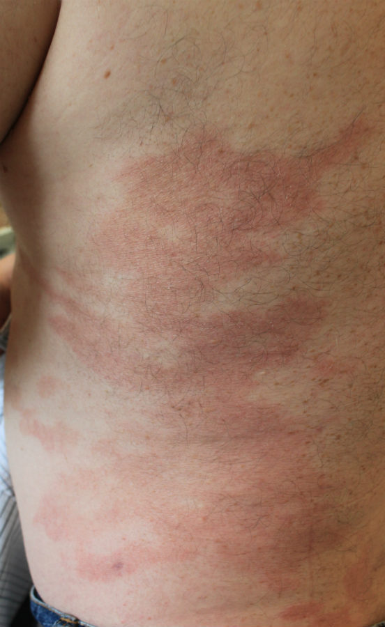
  • In other cases patches may be much larger and polymorphous in shape  
  • The colour is pink-red, brown or slightly yellow. The individual patches often have a slightly waxy appearance 
  • Fine scaling is common
  • After extending for a time most cases then remain static and with minor fluctuations persist throughout life. In a few patients the condition clears permanently

There is much debate as to whether or not some cases of chronic superficial scaly dermatitis are a precursor to mycoses fungoides (MF), or whether these cases were misdiagnosed and were MF from the onset. Indeed skin biopsies of early MF lesions may miss the diagnosis. The current view is as follows:

  • Lesions more likely to run a benign course - smaller, more regular lesions, with histological features showing eczematous-like change and little or no dermal infiltrate 
  • Lesions of greater concern - larger lesions that are often polymorphic with bizarre or angulated shapes, involvement of the girdle areas or breasts, and histological features showing absence of eczematous change and presence of dermal infiltrate 

Clinical Images

Please refer to notes on image rights at bottom of the page with regards to individual image ownership.
Investigations

  • An incisional skin biopsy of both affected and adjacent unaffected skin is necessary to try and distinguish chronic superficial scaly dermatitis from other skin conditions, particularly MF
  • Histologically the changes can resemble mild eczema or can be non-specific - sometimes several biopsies are needed, and these may need to be repeated if the rash changes over time

Management

  • Emollients provide relief to dryness and scaling
  • Referral for diagnosis and consideration of phototherapy, which can clear the patches, although recurrence at the same sites is common
  • Progression of lesions to plaques, ulceration, or those showing poikilodermatous change (red-brown, atrophic patches with telangiectases) strongly suggest MF

Disclaimer - the author PCDS cannot accept responsibility for any misleading or incorrect statements, and the management of individual patients remains the direct responsibility of the individual doctor. We do however hope that visitors to this site can contact us regarding comments that are considered misleading or incorrect so that we can continue to improve the site.

Image Rights - The PCDS would like to thank Dermatoweb, DermQuest (Galderma), and others who have contributed images. All named individuals and organisations maintain copyright for the relevant images.

Quick Links

The following pharmaceutical companies have had no involvement in the content of this website or in our conference programmes


Almirall
Galderma
Glenmark
Johnson & Johnson
La Roche-Posay
LEO Pharma
Pierre Fabre
Schuco