Dermatomyositis (including subgroups, and the Antisynthetase syndrome)
LAST UPDATED: Jun 30, 2023
Introduction
Dermatomyositis is a rare, multisystem, autoimmune disorder mainly affecting the skin, muscle and blood vessels, which occurs in association with underlying malignancy in a subset of patients. Subgroups of dermatomyositis include amyopathic dermatomyositis (cutaneous features without clinical muscle weakness), juvenile dermatomyositis, necrotizing myopathy (syn. necrotizing autoimmune myopathy; immune-mediated necrotizing myopathy) and inclusion body myositis. The term polymyositis has largely been dropped. This chapter also discusses the Antisynthetase syndrome, in which myositis is found together with the antisynthetase antibodies.
This chapter is set out as follows:
Aetiology
-
While the exact cause is unknown, dermatomyositis (DM) is thought to be caused by a microangiopathy affecting skin and muscle
- Some patients have a genetic predisposition
- Triggers include:
- Medications - drugs reported include statins and HMF-CoA reductase inhibitors, penicillamine, hydroxyurea, cyclophosphamide, NSAIDs, anticonvulsants, and some immunisations
- Malignancy in a subset of patients
- Viral infections
History
- The condition affects adults and children
- Age - in adults, most forms of DM occurs in the late 40s to early 60s. In children, it most often appears between 5 and 15 years of age
- DM affects more females than males
- The rash of DM is sometimes described as stinging/burning
- Muscle weakness
- Most types of DM have proximal muscle weakness affecting the hips, thighs, shoulders, upper arms, and neck resulting in difficulties climbing stairs, standing up from a chair, and lifting arms over the head
- Speech and swallowing can also be affected
- The pattern of muscle weakness differs in inclusion body myositis
Clinical findings
Dermatomyositis (classical)
- Presents with characteristic skin changes along with proximal muscle weakness as described above
- Cases without muscle involvement are referred to as amyopathic dermatomyositis, which accounts for 5–20% of patients with DM
Characteristic cutaneous features of dermatomysositis
-
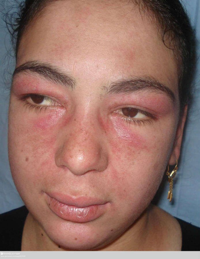
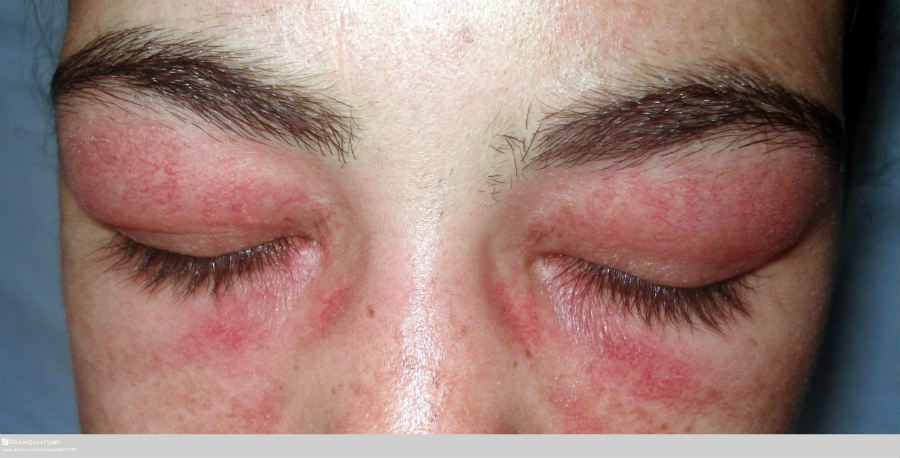
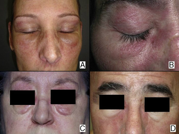
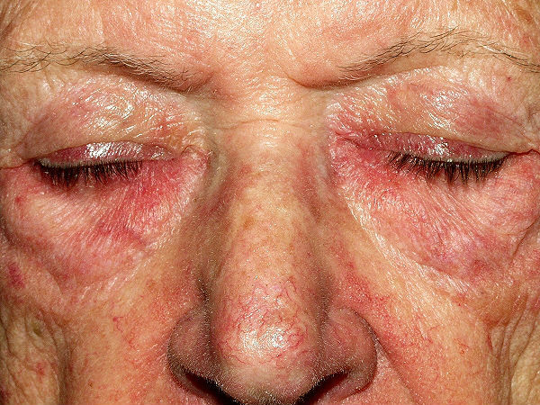
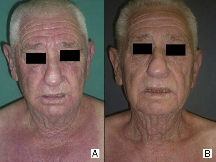
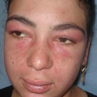
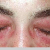
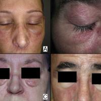
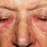
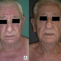
A purple-red ‘heliotrope’ facial rash, affecting the eyelids, upper cheeks, forehead and temples, frequently associated with oedema of the eyelids and periorbital skin. The lips can also be affected
- Facial erythema can sometimes be seen in a malar distribution with perioral sparing. The rash can involve the nasolabial folds, which can help to distinguish DM from cutaneous LE, which spares this site
-
Hands
-
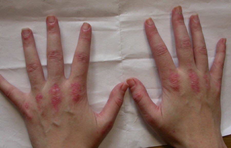
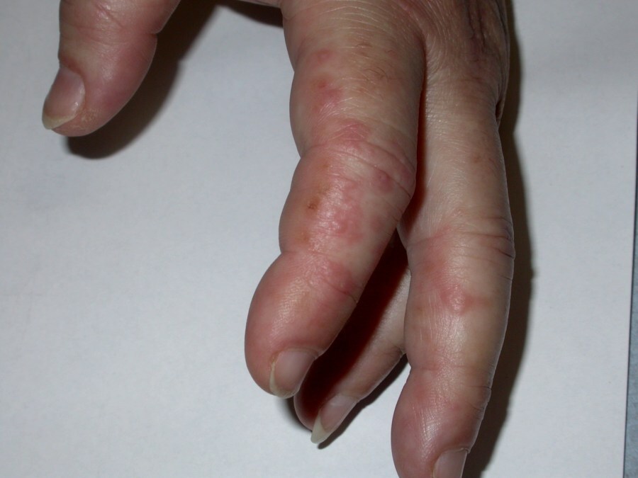
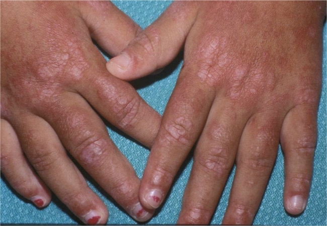
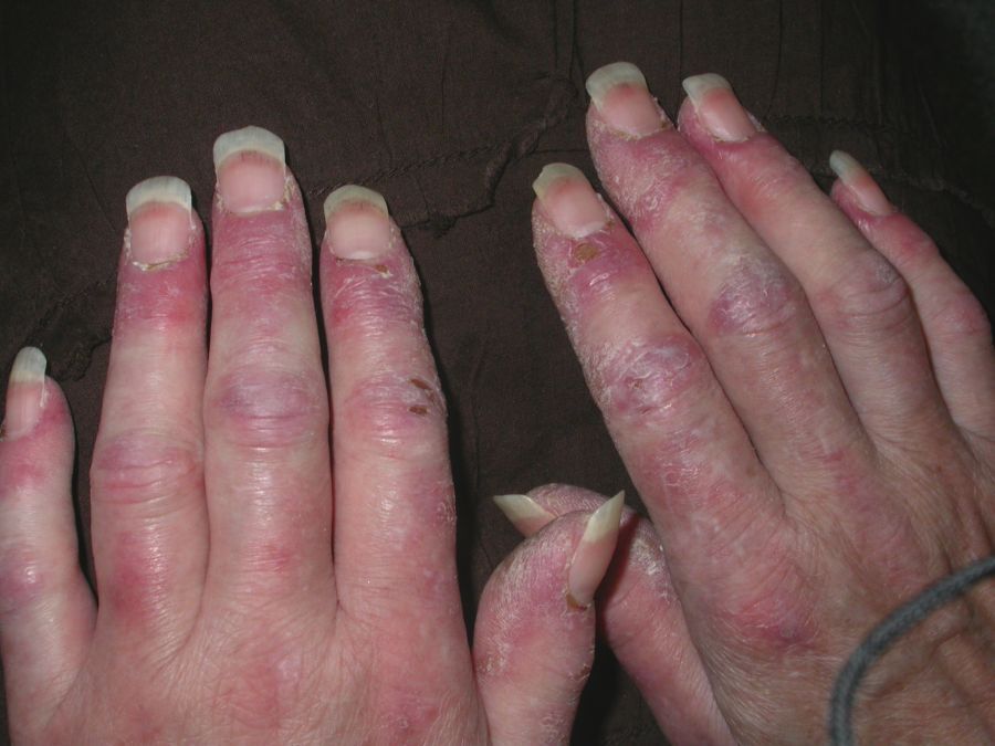
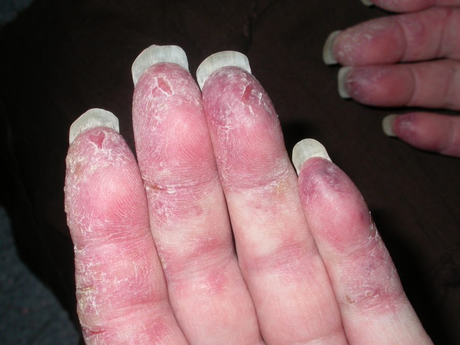
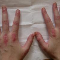
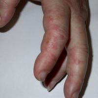
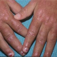
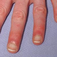
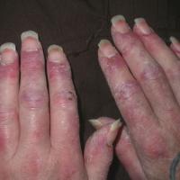
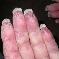
Gottron's papules - small red-purple, flat-topped papules, found on the extensor surfaces of the metacarpophalangeal and interphalangeal joints and around the nailfolds (they can also affect the feet and other extensor surfaces)
- Inverse Gottron's papules - very uncommon keratotic papules located on the flexor aspects of the fingers
-
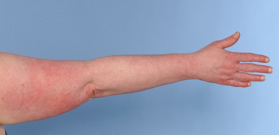
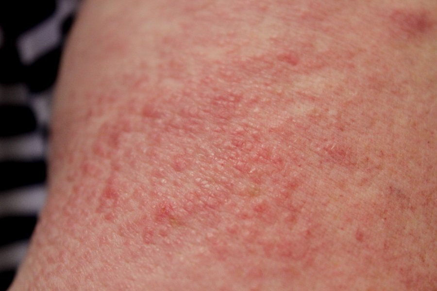
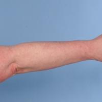
Linear erythema - affects the dorsal fingers and dorsal hands (linear erythema can be found at other sites)
-
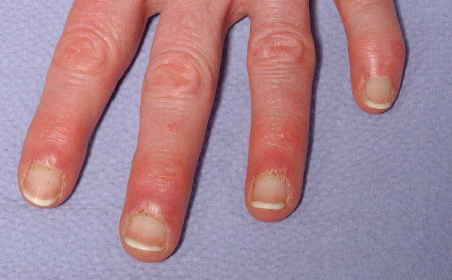
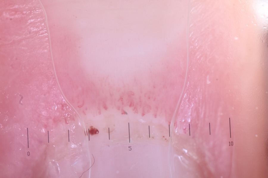
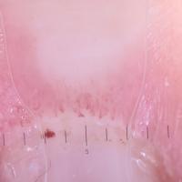
Nailfolds - shiny red nailfolds with ragged cuticles, and dilated-irregular capillaries seen dermoscopically
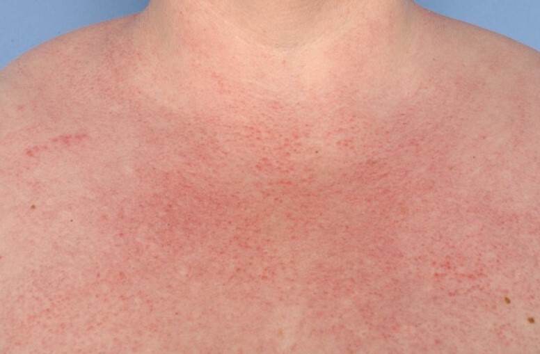
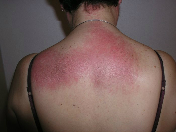
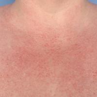
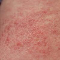
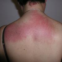
- A maculopapular violaceous erythema of the nape of neck, shoulders, and upper arms (the Shawl sign), which can also affect the upper chest (the ‘V’ sign). Other sites can be affected
- Photosensitivity is common, as is hyperpigmentation in skin of colour
- The Holster sign - erythema affecting the buttocks, hips, and lateral thighs
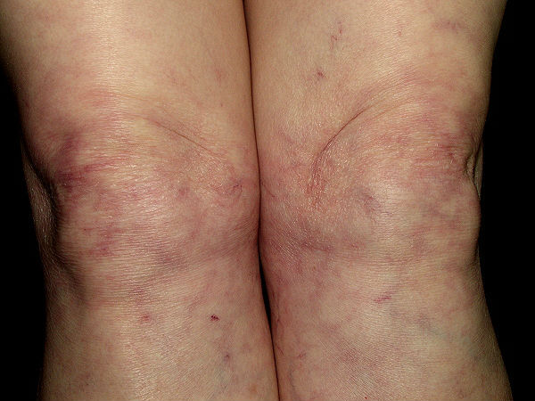
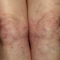
- Poikiloderma - patches of red-brown atrophic skin with prominent dilated blood capillaries
- Scalp - itchy and scaly scalp with inflammation and diffuse, non-scarring alopecia
- Other cutaneous features can include - Raynaud’s phenomenon, a flagellate erythema (erythematous streaks), urticarial lesions, vasculitis, bullous lesions of the dorsal hands and forearms, panniculitis, livedo reticularis, oral changes, and erythroderma especially if associated with malignancy
Extra-cutaneous features of dermatomyositis
- Malignancy in certain subsets - the most commonly affected organs are the cervix, lungs, pancreas, breasts, ovaries and gastrointestinal tract. There is also an association with non-Hodgkin’s lymphoma. In terms of who to screen refer below to the section on investigations
- Non-erosive polyarthritis or arthralgia of the small joints of the hands
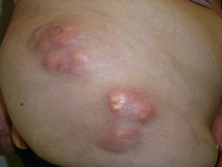
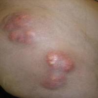
- Calcinosis (the deposition of calcium) in muscles or areas of panniculitis, which occurs in 50% of childhood cases and 15% of adult cases
- Interstitial pneumonitis affects up to 40 % of cases and typically presents with progressive shortness of breath and a dry cough
- Cardiac complications occur in 50% of cases but are rarely symptomatic unless disease is advanced. Conduction defects and atrial or ventricular dysrhythmias are the most common cardiac manifestations. Cardiac failure due to cardiac myositis is rare
Other types of dermatomyositis
Juvenile dermatomyositis
- The most common inflammatory myopathy of childhood
- Characterised by the presence of the typical rash and proximal muscle weakness
- Calcinosis is a common finding (up to 40% of patients)
- Not associated with malignancy
Inclusion body myositis
- The most commonly acquired myopathy in patients aged over 50
- Men more than women
- Very slowly progressive symptoms, which can lead to a delay in diagnosis
- Distal muscle weakness leads to frequent falls (including from foot drop), difficulty walking, a weakened hand grip, and difficulty flexing the fingers
Necrotising myopathy - also known as necrotising autoimmune myopathy (NAM) or immune-mediated necrotising myopathy (IMNM)
- Characterised by signs of necrosis, or cell death, in the muscles, which causes weakness and fatigue in the proximal muscles
- There are different categories of necrotizing myopathy that have different risk factors and treatments. These distinctions are based on the presence of different autoantibodies:
- HMGCR antibody - associated with statin-induced necrotising myopathy
- SRP antibody - this group typically experiences sudden and extreme muscle weakness. Some patients have cardiac involvement
The Antisynthetase syndrome
- A form of myositis, the hallmark of which is is the presence of serum autoantibodies directed against aminoacyl-tRNA synthetases, the most common of which is the anti-Jo-1 antibody
- Typically occurs in patients whose average age at onset is 50
- Clinical features include:
- Myositis affecting the proximal muscles
- Periodic fever in up to 30% of cases
- Non-erosive arthritis, most commonly a symmetrical arthritis affecting the small joints of the hands and feet
- Up to 75% have interstitial lung disease
- 'Mechanic's hands' in approximately 30% of patients causing hyperkeratosis and fissuring of the tips and margins of the fingers
- Raynaud's phenomenon in 40% of cases
- Not associated with malignancy
Clinical Images
Please refer to notes on image rights at bottom of the page with regards to individual image
ownership.
1
2
3
4
5
6
7
8
9
10
11
12
13
14
15
16
17
18
Investigations
-
Autoantibodies and other blood tests
- A high ESR with a normal-low CRP
- The CK is often elevated unless muscles are not involved
- ANA and ENA (Jo-1) can be positive - refer to the chapter on investigations for more information
- Myositis specific antibodies help predict different clinical patterns of DM
- Malignancy screen
- Malignancy is only considered in adult dermatomyositis
- The risk is lower for amyopathic dermatomyositis
- Clinical features associated with an increased risk include age > 45 at diagnosis, male gender, dysphagia, cutaneous necrosis, cutaneous vasculitis, and rapid onset of skin/muscle symptoms
- Patients with TIF1 or NXP-2 antibodies (both myositis specific antibodies) have a high-risk of malignancy requiring thorough investigations including radiology (some centres recommend PET scans)
-
Muscle biopsy and electromyography may be required for muscle involvement
Management
Management
-
Patients with suspected DM require urgent referral to Secondary Care
-
The main aim of treatment is to control the cutaneous changes and muscle involvement
-
Treatment of cutaneous features
-
Topical steroids, tacrolimus ointment, and antimalarial therapy (eg hydroxchloroquine) have been shown to be beneficial
-
Muscle / systemic involvement
-
Rest is required in the acute phase
- Treatments include conventional immunosuppressive drugs such as methotrexate and azathioprine, as well as tacrolimus, biologic therapies cyclophosphamide, and IVIG
- Physical therapy is required for calcinosis
Prognosis
- The prognosis is worse for cases associated with malignancy as well as those who have the MDA-5 antibody (a myositis specific antibody) - untreated this has a very poor prognosis with rapidly progressing lung disease and ulcerating skin lesions
-
Patients without muscle involvement or calcinosis have a better prognosis
Disclaimer - the author PCDS cannot accept responsibility for any misleading or incorrect statements, and the management of individual patients remains the direct responsibility of the individual doctor. We do however hope that visitors to this site can contact us regarding comments that are considered misleading or incorrect so that we can continue to improve the site.
Image Rights - The PCDS would like to thank Dermatoweb, DermQuest (Galderma), and others who have contributed images. All named individuals and organisations maintain copyright for the relevant images.