Erythema elevatum diutinum

LAST UPDATED: Nov 22, 2021
Introduction

Erythema elevatum diutinum (EED) is a rare form of cutaneous small vessel vasculitis characterised by red, purple, brown, or yellow papules, plaques, or nodules, presenting symmetrically on extensor surfaces, most commonly the hands, elbows, knees, and Achilles tendons. The term 'diutinum' is the latin word for long lasting.

This chapter is set out as follows: 


Aetiology

  • EED is classified as a small vessel vasculitis
     
  • The cause is unknown but it has been associated with a number of conditions:
    • The association with haematological abnormalities, such as multiple myeloma, is strong; however, EED may precede the haematological disease by several years
    • Viral infections eg hepatitis B and HIV
    • Rheumatoid arthritis
    • Inflammatory bowel disease and Coeliacs disease   

History

  • Although EED can occur at any age, it is most commonly seen between the ages of 30-60 years
  • It affects males and females equally
  • The lesions can be completely asymptomatic, painful, or cause a sensation of burning or itching. These symptoms can be exacerbated by cold

Clinical findings

Distribution

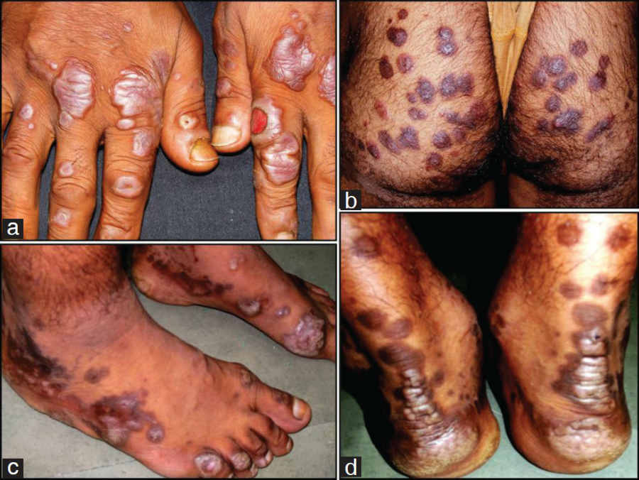
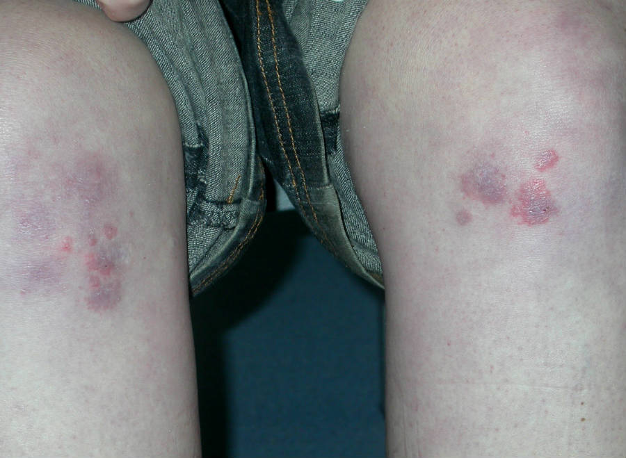
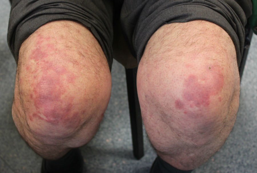
  • Lesions tends to appear in a symmetrical fashion over the dorsa of the hands, elbows, knees, and feet
  • The buttocks, face, and ears as well as the palms, soles, legs, forearms, and genitalia may also be involved; however, the trunk is usually spared

Morphology

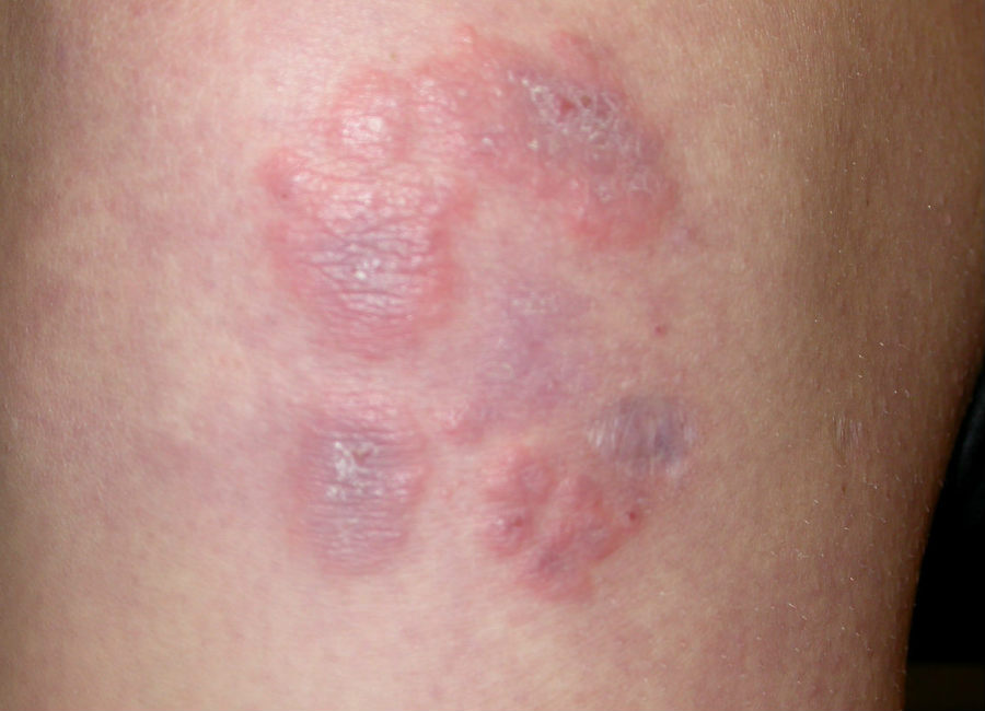
  • Lesions are red, purple, brown, or yellow papules, plaques, or nodules. Initially the lesions are soft, but eventually they fibrose and later leave atrophic scars
  • Rarely there are bullae and ulcers 

Clinical Images

Please refer to notes on image rights at bottom of the page with regards to individual image ownership.

Investigations

  • Blood tests
    • FBC, U&E, LFT, along with immunoglobulins and plasma electrophoresis should be tested for haematological abnormalities
    • Antineutrophil cytoplasmic antibodies (ANCA) may be positive
    • Other blood tests can be done if appropriate eg for viral infections (refer to aetiology)
       
  • Histology (reference: Rook's Textbook of Dermatology)
    • Acute lesions of EED are characterised by leukocytoclastic vasculitis
    • Chronic lesions demonstrate angiocentric eosinophilic fibrosis, capillary proliferation and infiltration of macrophages, plasma cells and lymphocytes

Management

  • EED may last for many years, with crops of new lesions developing every few weeks to months
  • Treat any underlying cause
  • High potency topical, or intralesional, steroids may reduce the size of lesions. Dapsone (provided under specialist supervision) can be highly effective in controlling the condition - patients require the regular monitoring of FBC, U&E, LFT and reticulocyte count due to the risk of haemolytic anamia. Nicotinamide also appears to be effective

Disclaimer - the author PCDS cannot accept responsibility for any misleading or incorrect statements, and the management of individual patients remains the direct responsibility of the individual doctor. We do however hope that visitors to this site can contact us regarding comments that are considered misleading or incorrect so that we can continue to improve the site.

Image Rights - The PCDS would like to thank Dermatoweb, DermQuest (Galderma), and others who have contributed images. All named individuals and organisations maintain copyright for the relevant images.

Quick Links

The following pharmaceutical companies have had no involvement in the content of this website or in our conference programmes


Almirall
Galderma
Glenmark
Johnson & Johnson
La Roche-Posay
LEO Pharma
Pierre Fabre
Schuco