
LAST UPDATED: Nov 22, 2021
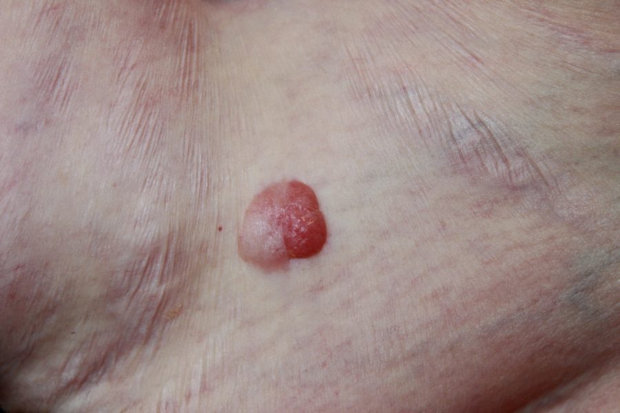
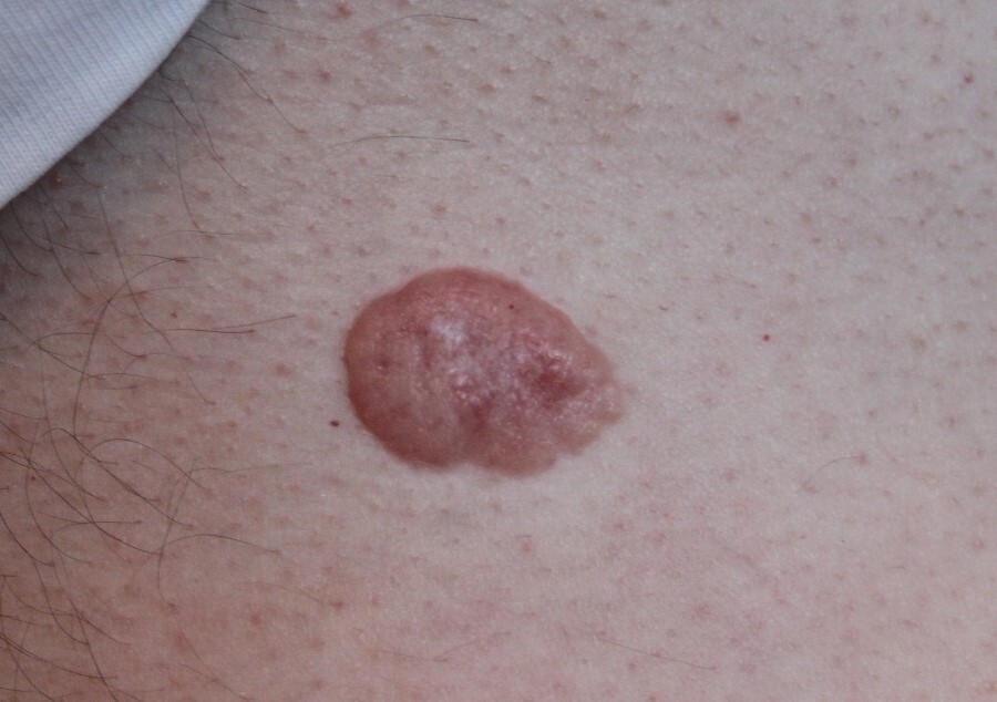
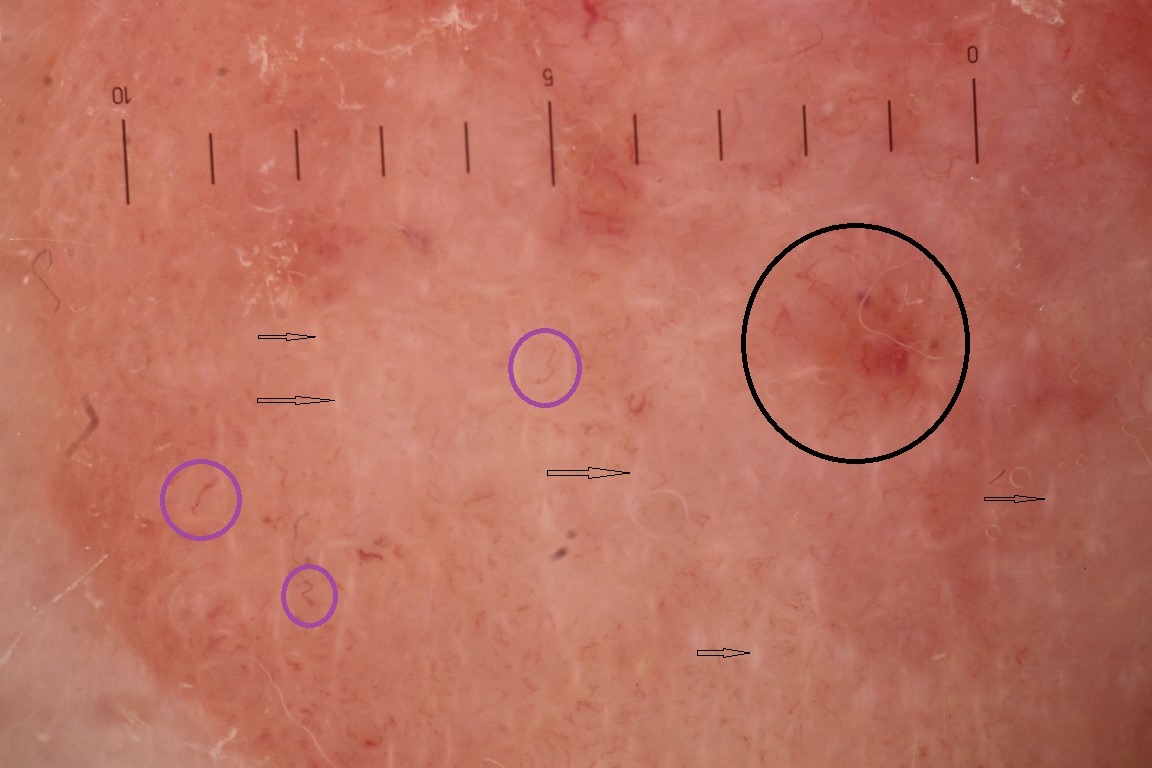
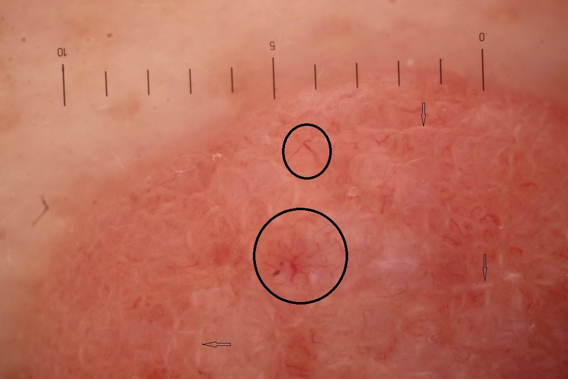
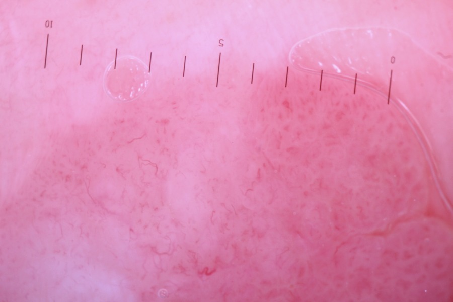
Fibroepithelioma of Pinkus was first described by Hermann Pinkus in 1953 as a rare, premalignant fibroepithelial tumour. Pinkus observed that some of the lesions had a peculiar histologic appearance that resembled both a reticulated seborrhoeic keratosis and a basal cell carcinoma (BCC). Pinkus thus considered the tumour to be a variant of basal cell carcinoma, which illustrated the interaction and interdependence of stromal and epithelial components of basal cell carcinoma.
Lesions are often diagnosed retrospectively, mostly commonly being mistaken clinically for a skin tag.
This chapter is set out as follows:
Please refer to notes on image rights at bottom of the page with regards to individual image ownership.
Disclaimer - the author PCDS cannot accept responsibility for any misleading or incorrect statements, and the management of individual patients remains the direct responsibility of the individual doctor. We do however hope that visitors to this site can contact us regarding comments that are considered misleading or incorrect so that we can continue to improve the site.
Image Rights - The PCDS would like to thank Dermatoweb, DermQuest (Galderma), and others who have contributed images. All named individuals and organisations maintain copyright for the relevant images.
It is important for the PCDS to build its own image bank, as such we welcome original images from our readers, especially in skin of colour:
Please start typing below the word of any medical term that you do not understand.
The following pharmaceutical companies have had no involvement in the content of this website or in our conference programmes







