Halo naevus (syn. Sutton naevus)
LAST UPDATED: Oct 09, 2025
Introduction
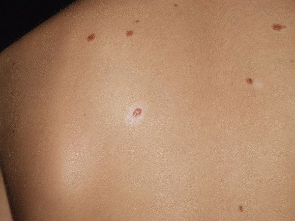
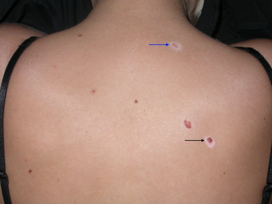
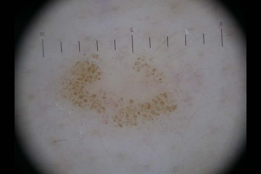
A halo naevus is essentially a benign melanocytic naevus with a white ring or 'halo' around it. It is a benign skin lesion that represents a melanocytic naevus in which an inflammatory infiltrate develops, resulting in a zone of depigmentation surrounding the naevus.
This chapter is set out as follows:
Aetiology
- The cause of the spontaneous loss of pigmentation is unknown but appears to be related to an immunologic destruction of melanocytes. Adding support to this hypothesis is the fact that several patients with halo naevi have a tendency toward the development of vitiligo. Halo naevi may follow the Koebner phenomenon, arising within a mole that has been traumatised
History
- They present most frequently in older children and young adults. Halo naevi occasionally arise in older individuals
- In fair-skinned individuals a halo naevus is most often recognised during summer months when the surrounding normal skin tans, whereas the halo of depigmentation does not
- Although common melanocytic naevi are those most frequently associated with the halo phenomenon, it may also occur in other naevi such as blue naevi and Spitz naevi
- Natural history - the central naevus gradually disappears over several months, although sometimes this process can take a number of years
Clinical findings
Distribution
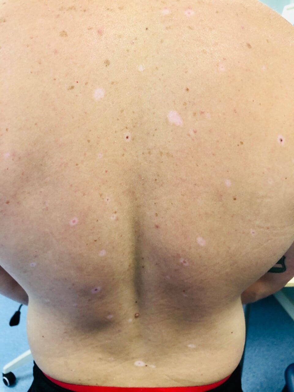
- Lesions can be single, but are frequently multiple
- They can develop anywhere on the body but are seen most frequently on the trunk, especially the back
Morphology - there are four stages
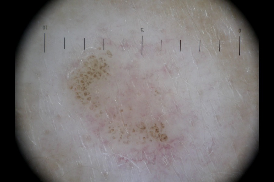
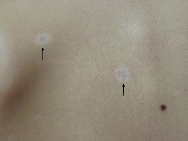
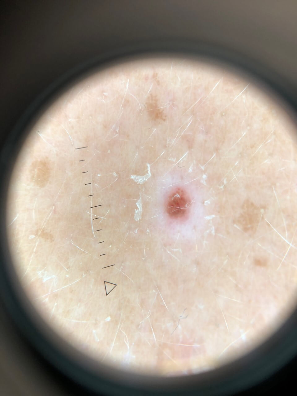
- A rim of pale skin surrounds the naevus - the width of the halo is variable but is generally of uniform radial distance from the central naevus
- The naevus may become pinker or less pigmented, and eventually disappears
- A circular or oval area of depigmentation persists
- The affected skin gradually returns to its normal colour
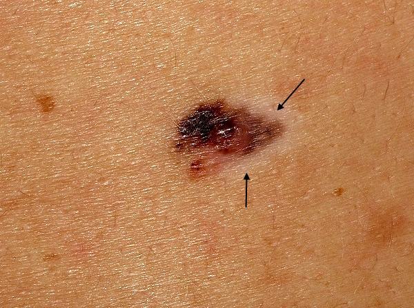
Clinical Images
Please refer to notes on image rights at bottom of the page with regards to individual image
ownership.
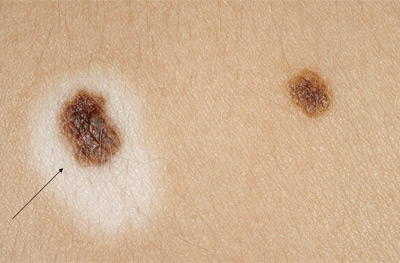
Management
- Halo naevi are harmless - no treatment is required
- Differentiating a halo naevus from a regressing melanoma is usually straight forward, although a regressing melanoma can have white areas. A melanoma should be suspected in the following:
- A solitary lesion with an irregular area of depigmentation surrounding/within an 'odd' looking mole
- A new 'halo naevus' in an older patient
- Patients with suspicious lesions should be referred on an urgent skin cancer pathway to Secondary Care
Disclaimer - the author PCDS cannot accept responsibility for any misleading or incorrect statements, and the management of individual patients remains the direct responsibility of the individual doctor. We do however hope that visitors to this site can contact us regarding comments that are considered misleading or incorrect so that we can continue to improve the site.
Image Rights - The PCDS would like to thank Dermatoweb, DermQuest (Galderma), and others who have contributed images. All named individuals and organisations maintain copyright for the relevant images.