Langerhans cell histiocytosis
LAST UPDATED: Jul 14, 2021
Introduction
Langerhans cell histiocytosis (LCH) is a rare condition, most commonly arising in young children, and which is characterised by the reactive proliferation of CD1a + Langerhans cells in various sites, leading to tissue damage. LCH is usually a multisystem disorder, but occasionally affects only the skin. There is a wide range of clinical features that range from mild, to severe and life threatening.
This chapter, which is set out as below, provides a brief overview of this complex condition:
Aetiology
-
LCH affects 2–5 children per million per year with an estimated prevalence of 1:50 000 children under 15 years of age
History
-
LCH is slightly more common in boys than girls
-
It may develop at any age but most commonly occurs between the neonatal period and the age of four years
-
In adults, the mean age at diagnosis is 35 years, with 10% being older than 55 years
Clinical findings
-
Most of the old terminology (eg Letterer-Siwe disease) has now been replaced by a classification based on the number of organ systems involved with an initial subdivision into single system (SS-LCH) and multisystem (MS-LCH) disease
-
SS-LCH
-
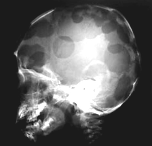
SS-LCH of the bone is the most common form of LCH, with the skull the most frequent site of involvement
-
SS-LCH affecting the skin only occurs in approximately 10% of children and adult cases - in children the skin is the second most commonly affected organ
-
MS-LCH
-
Refers to the involvement of any organ, although the gonads and kidneys are usually spared
-
It is divided into ‘low‐risk’ disease and ‘high-risk’ disease
-
Morphology
-
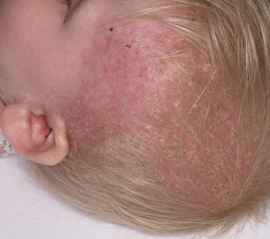
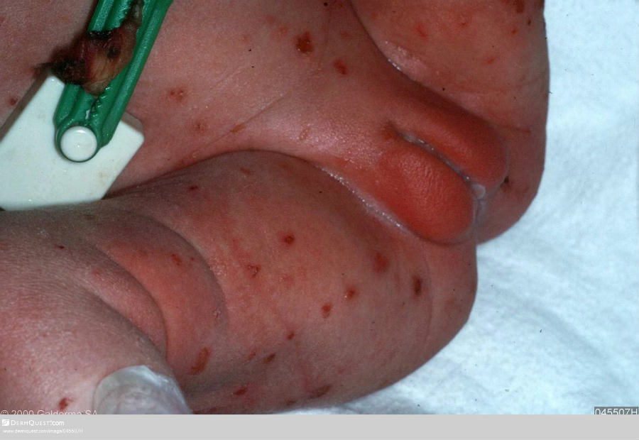
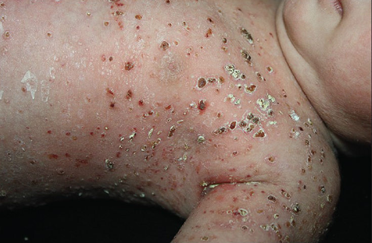
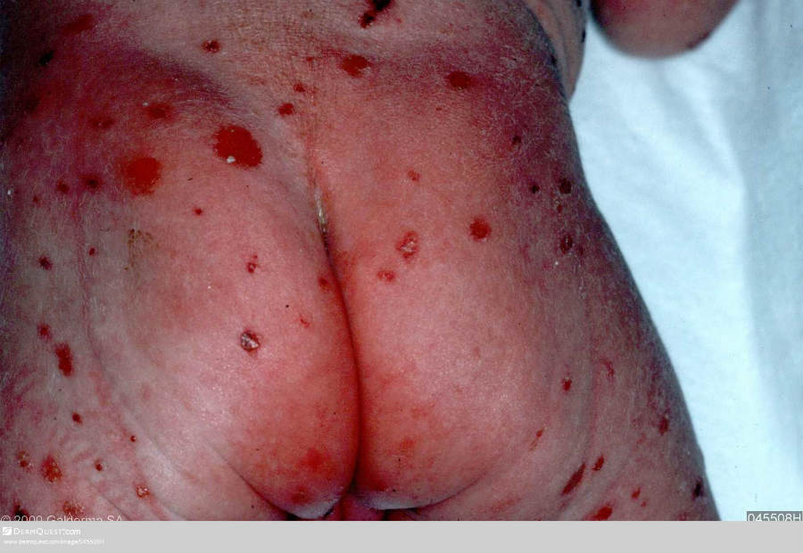
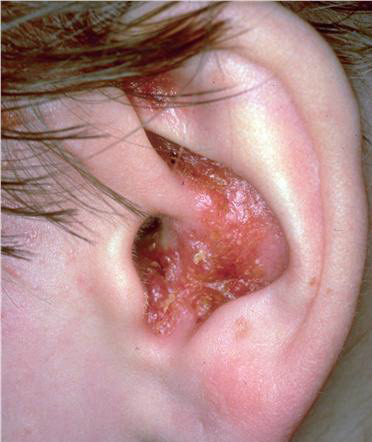
The appearance of skin lesions is variable and includes many small pinkish or reddish-brown macules-papules, or plaques, that may erode and become crusted
-
The most characteristic cutaneous lesion of LCH in children consists of papulosquamous lesions with greasy scales, affecting the scalp, resembling seborrhoeic eczema. Other sites include skin folds such as the gluteal cleft, and the midline of the trunk, but any area can be involved including the nails and genital mucosa
-
A persistent eruption of the scalp and in skin flexures beyond infancy should raise the suspicion of LCH even in the absence of other signs and symptoms
-
In more severe cases there can be widespread cutaneous involvement. Extensive ulceration, superinfection, petechia and purpura may accompany skin lesions. Multiple organs are involved including the bones, liver, spleen, lungs, central nervous system and bone marrow. This form carries the worst prognosis, is the least likely to resolve spontaneously and always requires systemic therapy
Clinical Images
Please refer to notes on image rights at bottom of the page with regards to individual image
ownership.
Investigations
-
The histologic picture unifies the varied presentations of LCH, which are influenced by the location and age of the lesions. Although lesions typically appear granulomatous, with a reactive background of macrophages, eosinophils, multinucleated giant cells, and T-cells, the key to diagnosis is to identify the pathologic Langerhans cell, which resembles the normal Langerhans cell of the skin, except that it is not dendritic
-
Further investigations will be needed to look for possible systemic involvement
Management
-
The mortality from SS-LCH and low‐risk MS‐LCH is extremely low, however, complications include diabetes insipidus, and some cases may progress to high-risk disease, which can be fatal
-
The predictors of a poor outcome are:
-
Age less than two years at onset
-
Multi-organ involvement
-
Involvement of a vital organ (blood, liver, lungs or spleen)
-
Treatment depends on the severity of the disease and the number of organs involved. Evidence of damage to the organs is more important than involvement of the organ as such. For more severe disease treatments include oral corticosteroids and chemotherapy
Disclaimer - the author PCDS cannot accept responsibility for any misleading or incorrect statements, and the management of individual patients remains the direct responsibility of the individual doctor. We do however hope that visitors to this site can contact us regarding comments that are considered misleading or incorrect so that we can continue to improve the site.
Image Rights - The PCDS would like to thank Dermatoweb, DermQuest (Galderma), and others who have contributed images. All named individuals and organisations maintain copyright for the relevant images.