Cutaneous leiomyoma
LAST UPDATED: Nov 21, 2021
Introduction
A leiomyoma is an uncommon, benign tumour of smooth muscle. There are three types of cutaneous leiomyomas: piloleiomyomas, angioleiomyomas and genital leiomyomas. This classification reflects the most logical origin of the tumour, corresponding to the histologic or anatomic site where the leiomyomas are found.
Female patients with cutaneous leiomyomas may also develop uterine leiomyomas (fibroids), and very rarely leiomyomas can be associated with renal cell carcinoma (hereditary leiomyomatosis and renal cell cancer).
This chapter is set out as follows:
Clinical findings
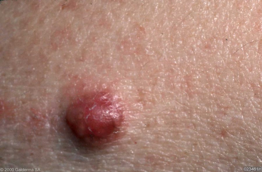
Piloleiomyoma (syn. leiomyoma cutis)
-
Originate from the arrector pili muscle of the pilosebaceous unit in the skin
-
Although they can occur at any age, lesions most commonly present in early adult life
-
Tend to present as a single lesion but often become multiple
-
Lesions can be painful / tender, especially in the cold and when touched
-
Distribution - the limbs are the most common site
-
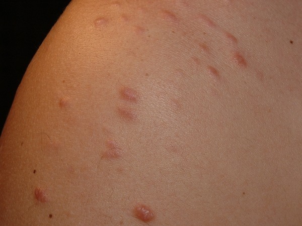
Morphology - red to brown papules / small nodules, often distributed in a linear manner. Adjacent tumours sometimes coalesce to form a plaque
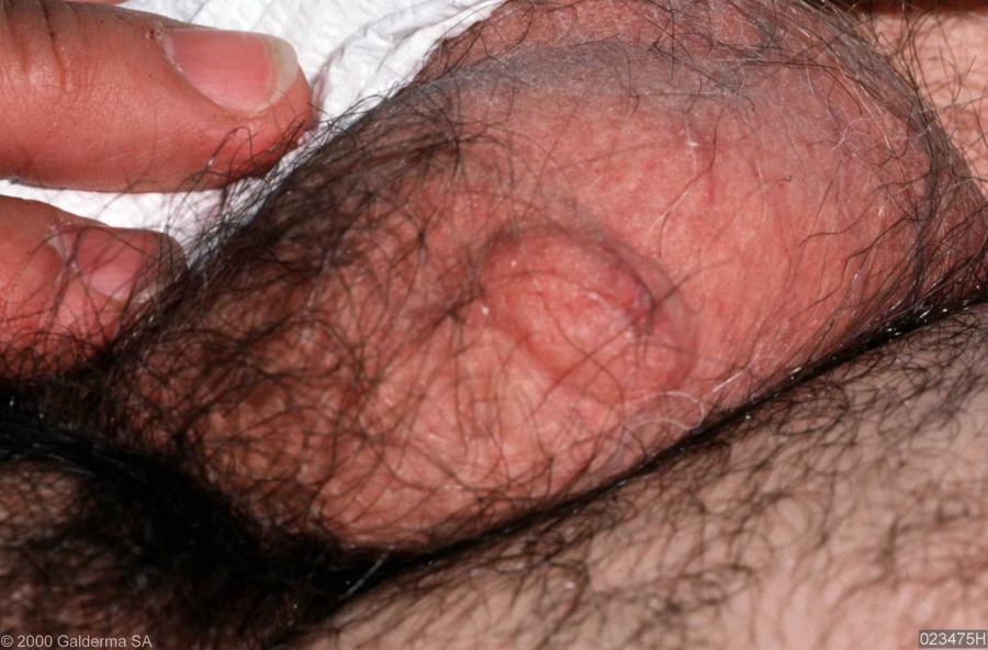
Genital leiomyoma (syn. dartoic myoma)
-
Originate from the smooth muscle of the genitalia and nipple
-
Can occur at any age
-
Pain is less of a feature
-
Distribution - most commonly affect the scrotum and labia majora
- Morphology - a solitary dermal nodule
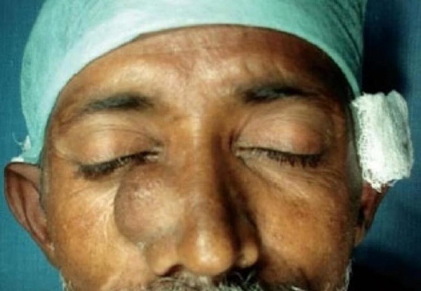
Angioleiomyoma
-
Originate from the smooth muscle of blood vessels, the tunica media
-
Occur in adults
-
Pain, tenderness and temperature sensitivity are common findings
-
Distribution - most commonly affect the lower extremities
-
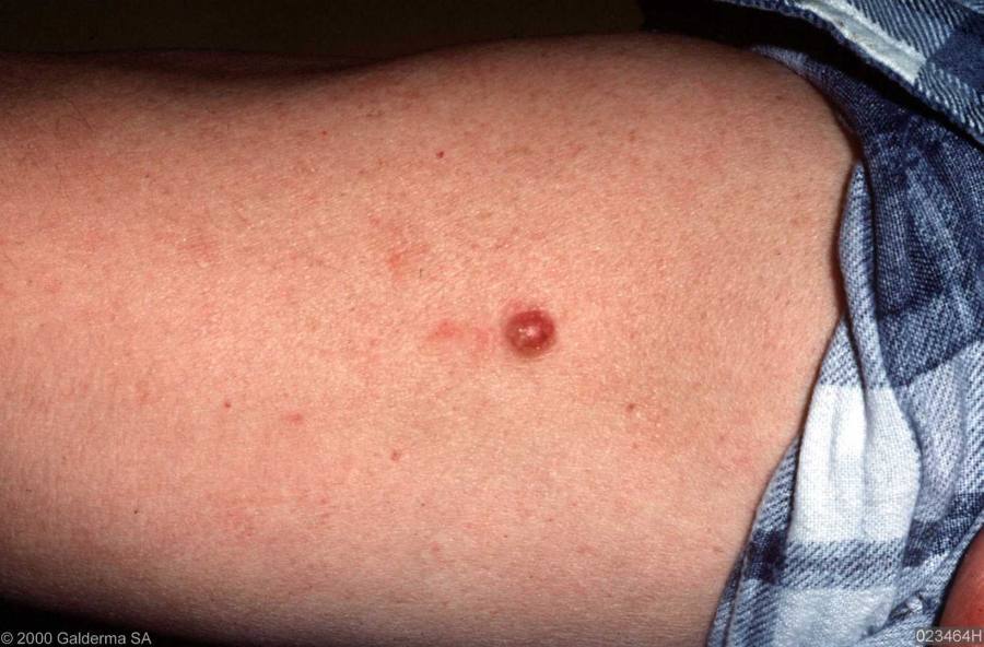
Morphology - a solitary subcutaneous tumour, which can grow up to 4 cm in diameter
Hereditary leiomyomatosis and renal cell cancer (HLRCC)
-
This is a rare autosomal dominant disorder, which has been reported in approximately 100 families worldwide
-
Patients develop cutaneous leiomyomas, and female patients often develop uterine leiomyomas (fibroids), which tend to be large and often appear earlier than in the general population. Patients have an increased risk of renal cancer, occuring in about 10%-16% of individuals with HLRCC with a median age of detection of 44 years
-
All family members should be informed and referred to a geneticist for DNA testing
Clinical Images
Please refer to notes on image rights at bottom of the page with regards to individual image
ownership.
Management
Piloleiomyomas
- Although lesions are benign, solitary lesions are often excised for histological confirmation
- Multiple lesions:
- If lesions are painful, medical treatment with calcium-channel blockers or gabapentin may improve symptoms
- Excision of an area containing multiple tumours can be considered, however, surgery is often followed by the development of further lesions in the surrounding skin
Angioleiomyomas and genital leiomyomas
- A biopsy is often needed for diagnosis
-
Patients with larger lesions may choose to have them excised
Disclaimer - the author PCDS cannot accept responsibility for any misleading or incorrect statements, and the management of individual patients remains the direct responsibility of the individual doctor. We do however hope that visitors to this site can contact us regarding comments that are considered misleading or incorrect so that we can continue to improve the site.
Image Rights - The PCDS would like to thank Dermatoweb, DermQuest (Galderma), and others who have contributed images. All named individuals and organisations maintain copyright for the relevant images.