Lymphoma and leukaemia - cutaneous presentations

LAST UPDATED: Feb 03, 2022

Introduction

Lymphoma and leukaemia can present in the skin as a primary cutaneous disorder (ie occur in the skin with no evidence of disease elsewhere at the time of diagnosis), a secondary infiltration, or indirectly eg as pruritus secondary to Hodgkin’s lymphoma. The remit of this chapter, which is set out as below, is to provide a brief overview into some of the more commonly recognised conditions within this group of disorders. 


Clinical findings

The conditions discussed in this chapter are classified into the following groups:

  • Primary cutaneous T-cell lymphoma (including mycosis fungoides)
  • Primary cutaneous CD30+ lymphoproliferative disorders (including lymphomatoid papulosis) 
  • Primary cutaneous B-cell lymphoma
  • Leukaemia - cutaneous presentations (including leukaemia cutis) 

Primary cutaneous T-cell lymphoma

The majority of primary cutaneous lymphomas are T-cell in type, for example mycosis fungoides, run a relatively indolent course and the time between onset and diagnosis can be considerable. A few, such as the Sézary syndrome, are more aggressive. The different disorders are described below. 

Mycosis fungoides (MF) 
  • Is the most common form
  • It is often characterised by typical cutaneous stages:
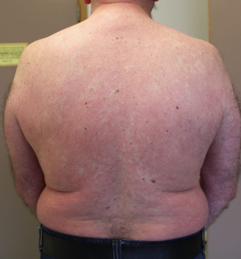
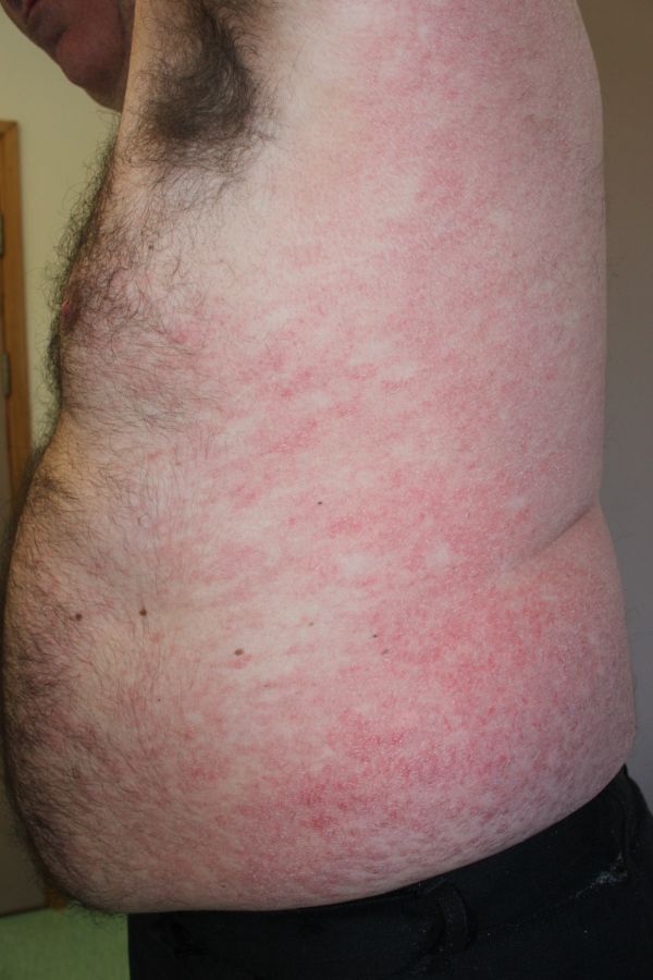
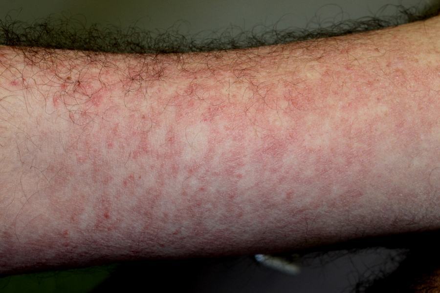
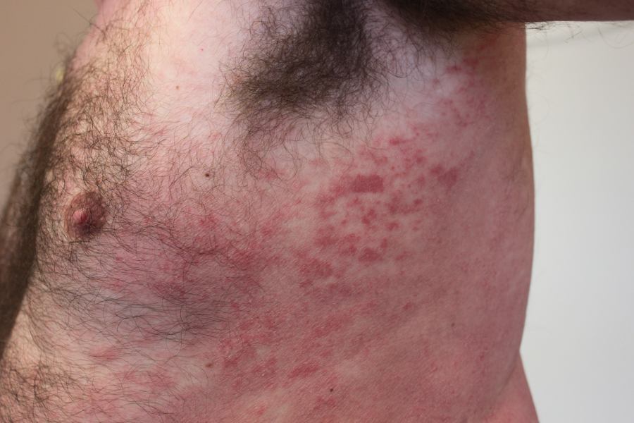
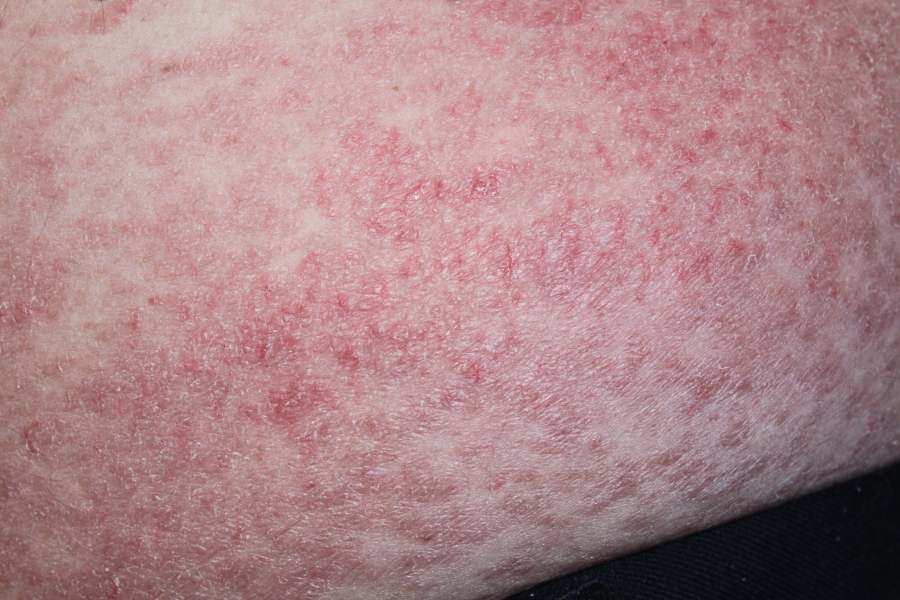
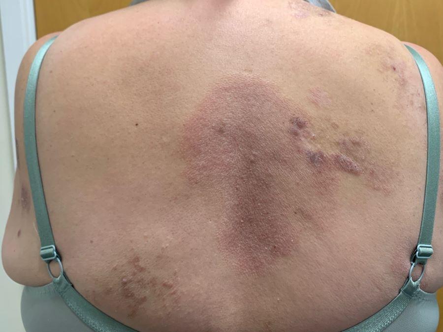
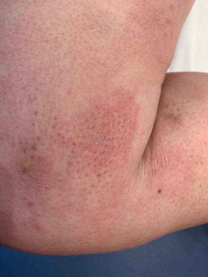
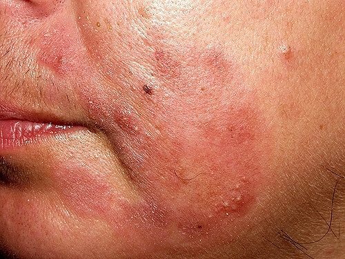
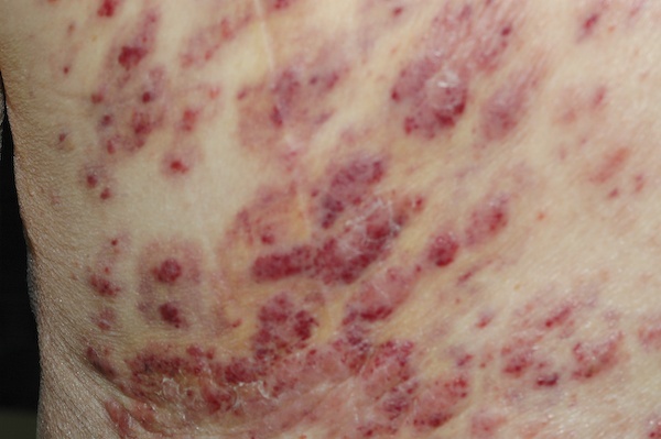
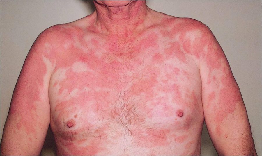
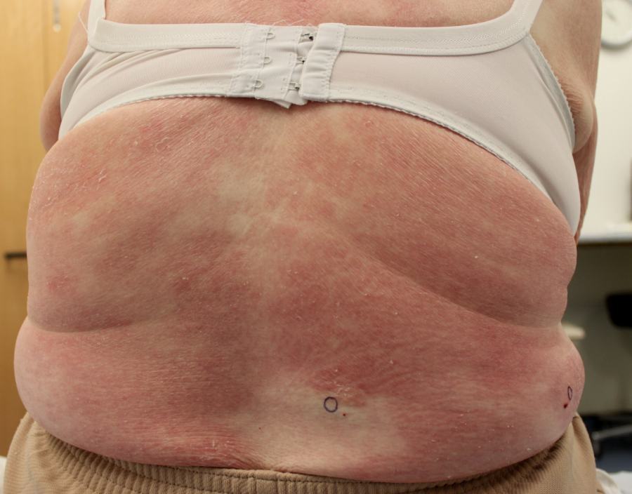
    • The majority of cases arise as multiple, non-itchy, erythematous patches, which have fine scale, and occur most frequently on the trunk, buttocks and limb girdle areas. Patients may have mild pruritus but are often asymptomatic
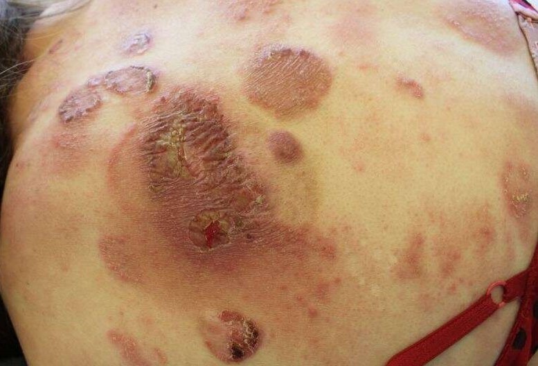
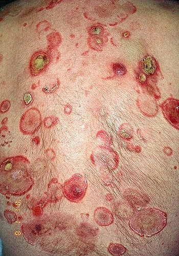
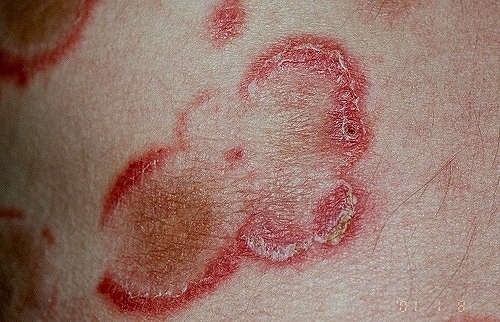
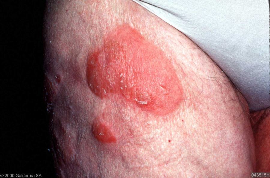
    • Over time well-demarcated plaques develop, which are polymorphous often having an annular or serpiginous pattern. Central clearing of the plaques can lead to arciform-shaped lesions. Lesions commonly have mild scale, although sometimes much more pronounced psoriasiform scaling can be a feature. Rarely, plaques can be hyperkeratotic or verrucous
    • Unlike pityriasis rosea, tinea or psoriasis, lesions do not improve over time or respond to topical therapy 
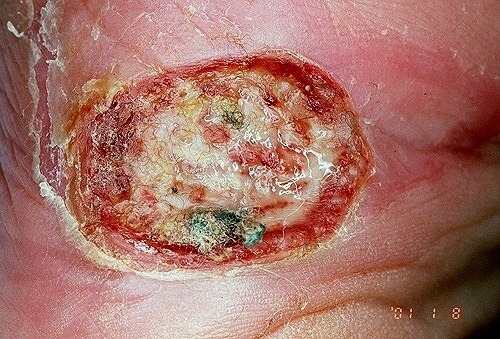
    • The natural history of MF is considerable. Most patients with patch / plaque stage MF progress little over many years. A few progress more quickly and may develop ulceration, nodules and rarely erythroderma 
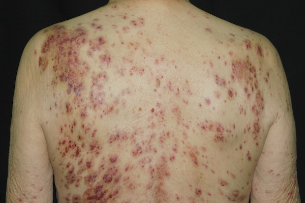
  • There are a large number of clinical variants of MF including follicular (see below), purpuric (not unlike the pigmented purpuric dermatoses), poikilodermatous (atrophy, pigmentation and telangiectasia) and hypopigmented MF (more common in younger patients with darker skin types. Lesions, which can occur at any site, are predominantly distributed on the trunk, buttocks, and proximal extremities)

Follicular mucinosis 
  • There are two types of follicular mucinosis:
    • One type is associated with MF (syn. folliculotropic MF)
    • The other is a benign, inflammatory condition, not associated with MF. This type is sometimes referred to as alopecia mucinosa
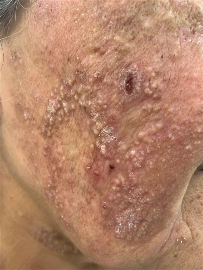
  • Both types have a similar presentation with follicular papules and plaques with a predilection for the face and scalp, marked pruritus is common, and other features can include alopecia and acneiform lesions. The folliculotropic type may be characterised by a more generalised chronic form in a slightly older age group, with larger and more numerous plaques on the extremities, trunk and face
  • Folliculotropic MF has a worse prognosis than other forms of MF, with disease specific survival rates of 81% at five years and 36% at ten years 

Pagetoid reticulosis
  • A rare, localised, solitary variant of cutaneous T-cell lymphoma 
  • Affects younger adults
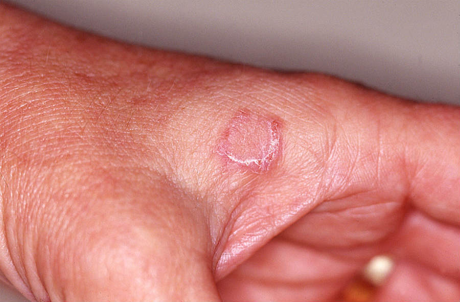
  • It presents as an isolated, persistent, scaly plaque often affecting acral sites 
  • The lesion is asymptomatic and slowly expands, but no further plaques develop

Sézary syndrome
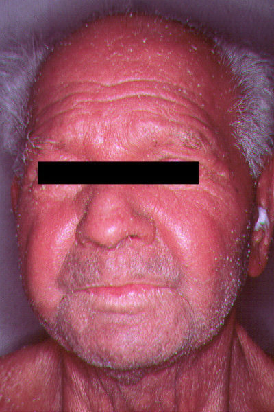
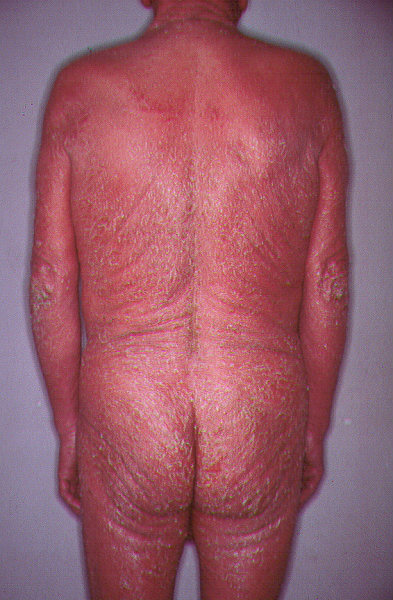
  • Defined by the triad of erythroderma, peripheral lymphadenopathy and atypical mononuclear cells (Sézary cells) 
  • Most patients are elderly males
  • It can present de-novo or as a progression from classical MF 
  • There may be associated palmoplantar hyperkeratosis, gross subungual hyperkeratosis, scalp alopecia and ectropion 

Primary cutaneous CD30+ lymphoproliferative disorders 

Primary cutaneous CD 30+ lymphoproliferative disorders consist of a spectrum of conditions; lymphomatoid papulosis and primary cutaneous (anaplastic) CD30+ large cell lymphoma. It is the second most common group of cutaneous t-cell lymphomas, accounting for about 30% of all cases. CD30 antigen, originally identified as a cell surface marker of the malignant Hodgkin and Reed-Sternberg (HRS) cells in Hodgkin’s lymphoma, is found in variable amounts in different lymphomas of B-cell or T-cell derivation, and in several reactive conditions. However, strong and homogeneous CD30 expression in most neoplastic cells is restricted to fewer entities, mainly Hodgkin’s lymphoma, lymphomatoid papulosis and primary cutaneous (anaplastic) CD30+ large cell lymphoma.

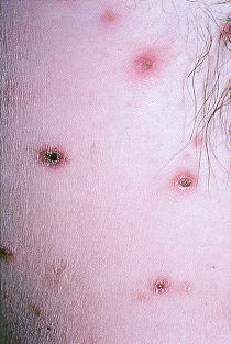
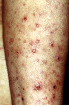
Lymphomatoid papulosis
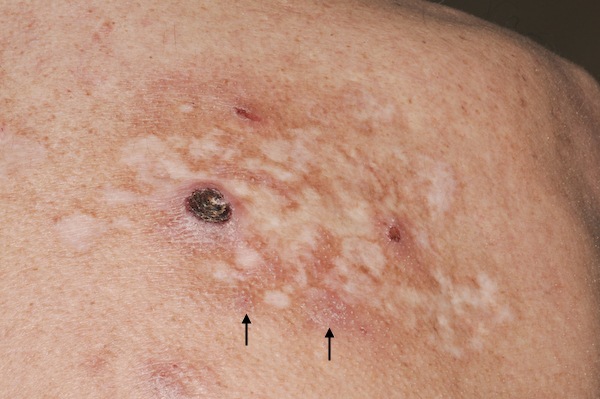
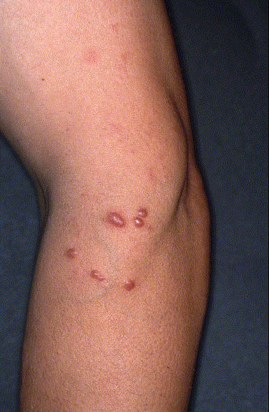
  • Presents with recurrent crops of papular, papulonecrotic or nodular lesions, generally in adult life 
  • The trunk is the most common site, although any body site can be affected
  • Lesions grow rapidly over a few days and develop ulcerated necrotic centres. Healing occurs slowly over 1-3 months, leaving scars. The cycle recurs every few months
  • Although the condition generally runs a benign course, occasionally transformation into other forms of lymphoma such as CD30+ anaplastic large cell lymphoma or Hodgkin's lymphoma occurs

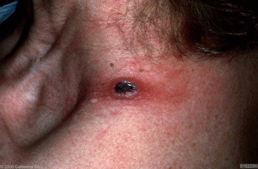
Primary cutaneous (anaplastic) CD30+ large cell lymphoma
  • Usually occurs in adults 
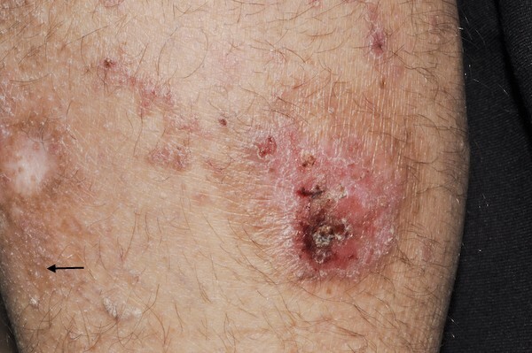
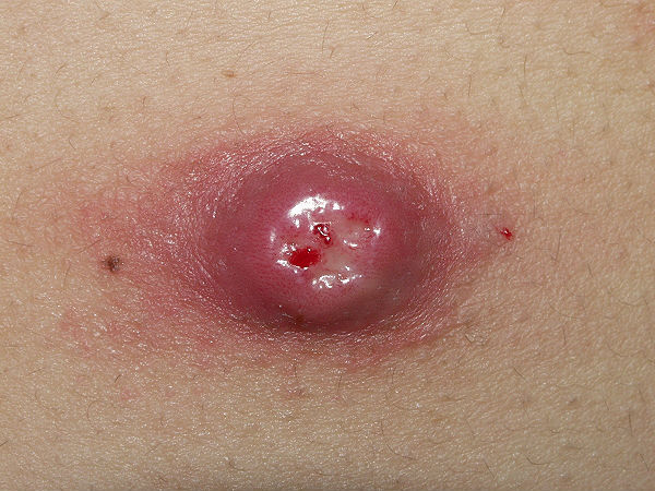
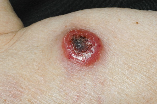
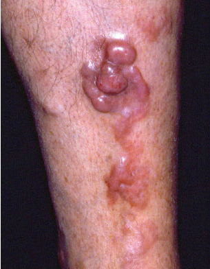
  • Lesions present as large, solitary or multiple and often ulcerated nodules, most commonly on the trunk
  • Extracutaneous involvement occurs in approximately 10% of cases
  • 5-year survival rates range from 90% for localised disease to 50% with more widespread tumour involvement 

Primary cutaneous B-cell lymphoma

The primary cutaneous B-cell lymphomas constitute approximately one-quarter of all primary cutaneous lymphomas. There are three subtypes: marginal zone, follicle centre cell and diffuse large B-cell lymphomas. Full staging investigations are essential to exclude a primary nodal lymphoma. Most primary cutaneous B-cell lymphomas, with the exception of diffuse large B-cell lymphoma, have an excellent long-term prognosis.

Marginal zone lymphoma
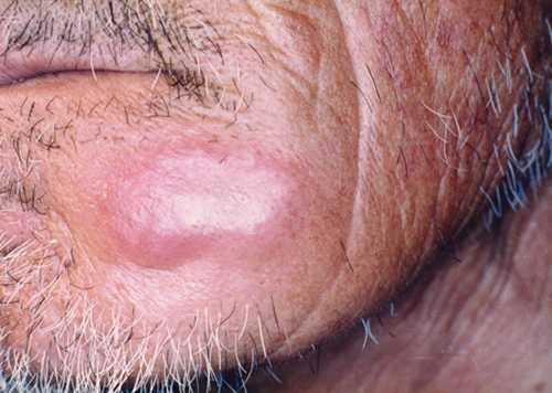
  • Present as asymptomatic, solitary or multiple dermal papules, plaques or nodules. The colour ranges from pink to purple 
  • Lesions occur on any body site, although the trunk is most often involved

Follicle centre cell lymphoma
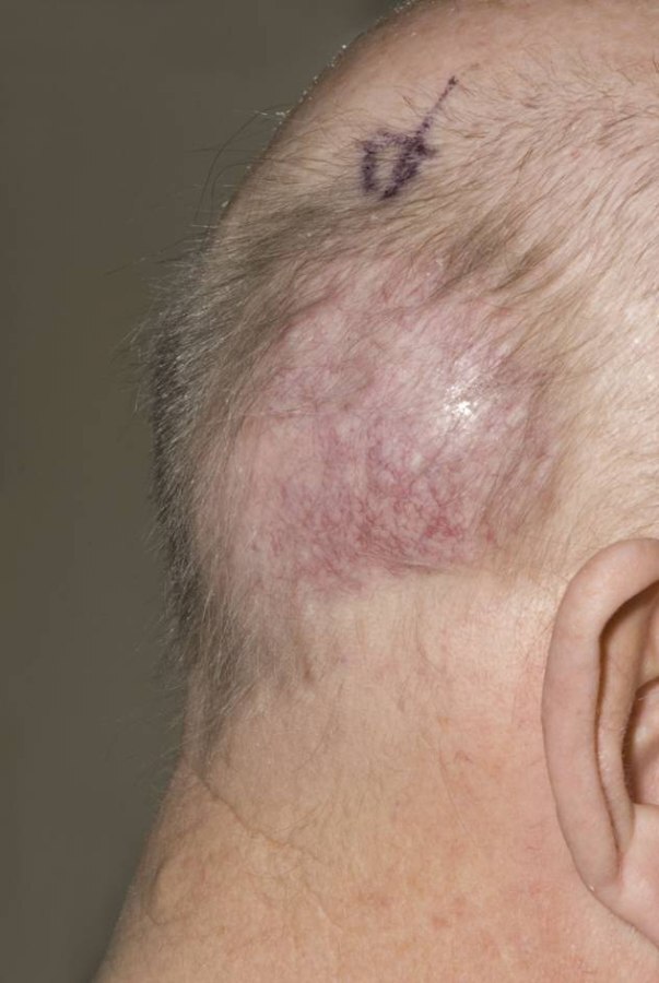
  • Present with non-specific solitary or grouped papules, nodules or tumours
  • The head and neck or trunk are the most commonly affected sites, although any body site can be involved
  • A gradual increase in the size and / or number of lesions may occur over time

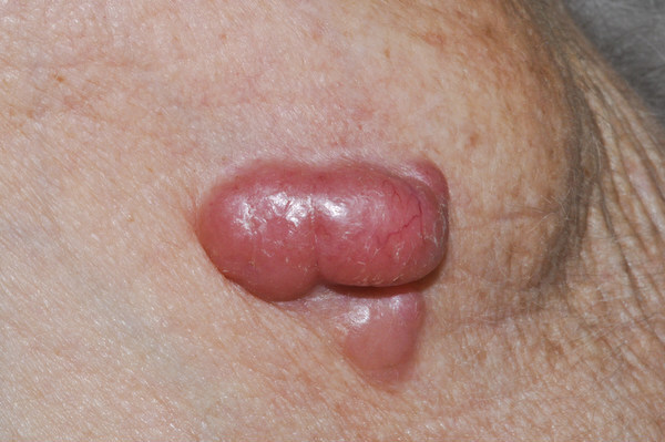
Diffuse large B-cell lymphoma
  • Rare
  • Affects older patients with a female predominance 
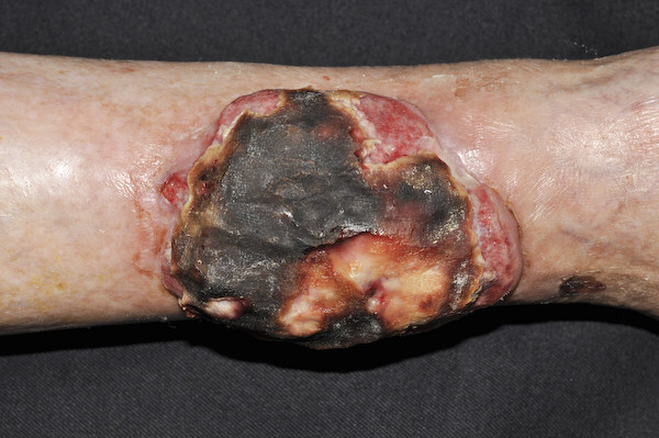
  • Lesion present as large dermal nodules or tumours, which are either solitary or multifocal and rapidly enlarging 
  • The lower limbs are the most common site
  • The 5-year survival rate is approximately 50% 

Leukaemia - cutaneous presentations 

Leukaemia can present in the skin either directly, known as leukaemia cutis, or indirectly eg purpura caused by thrombocytopenia, disseminated herpes zoster secondary to immunosuppression, or as paraneoplastic phenomena such as some cases of Sweet’s syndrome or vasculitis.

Leukaemia cutis
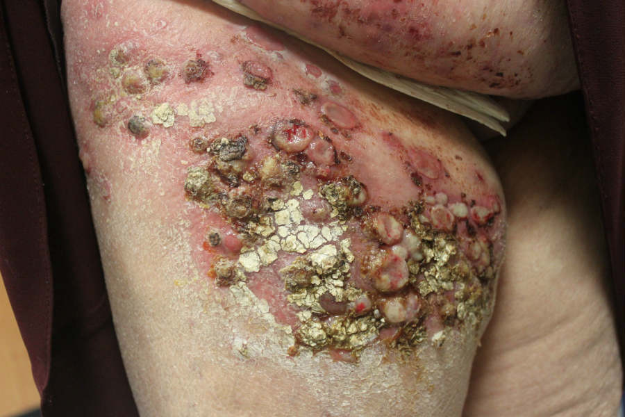
  • Leukemia cutis is the infiltration of neoplastic leukocytes or their precursors into the epidermis, the dermis, or the subcutis, resulting in clinically identifiable cutaneous lesions
  • It may follow, precede or occur concomitantly with the diagnosis of systemic leukaemia
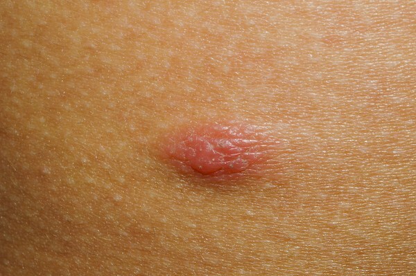
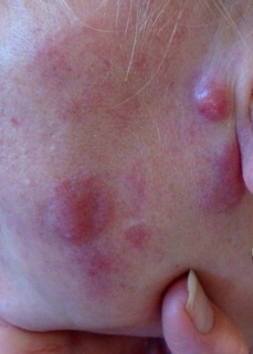
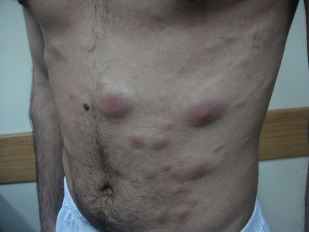
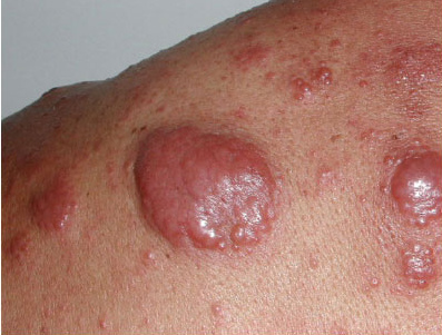
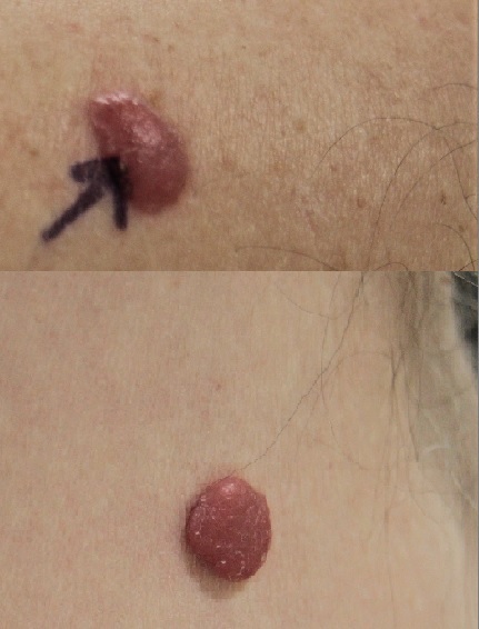
  • The infiltrate takes on a number of presentations including red to purple papules or nodules, which may be persistent or fleeting
  • Treatment is based on the management of the underlying disease 

Clinical Images

Please refer to notes on image rights at bottom of the page with regards to individual image ownership.
Management

  • Management is based in Secondary Care and may require a multi-disciplinary team approach
     
  • With specific regards to cutaneous T-cell lymphoma:
    • Both clinically and histologically the diagnosis can be difficult, and biopsies can miss the early stages of some cutaneous lymphomas such as MF. Accordingly patients presenting with unusual rashes, and not responding to topical therapy, should be referred to a dermatologist rather than doing a biopsy in Primary Care 
    • Therapy is generally seen as being palliative rather than curative, and is based on the stage of the disease. Patients with milder disease can simply be observed or treated with phototherapy. Patients with more advanced disease may require radiotherapy or chemotherapy 
       
  • With specific regards to cutaneous B-cell lymphoma further investigation is required to rule out systemic involvement 

Other resources


Disclaimer - the author PCDS cannot accept responsibility for any misleading or incorrect statements, and the management of individual patients remains the direct responsibility of the individual doctor. We do however hope that visitors to this site can contact us regarding comments that are considered misleading or incorrect so that we can continue to improve the site.

Image Rights - The PCDS would like to thank Dermatoweb, DermQuest (Galderma), and others who have contributed images. All named individuals and organisations maintain copyright for the relevant images.

Quick Links

The following pharmaceutical companies have had no involvement in the content of this website or in our conference programmes


Almirall
Galderma
Glenmark
Johnson & Johnson
La Roche-Posay
LEO Pharma
Pierre Fabre
Schuco