Mastocytosis
LAST UPDATED: Nov 18, 2021
Introduction
Mastocytosis is a term used to describe a group of conditions characterised by a local or diffuse increased growth and accumulation of mast cells in the skin and/or internal organs, especially the bone marrow and gastrointestinal tract. This chapter considers the terminologies of mastocytoma, maculopapular cutaneous mastocytosis (previously referred to as urticaria pigmentosa), and telangiectasia macularis eruptiva perstans. These conditions generally run a benign course, but rarely, those with systemic involvement can evolve into a more serious haematological disorder.
This chapter is set out as follows:
Aetiology
-
Population estimates suggest a lifetime prevalence in the region of 1:10 000 to 1:30 000 including children and adults although reliable data are not available
-
Most adults with cutaneous mastocytosis will have systemic mastocytosis, whether or not they are symptomatic
-
The World Health Organization (WHO) uses the term maculopapular cutaneous mastocytosis to cover what was previously referred to as urticaria pigmentosa, and in addition telangiectasia macularis eruptiva perstans (TMEP) - this is logical because some patients with extensive urticaria pigmentosa have a marked telangiectatic component. Although for reasons described in the clinical section, the term TMEP can still be of use
-
Progression into an aggressive form of systemic mastocytosis and mast cell leukaemia is rare, and there are no reliable markers to identify subgroups of patients at greatest risk of this
History
-
Most cases of maculopapular cutaneous mastocytosis occur in children with 75% arising during infancy and early childhood
-
The most common age of onset for adult disease is 20–40 years
-
Males and females are equally affected
-
Most of the reported cases of mastocytosis have been in white people
-
Flushing occurs in about 50% of patients
-
Not all patients with systemic involvement will be symptomatic, but in those who are symptoms can include nausea, diarrhoea, palpitations, hypotension, syncope, headache, bone pain, and rarely wheeze
Clinical findings
Mastocytoma
-
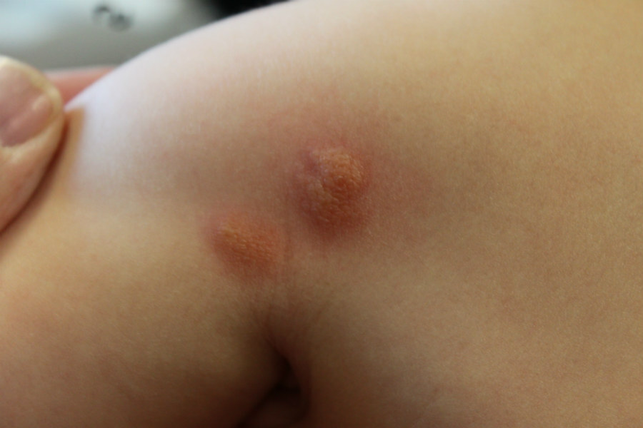
A mastocytoma is a red-pink or yellowish nodule or plaque, measuring up to 3–4 cm in diameter, that usually arises in infancy or early childhood
-
Lesions are usually solitary and tend to swell or blister if rubbed
-
Almost all mastocytomas involute over the first few years of childhood
-
Refer to the chapter Mastocytoma for more information
Maculopapular cutaneous mastocytosis (syn. urticaria pigmentosa)
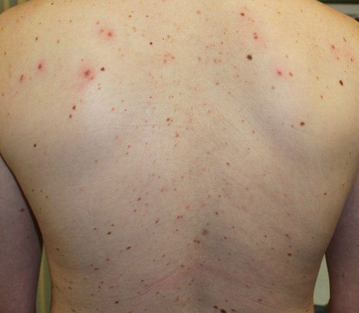
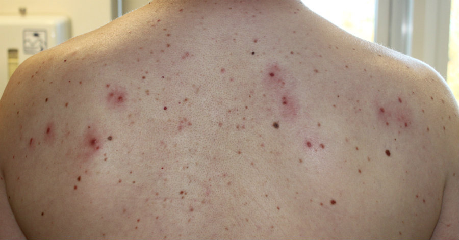
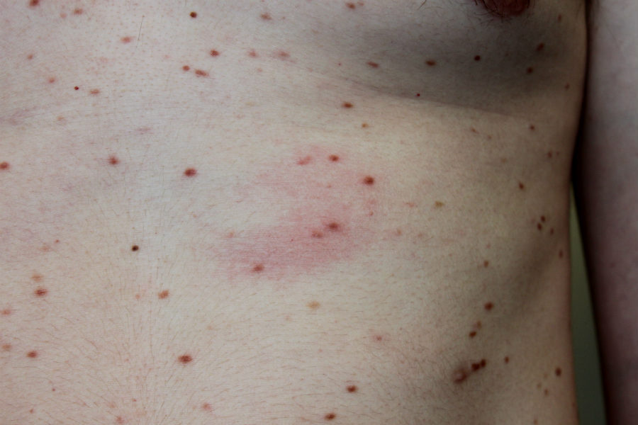
- The trunk and thighs have the highest concentration of lesions, the face, palms and soles are usually spared
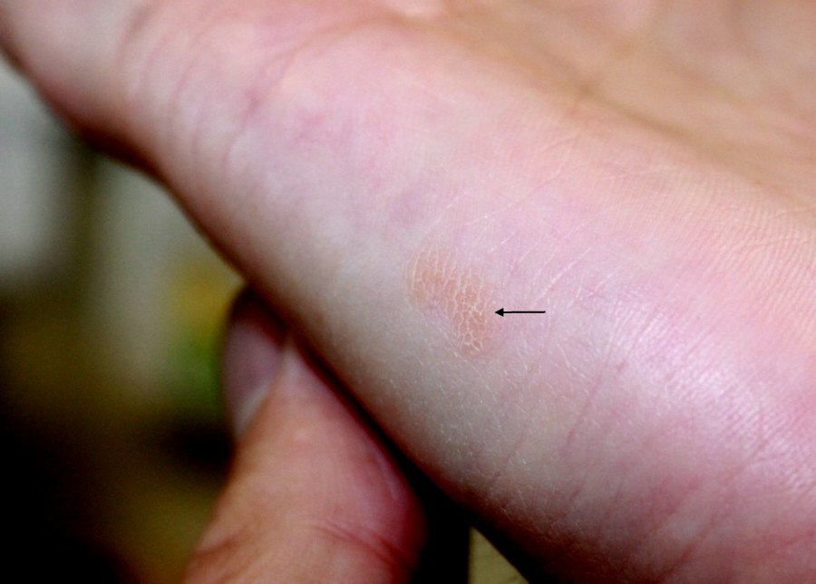
- Red-brown macule and papules, with occasional nodules or plaques
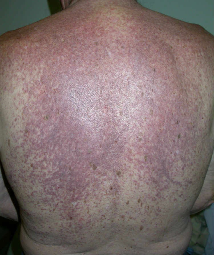
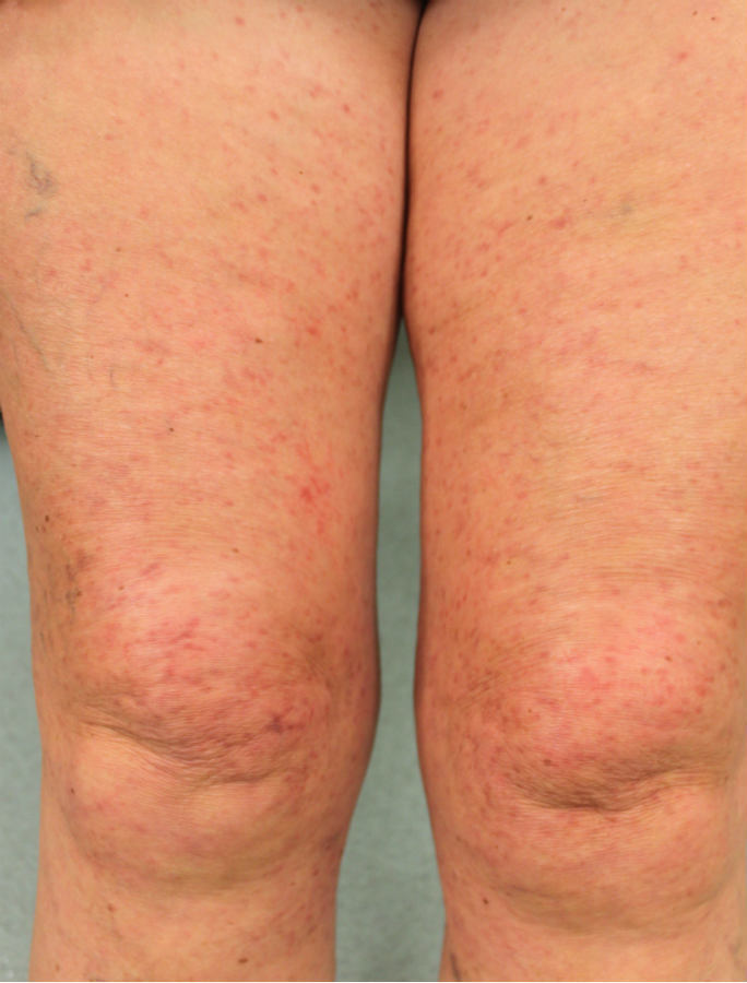
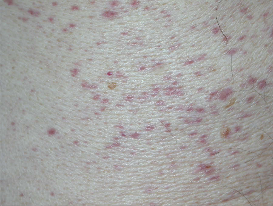
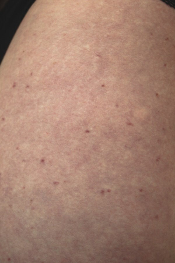
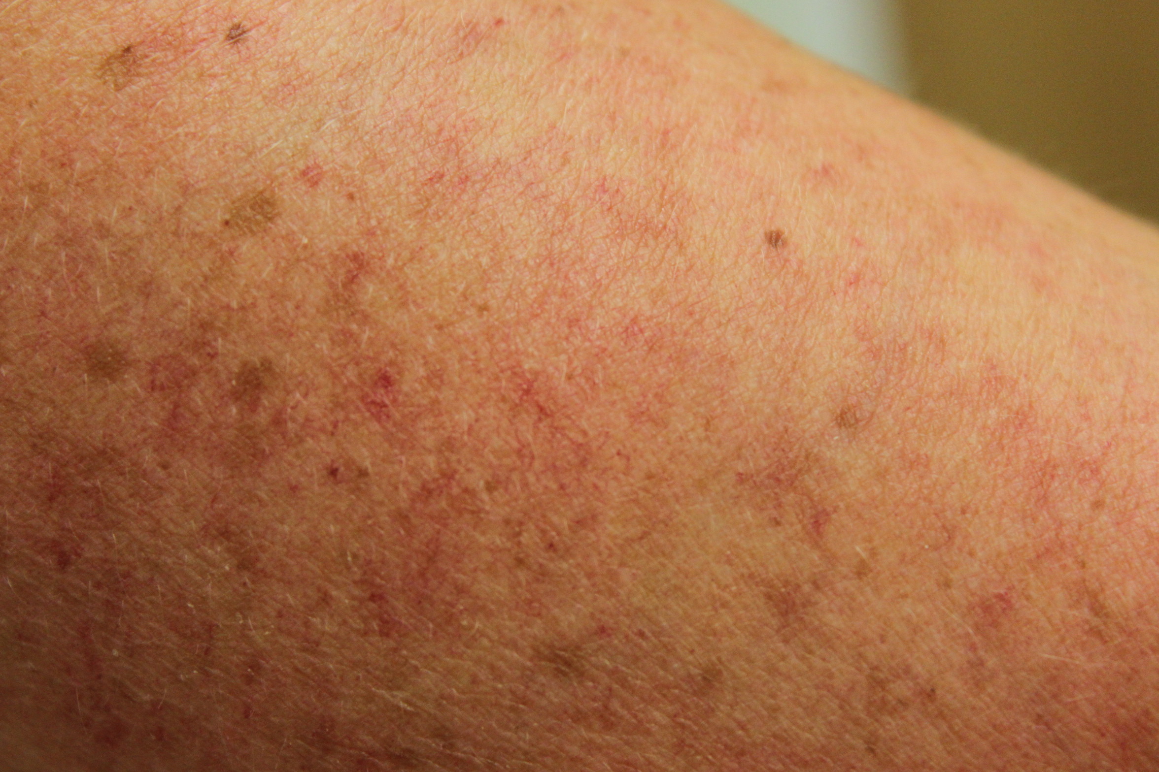
- Some patients have telangiectases
- Darier's sign - in 90% of patients lesions urticate (become red and swollen) within a few minutes of gentle rubbing
Telangiectasia macularis eruptiva perstans (TMEP)
-
As previously discussed TMEP is increasingly referred to as part of the maculopapular cutaneous mastocytosis spectrum
-
However, the term TMEP may still be useful when describing patients with extensive telangiectatic change as this group of patients usually lack any systemic involvement
Clinical Images
Please refer to notes on image rights at bottom of the page with regards to individual image
ownership.
Investigations
-
The diagnosis may be made on clinical grounds
-
Sometimes a skin biopsy is needed, although the histology is not always conclusive. At the time of biopsy the use of local anaesthetic without adrenaline, and injected around the biopsy site as opposed to into it may be more likely to yield a conclusive report. A histological diagnosis should demonstrate mast cell granules using the Giemsa stain or toluidine blue stain
Management
Management overview for maculopapular cutaneous mastocytosis
Children
-
Most patients presenting to dermatology clinics with maculopapular cutaneous mastocytosis will have an excellent prognosis, particularly children, thus it is important to provide reassurance about the nature and prognosis of the condition. A retrospective study from the Mayo Clinic of 159 adult patients showed no difference in lifetime survival between those with and those without systemic involvement
-
In many cases the rash resolves during adolescence
Adults
-
The behaviour of maculopapular cutaneous mastocytosis in adults differs to that in children, with resolution being observed in only about 10% of adults
-
The recommendation is that adults require a FBC, LFT and serum tryptase levels, and patients found to have two readings of tryptase of 20 μg/L or greater require referral to haematology for further investigations and long-term follow-up. This view is not shared by all as it may increase anxiety unnecessarily, especially given that the experiences of clinics with larger numbers of patients under review that progression to significant haematological disorders is rare, even with proven bone marrow involvement
All patients
-
Any patient that becomes unwell, eg with bone pain, weight loss, other increasing systemic symptoms, lymphadenopathy, hepatosplenomegaly, or is found to have an abnormal blood film, needs to be referred to haematology
-
It is also important to be aware that patients are at an increased risk of anaphylaxis and osteoporosis
Specific measures
-
Anaphylaxis
-
Up to 50% of adults and 20% of children experience one or more episodes of anaphylaxis so patients should be provided with Epipens ® and taught how to use them
-
Reduce exposure to triggers that cause mast -cell destabalisation such as:
-
Physical triggers (especially rubbing, heat or exertion)
-
Alcohol
-
Non‐steroidal anti‐inflammatory drugs (eg aspirin, ibuprofen, diclofenac)
-
Various general anaesthetic drugs - patient's must be made aware of this
-
Opiates
-
Insect and snake venoms
-
Other allergens eg latex
-
Medical treatment
-
Is generally disappointing
-
Phototherapy has been used to try and improve the appearance of maculopapular cutaneous mastocytosis, but for many, improvement will be minimal
-
Antihistamines, sometimes at high doses, may be used for itch, but again often lack efficacy
Other resources
Disclaimer - the author PCDS cannot accept responsibility for any misleading or incorrect statements, and the management of individual patients remains the direct responsibility of the individual doctor. We do however hope that visitors to this site can contact us regarding comments that are considered misleading or incorrect so that we can continue to improve the site.
Image Rights - The PCDS would like to thank Dermatoweb, DermQuest (Galderma), and others who have contributed images. All named individuals and organisations maintain copyright for the relevant images.