Multicentric reticulohistiocytosis
LAST UPDATED: Jul 27, 2021
Introduction
A rare histiocytic proliferative disorder, first described in 1954 by Goltz and Laymon, in which joints, skin and mucous membranes are affected. Other organs may be involved and 20% of patients have an internal malignancy.
This chapter is set out as follows:
History
-
Women are affected more than men
-
Tends to affect middle-aged individuals
-
Joint involvement usually preceded skin and mucosal involvement
Clinical findings
-
Distribution
-
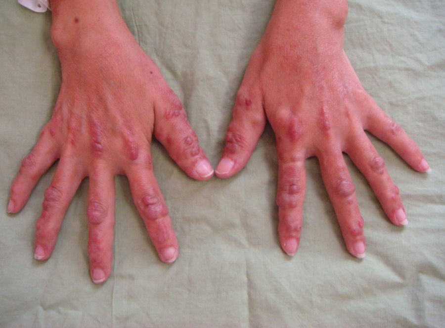
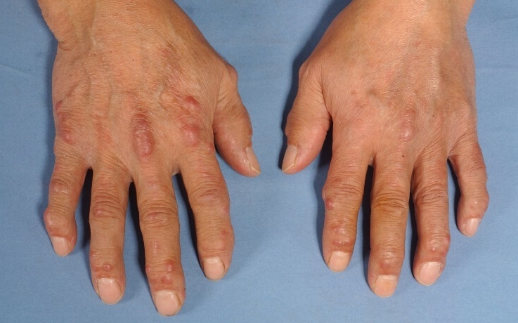
Extensor surfaces, especially of the hands and forearms
-
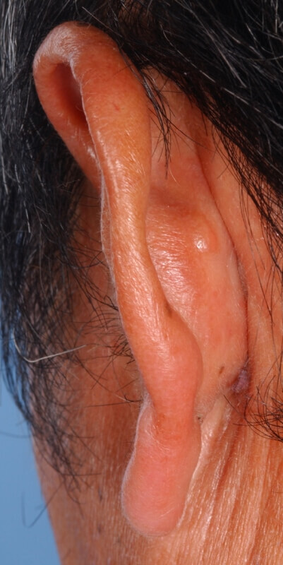
The face, ears and scalp are often affected
-
Involvement of the lower trunk and legs is rare
-
Morphology
-
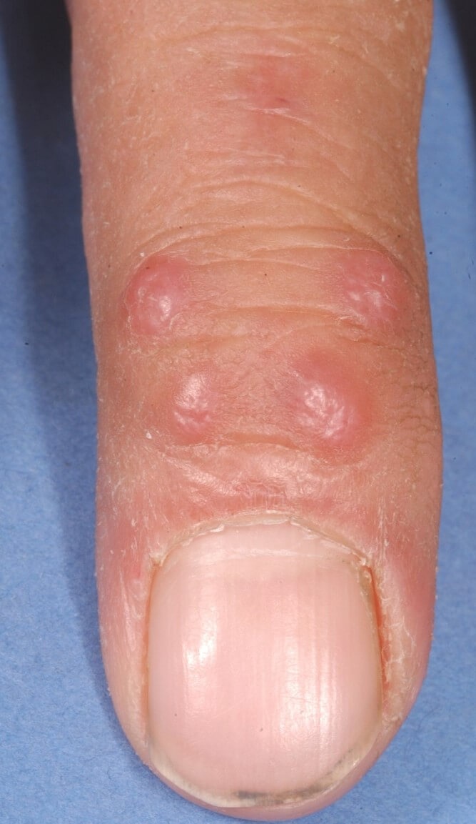
Non-tender, firm, brown-yellow papules
-
Lesions vary in size from 1-10 mm and can coalesce to form plaques with a cobblestone surface. This may give a characteristic “coral-bead” appearance, which is considered pathognomonic. The nodules grow slowly, and rarely ulcerate
-
Nail involvement can lead to dystrophic change
-
Lesions on the ears and nose may cause destruction of cartilage resulting in a disfigured appearance
-
Other clinical features
-
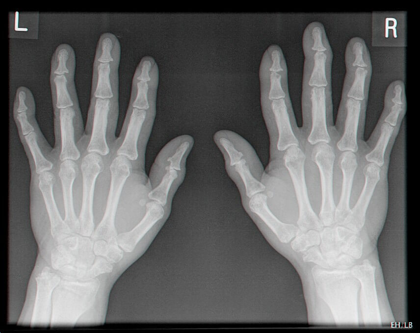
Joint involvement is characterised by a destructive and deforming symmetrical polyarthritis with a predilection for the distal interphalangeal joints
-
50% have mucosal involvement, especially the lips and tongue
-
30% have lipid abnormalities
-
25% have pruritus
-
Internal organs can be affected
-
Internal malignancy
-
Affects 20% of patients
-
The most common tumours are gastric, ovarian, breast and uterine carcinomas, myeloma, melanoma and lymphoma
Clinical Images
Please refer to notes on image rights at bottom of the page with regards to individual image
ownership.
Management
-
Because up to 20% of cases may stem from an underlying malignancy, evaluation for a possible malignancy is essential. Thorough history taking and physical examination with age-appropriate cancer screening is recommended
-
If there is no systemic malignancy the disease will often remit within 7-8 years, however, by this time considerable joint destruction may have occurred
-
Management is in Secondary Care, although no treatment appears to be of consistent value
Disclaimer - the author PCDS cannot accept responsibility for any misleading or incorrect statements, and the management of individual patients remains the direct responsibility of the individual doctor. We do however hope that visitors to this site can contact us regarding comments that are considered misleading or incorrect so that we can continue to improve the site.
Image Rights - The PCDS would like to thank Dermatoweb, DermQuest (Galderma), and others who have contributed images. All named individuals and organisations maintain copyright for the relevant images.