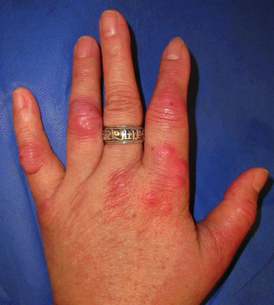
Mycobacterial infections
LAST UPDATED: Jun 08, 2021
Introduction
Tuberculosis is a mycobacterial infection that most frequently occurs due to infection with Mycobacterium tuberculosis, an acid-fast bacillus. The high prevalence of tuberculosis worldwide (one-third of the world population), its transmissible nature, and the significant morbidity and mortality associated with this infection account for the status of tuberculosis as a major public health concern.
Tuberculous infection of the skin is rare, accounting for approximately 1-2% of all reported cases of tuberculosis. This chapter, which is set out as below, refers to the pathogenic mycobacterial tuberculoses, and non-tuberculous mycobacteria (also referred to as atypical mycobacteria). Mycobacterium leprae (leprosy) is discussed in the related chapter.
Aetiology
Introduction
-
The genus Mycobacterium contains more than 100 species, most of which are harmless
-
Cutaneous tuberculosis was first documented in 1826, when Laennec reported his own "prosector's wart," a lesion that likely represented tuberculosis verrucosa cutis, a variant of tuberculosis that results from direct entry of the organism into the skin. However, the causative organism of tuberculosis was unknown until Robert Koch discovered Mycobacterium tuberculosis in 1882. Subsequently, the bacillus was detected in cutaneous lesions
-
Mycobacterium tuberculosis is a worldwide, problematic, communicable pathogen. The recent increase in incidence has been related to such factors as the association of tuberculosis with HIV, increased immigration from endemic countries, and the transmission of tuberculosis in crowded settings, such as healthcare facilities, prisons, and homeless shelters. Most often tuberculosis is an airborne transmissible disease with skin manifestations presenting as a result of haematogenous spread or direct extension from latent or active foci of infection. However, primary inoculation may occur as a direct introduction of the mycobacterium into the skin or mucosa of a susceptible individual by trauma or injury. Increased risk of acquiring disease occurs with HIV infection, intravenous drug abuse, diabetes mellitus, immunosuppressive therapy, malignancies, end-stage renal disease, and in infancy
-
Once infected, the spread of tuberculosis to other organs depends on the virulence of the organism, host resistance and other immunological factors
Mycobacterium and HIV
-
The lifetime risk of developing active infection in patients with both HIV and mycobacteria is at least 50%
-
Two-thirds of the organisms isolated are Mycobacterium avium-intracellulare, 10% are Mycobacterium tuberculosis
-
Up to 25% of HIV patients die from mycobacterial infection
-
The cutaneous features, and radiographic appearance of pulmonary tuberculosis, are often atypical, and extra-pulmonary involvement, including the skin, is frequent
History
The terminology used in cutaneous mycobacterial infection can be confusing as the diagnosis can be labelled in a number of ways. The following section below provide a practical guide and is set out as follows:
- Type of infection
- The Mycobacterium tuberculosis complex (MTBC)
- Non-tuberculous mycobacteria (syn. atypical mycobacteria; opportunistic mycobacteria; environmental mycobacteria)
- Primary or secondary cutaneous infection
- Lupus vulgaris
- Miliary tuberculosis
- The tuberculids
Type of infection
-
The Mycobacterial tuberculoses (refer underneath to the Mycobacterium tuberculosis complex) - the most common of which is Mycobacterium tuberculosis (M. Tuberculosis)
-
The non-tuberculous mycobacteria, eg Mycobacterium avium-intracellulare and Mycobacterium marinum, are also referred to as atypical mycobacteria
The Mycobacterium tuberculosis complex (MTBC)
-
Includes Mycobacterium tuberculosis, Mycobacterium bovis, Bacillus Calmette-Guérin (BCG, the vaccine strain), Mycobacterium africanum, and Mycobacterium microti
-
Transmission of infection within and between species is mainly by inhalation of airborne droplets, which results in pulmonary infection. Others routes of infection include direct inoculation of the skin by Mycobacterium tuberculosis and penetration of the GI tract by Mycobacterium bovis following consumption of infected cow's milk products
-
Mycobacterium tuberculosis
-
Is the cause of most human tuberculosis
-
The incidence is rising, with the highest incidence in Asia and sub-Saharan Africa
-
About one-third of the world's population have latent infection, with 5-10% of these patients likely to develop symptomatic tuberculosis. The risk of developing symptomatic tuberculosis is greatly increased in cases of HIV
-
In Western Europe, tuberculosis of the skin had become relatively uncommon by the late 1980s. Since then the incidence is increasing, especially in urban areas, HIV co-infection and in certain ethnic groups, especially those of the Indian subcontinent and black African origin
-
Mycobacterium bovis
-
Is the main cause of tuberculosis in cattle, deer, and other mammals
-
It is an uncommon human pathogen, accounting for approximately 1% of all isolates
-
Mycobacterium tuberculosis may have evolved from Mycobacterium bovis in the setting of animal domestication
Non-tuberculous mycobacteria (syn. atypical mycobacteria; opportunistic mycobacteria; environmental mycobacteria)
- There are at least 30 species of mycobacteria that do not cause tuberculosis or leprosy. They are often found in soil and water. Many are not pathogenic to humans, of those that are:
- Mycobacterium avium-intracellulare and Mycobacterium kansasii primarily cause lung disease similar to pulmonary tuberculosis
- Mycobacterium marinum, Mycobacterium ulcerans, Mycobacterium fortuitum and Mycobacterium chelonae cause cutaneous infections
- The Mycobacterium avium complex (MAC) is the atypical mycobacterium most commonly associated with human disease. MAC consists of two species: M. avium and M. intracellulare; because these species are difficult to differentiate, they are also collectively referred to as Mycobacterium avium-intracellulare (MAI)
- MAI is primarily a pulmonary pathogen that affects individuals who are immunocompromised (eg, from HIV, haematological disease, immunosuppressive chemotherapy)
- There are many environmental sources of MAI including soil, hot water systems, birds and farm animals
- Transmission to humans is via the respiratory and the gastrointestinal routes
Primary or secondary cutaneous infection
-
Primary infection - refers to direct inoculation of the skin usually through a wound, the types described are:
-
A tuberculous chancre
-
Warty tuberculosis (syn. verruca cutis)
-
Rarely, following BCG vaccination. Signs of infection usually develop a few months after immunisation, but have been known to occur up to three years after
- Secondary infection - refers to cutaneous infection that has resulted from spread from a primary focus, eg in the lungs, the types described are:
- A tuberculous gumma (syn. metastatic tuberculous abscess; cold abscess) describes single or multiple subcutaneous nodules or abscesses, which result from haematogenous spread, usually in immunosuppressed patients
- Lupus vulgaris arises mainly as a result of haematogenous spread in immunocompetent patients
- Miliary tuberculosis arising from haematogenous spread usually in immunosuppressed patients
- Scrofuloderma is a term used to describe a cutaneous ulcer caused by direct spread from an underlying lymph node, bone or joint
- Orofacial tuberculosis is a term reserved for tuberculous infection of mucosal and adjacent tissues, predominantly the mouth (anogenital involvement is much less common). It arises in patients with advanced systemic involvement, and is usually associated with immunosuppression
Lupus vulgaris
-
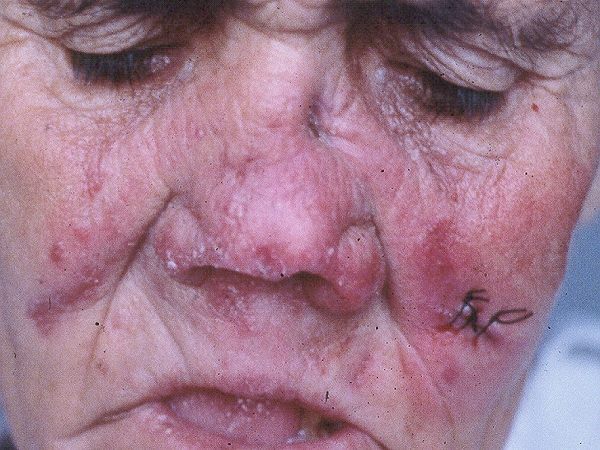
Lupus vulgaris is the most common cutaneous presentation of tuberculosis worldwide
-
It is a chronic, progressive and tissue-destructive form of cutaneous tuberculosis arising mainly in immunocompetent patients
-
The head and neck are the most commonly affected sites in European countries, whereas in India it is the buttocks and trunk
-
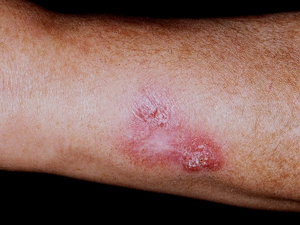
The classical lesion consists of a reddish-brown plaque with “apple-jelly” colour on diascopy, although the latter features may be absent. Lesions progress by peripheral extension and central healing with atrophy and scarring
Miliary tuberculosis
-
Is the widespread dissemination of mycobacterium tuberculosis via haematogenous spread, usually occuring in immunosuppressed patients
-
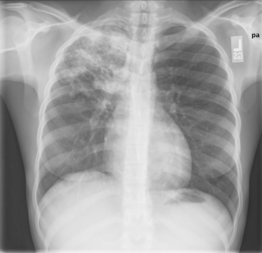
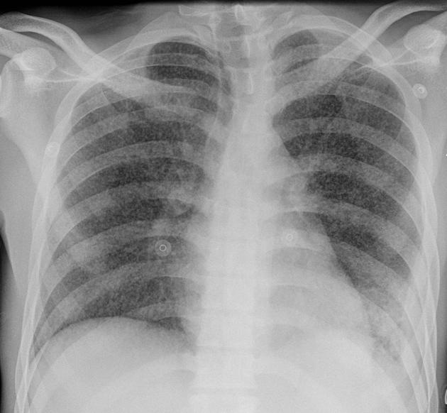
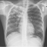
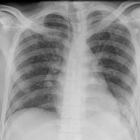
Classic miliary tuberculosis is defined as millet-like (1-5 mm) seeding of tuberculous bacilli in the lung, as evidenced on chest radiography
-
Miliary tuberculosis may occur in an individual organ (< 5%), in several organs, or throughout the entire body (>90%)
-
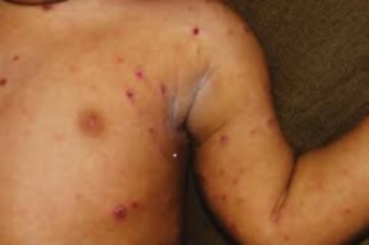
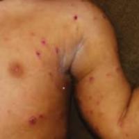
In the skin it presents with multiple papules, vesicles or haemorrhagic lesions
The tuberculids
-
These are a hypersensitivity reaction to Mycobacterium tuberculosis in immunocompetent patients
-
The bacilli are almost always absent in skin biopsies, although PCR can be positive (refer to the section on investigations in this chapter)
-
The tuberculid reactions described are lichen scrofulosorum, papulonectrotic tuberculid and a form of nodular vasculitis (syn. Erythema induratum of Bazin and Whitfield)
Clinical findings
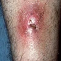
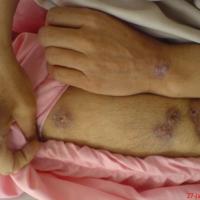
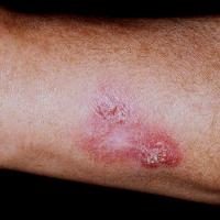
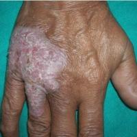
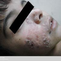
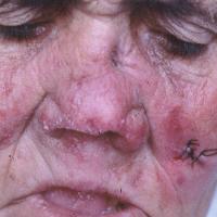
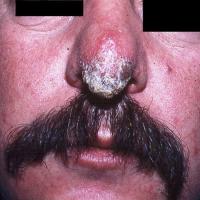
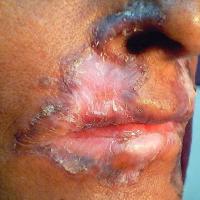
Cutaneous mycobacterial infection, most commonly caused by Mycobacterium tuberculosis, is one of the great mimickers and has many presentations. For practical purposes the presentations are grouped as follows:
- Any persistent, painless, and recalcitrant (ie poor response to treatment) plaque, nodule, abscess, or ulcer, especially in the presence of lymphadenopathy
- Involvement of the face
- Papules / pustules (with or without ulceration)
- Other pathogenic mycobacterial species
Any persistent, painless, and recalcitrant plaque, nodule, abscess, or ulcer, especially in the presence of lymphadenopathy
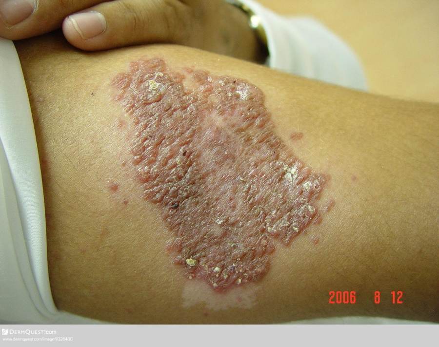
Plaques (with or without scale or verrucous change)
-
Is a secondary cutaneous tuberculous infection caused by haematogenous spread
-
Any site can be affected, although 80% of cases affect the head and neck, especially around the nose
-
The most common lesion is a red-brown plaque, which can be smooth or scaly, and may have papules / small nodules within, which show an ‘apple-jelly’ colour when pressed with a glass spatula (diascopy)
-
As plaques grow there can be central resolution / scarring such that the plaques become more annular or serpiginous
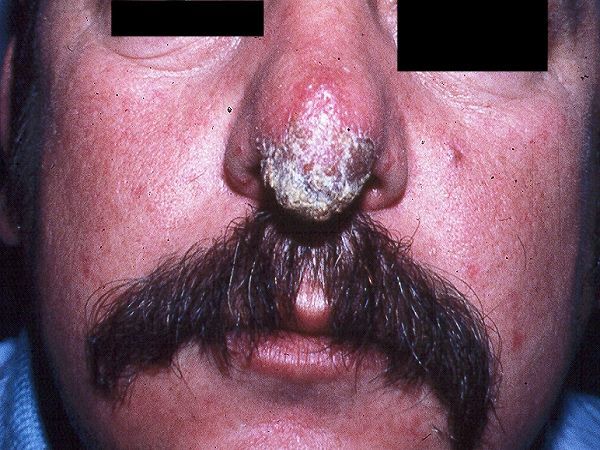
- Warty tuberculosis
-
Predominantly affects exposed sites
-
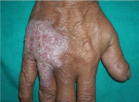
Presents a slow growing warty plaque
-
Lesions have irregular extensions at the edge
-
Central atrophic change may occur
Nodules / abscesses (with or without ulceration)
-
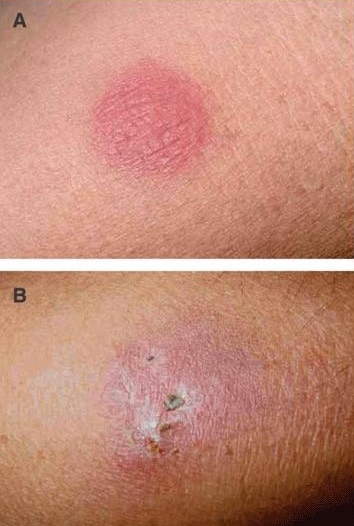
Tuberculous chancre
-
Is a primary tuberculous infection and usually follows skin trauma, with exposed sites being the most commonly affected
-
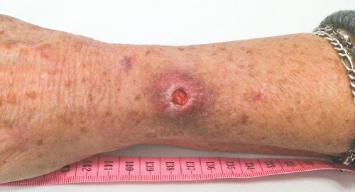
Lesions present as painless red-brown papules, nodules or ulcers with an undermined edge, crusting is common
-
Tuberculous gumma (syn. metastatic tuberculous abscess; cold abscess)
-
Arise from haematogenous spread
-
The extremities are the most commonly affected sites
-
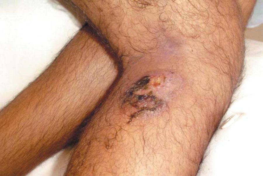
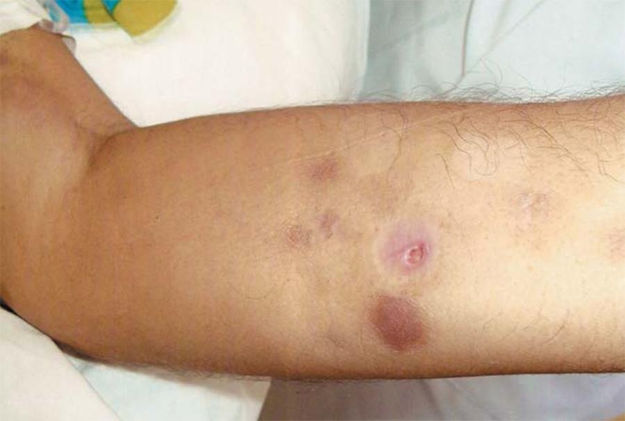
Lesions can be solitary or multiple, presenting as a subcutaneous nodule or fluctuant non-tender abscess, which may ulcerate
-
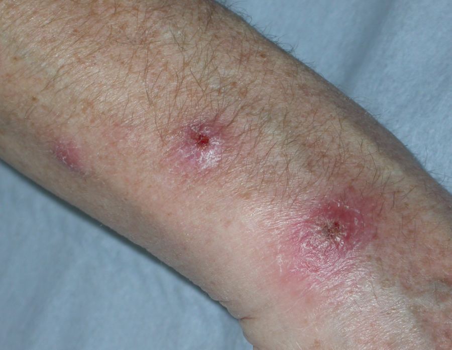
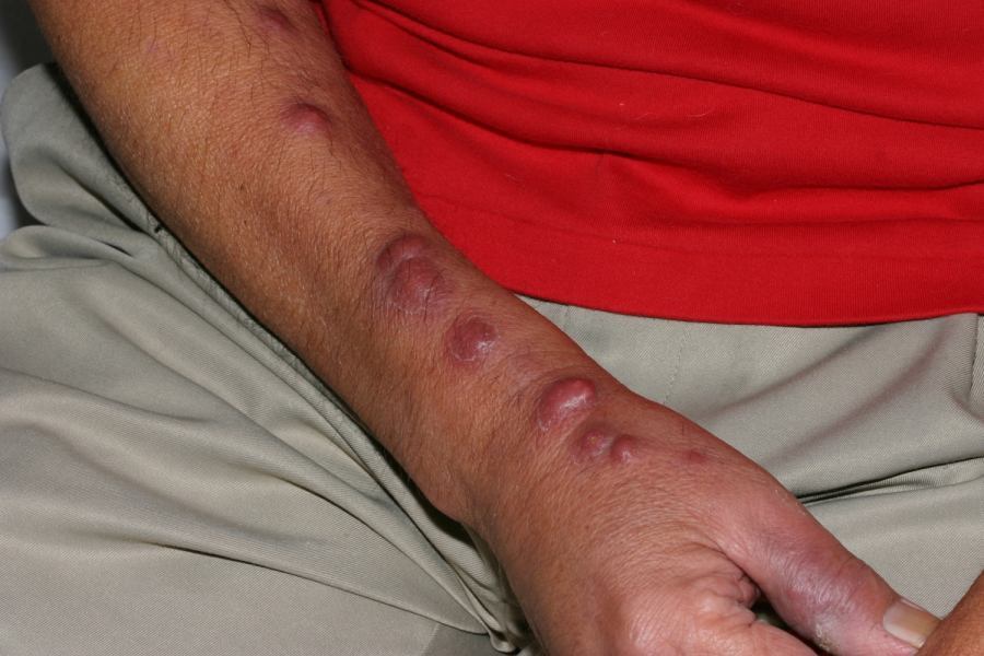
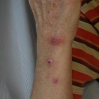
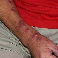
Mycobacterium marinum
-
Is a non-tuberculous mycobacteria
-
The organisms primary habitat is fresh and salt water. In fresh water mycobacterium marinum prefers warmer water, especially if not replenished, with tropical fish tanks being a frequent source of infection
-
The hands and forearms are the commonly affected sites if the infection is acquired from tropical fish tanks, the feet and lower legs are the predominant sites if the infection is acquired through swimming
-
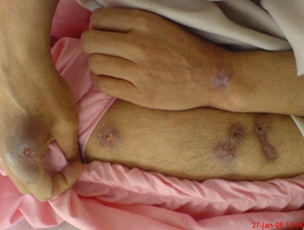
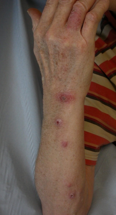
Mycobacterium marinum presents as a nodule or pustule, which may ulcerate. If untreated lesions often spread proximally
-
Unlike other mycobacterial infections, treatment is relatively straightforward. Localised infection may be treated with 3-4 months of doxycycline or clarithromycin, in more troublesome cases rifampicin may need to be added in
-
Nodular vasculitis (syn. Erythema induratum of Bazin and Whitfield)
-
Is a chronic relapsing lobular panniculitis with septal vasculitis, typically seen in healthy, middle-aged, sometimes obese women with venous stasis
-
Most cases of nodular vasculitis are idiopathic, only a minority are due to tuberculosis, which is more likely if lesions are ulcerated
-
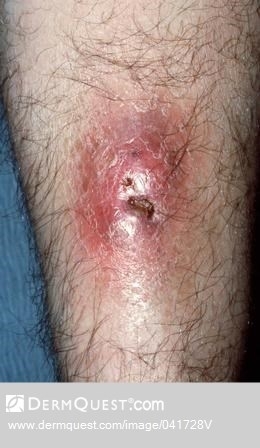
Lesions affect the posterolateral aspects of the lower third of one or both legs, presenting as tender, dusky, ill-defined nodules or plaques, which may ulcerate
-
Although this is a tuberculid reaction, PCR testing from the tissue culture of deep biopsies will often, but not always, confirm if tuberculosis is the cause
Ulcers
-
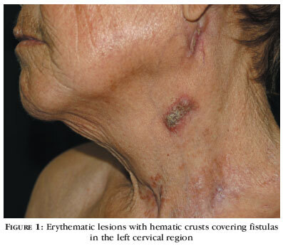
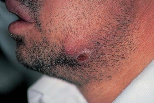
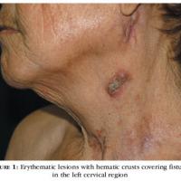
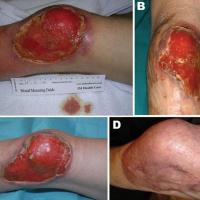
Scrofuloderma
-
Is a cutaneous tuberculous infection arising as a result of direct spread from an underlying infected lymph gland, bone or joint
-
The lesion presents as a red-blue nodule that soons breaks down to form an ulcer
-
Lesions may discharge, develop fistulae, or produce sizable amounts of granulation tissue
-
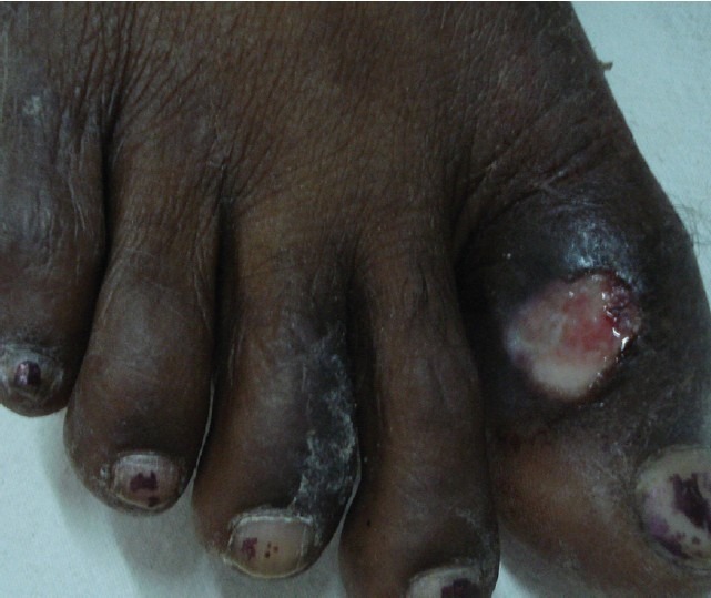
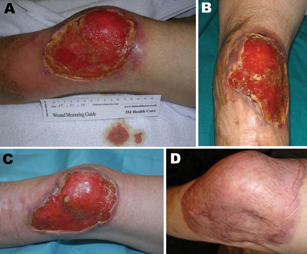
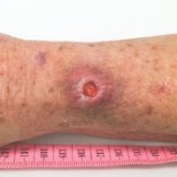
Mycobacterium ulcerans (syn. Buruli ulcer)
-
Is a non-tuberculous mycobacteria
-
Is the third most common mycobacteriosis worldwide
-
West Africa is the main endemic zone
-
Children are most frequently affected
-
The limbs are the most commonly affected sites
-
Lesions are painless, and present as nodules and plaques, which usually, but not always, develop into rapidly expanding, undermined, irregular ulcers. One of the main differential diagnosis is pyoderma gangrenosum
Involvement of the face
Lupus vulgaris - has several presentations:
-
The plaque form has already been described above
-
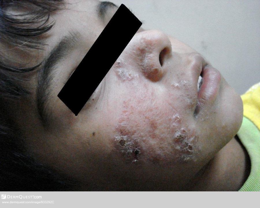
An ulcerating form on the face that scars to produce a similar appearance to that of discoid lupus erythematosus
-
Vegetating lesions that can affect mucous membranes and cartilage, and which can be very destructive if involving the nose or ears
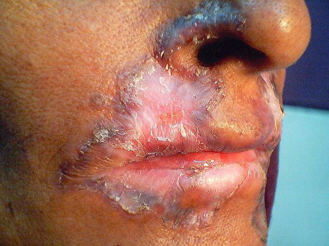
Orofacial tuberculosis
-
Arises in patients with advanced systemic involvement
-
Most commonly affects the oral mucosa / adjacent tissue
-
Multiple small red nodules develop into painful shallow ulcers
-
Crusting is common, and granulomatous enlargement of the lips may occur
-
Other sites occasionally affected include the genitalia and perianal skin
Papules / pustules (with or without ulceration)
Miliary tuberculosis
-
Results from haematogenous spread, usually in immunosuppressed patients
-
Presents with crops of red-blue papules, pustules, ulcers and occasionally nodules in an unwell patient. Lesions can be haemorrhagic
Lichen scrofulosorum
-
Is a tuberculid reaction ie a hypersensitivity reaction to Mycobacterium tuberculosis in an immunocompetent patient
-
The trunk and proximal limbs are the most commonly affected sites
-
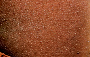
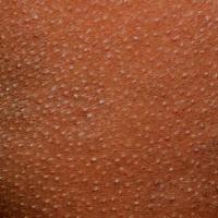
Lesions arise as asymptomatic, skin-coloured or red-brown, grouped papules. Lesions are often perifollicular
Papulonecrotic tuberculid
-
Is a tuberculid reaction
-
Predominantly affects the extensor surfaces of limbs, hands and feet, although any site can be affected, often symmetrically
-
Lesions arise as recurrent crops of crusted papules, some of which may ulcerate. Lesions heal to leave pigmented, and sometimes atrophic, scars
There are several other pathogenic mycobacterial species, and these can present in many of the ways described above, some of the more relevant infections are:
-
Mycobacterium kansasii is the second most common cause of disseminated mycobacterial infection in HIV
-
Rapidly growing mycobacterial infection eg Mycobacterium fortuitum and mycobacterium chelonae - these organisms caused localised cutaneous infections in immunocompetent patients, and disseminated infection in immunosuppressed patients
Clinical Images
Please refer to notes on image rights at bottom of the page with regards to individual image
ownership.
1
2
3
4
5
6
7
8
9
10
11
12
13
14
15
16
17
18
19
20
21
22
23
24

25
26
27
28
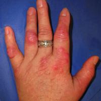
29
Investigations
Testing for latent infection
In the context of dermatology these tests are usually performed in patients with severe inflammatory dermatoses who require systemic therapies with immunosuppressive properties. It is important to check to see if the patient has latent tuberculosis before treatment is started, if they do then the latent infection needs treating first, otherwise the mycobacterial infection could be re-activated.
Interferon-γ assays
-
These measure the Interferon-γ release in antigens, which are highly specific for Mycobacterium tuberculosis but absent following BCG vaccination. The test is also of value in patients who are immunosuppressed
-
One example is the Quantiferon-TB Gold test
-
This testing is superior to that of the tuberculin skin test
The tuberculin skin test
-
Depends upon delayed-type hypersensitivity to mycobacterial antigens, it can be positive in the following circumstances:
-
Active or latent Mycobacterial tuberculosis infection
-
BCG vaccination
-
Contact with non-tuberculous mycobacteria
-
The tests also depends of a lymphocyte-induced reaction and so false negative reactions can be seen in the following circumstances:
-
Immunosuppressed patients including those with HIV, taking corticosteroids, or on immunosuppressive therapy
-
Other patients who are very unwell, including some patients with very active tuberculosis
Investigations used in the diagnosis of cutaneous mycobacteria
Skin biopsy for histopathology
- The characteristic lesion is a caseating (necrotic) granuloma, although in some cases, eg lupus vulgaris, caseation may be minimal
Acid-fast bacilli (AFB)
- Mycobacteria are rod-shaped, non-motile, aerobic organisms with a waxy coating that makes then resistant to most stains, however, the Ziehl-Neelsen acid stain is the most useful of all the various tests
- Samples used to look for AFB can be smears, the diagnostic yield of which is higher for wet or exudative lesions, or from skin biopsies
Mycobacterial culture
- Is the gold standard for determining the presence of active infection, and it can also distinguish mycobacteria subspecies and determine antibiotic susceptibility
- Culture is better from a biopsy than a swab - the sample needs to be sent in a plain white-capped universal bottle, with a small amount of normal saline or water
- However, results take 2-6 weeks, and there are high false-negative rates, in one study, culture was positive in only 23% of cases of cutaneous Mycobacterium tuberculosis
Polymerase chain reaction (PCR)
- The use of DNA amplification for detection of Mycobacterium tuberculosis and atypical mycobacterial infection provides rapid results, and significantly improve diagnostic accuracy, in many cases results can be positive even if all other tests are negative. Results can also be positive in some of the tuberculid reactions
- Testing for PCR can be from swabs of wet/exudative lesions and skin biopsies sent for tissue culture. PCR can also be tested from samples sent for histology (light microscopy), however, the process is more time consuming and may yield a higher level of false negative results
At times, mycobacterial infection can be extremely difficult to detect, as such, negative tests do not totally exclude the possibility of cutaneous mycobacterial infection
Management
-
The majority of cases of cutaneous mycobacterial infection should be referred to a respiratory physician to look for systemic involvement, and to advise on treatment
-
With regards to Mycobacterium marinum refer to the section on clinical findings
Disclaimer - the author PCDS cannot accept responsibility for any misleading or incorrect statements, and the management of individual patients remains the direct responsibility of the individual doctor. We do however hope that visitors to this site can contact us regarding comments that are considered misleading or incorrect so that we can continue to improve the site.
Image Rights - The PCDS would like to thank Dermatoweb, DermQuest (Galderma), and others who have contributed images. All named individuals and organisations maintain copyright for the relevant images.