Porokeratosis

LAST UPDATED: Dec 16, 2022
Introduction

The term porokeratosis refers to skin lesions with a thinned centre surrounded by a ridge-like border called the cornoid lamella. They arise from clones of cells with varying degrees of dysplasia. There are a number of different types of porokeratoses, those best defined are described below.

This chapter is set out as follows:


Clinical findings

The distribution depends on the type of porokeratosis 

  • Isolated lesions (syn. porokeratosis of Mibelli)
    • ​Familial lesions are autosomal dominant in inheritance and arise in childhood, sporadic lesions have a later onset
    • Lesions are single or a few in number and can be large
    • They are most common on the limbs, especially acral, but can affect any body site
  • Linear porokeratosis
    • Usually presents at birth or in childhood
    • Multiple lesions arise in a linear fashion along the lines of Blaschko, most commonly along a limb or one side of the trunk or head and neck 
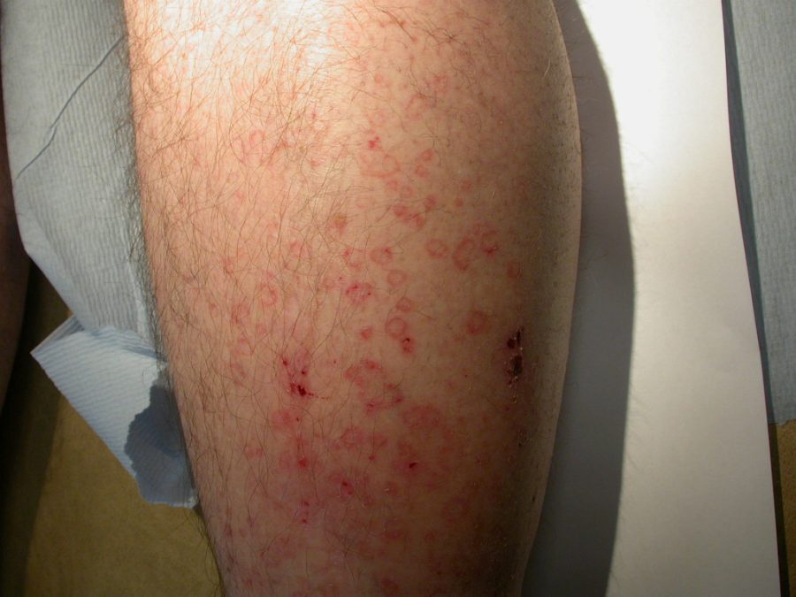
  •  Disseminated superficial actinic porokeratosis (DSAP)
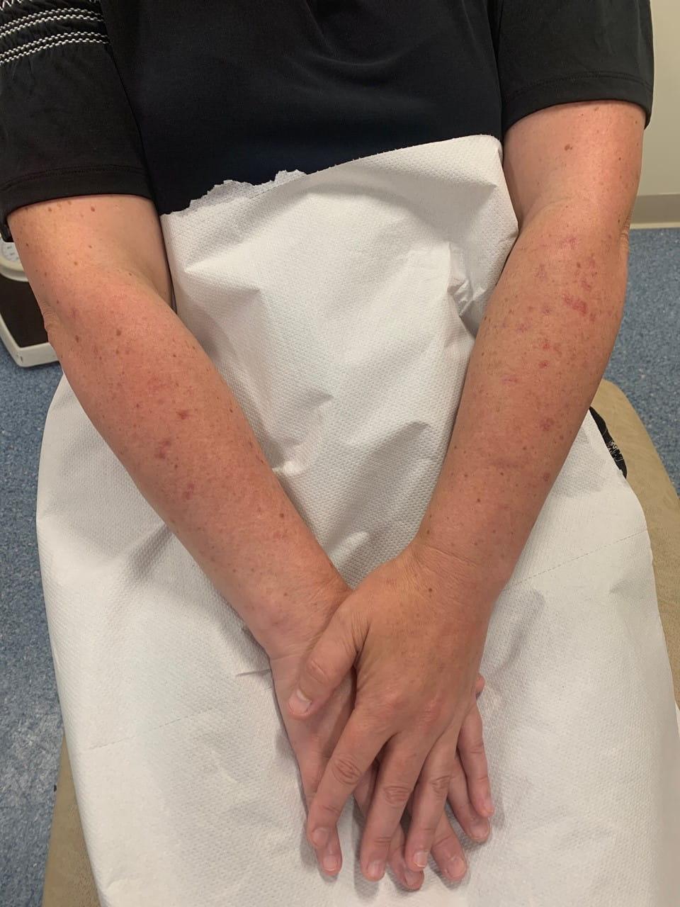
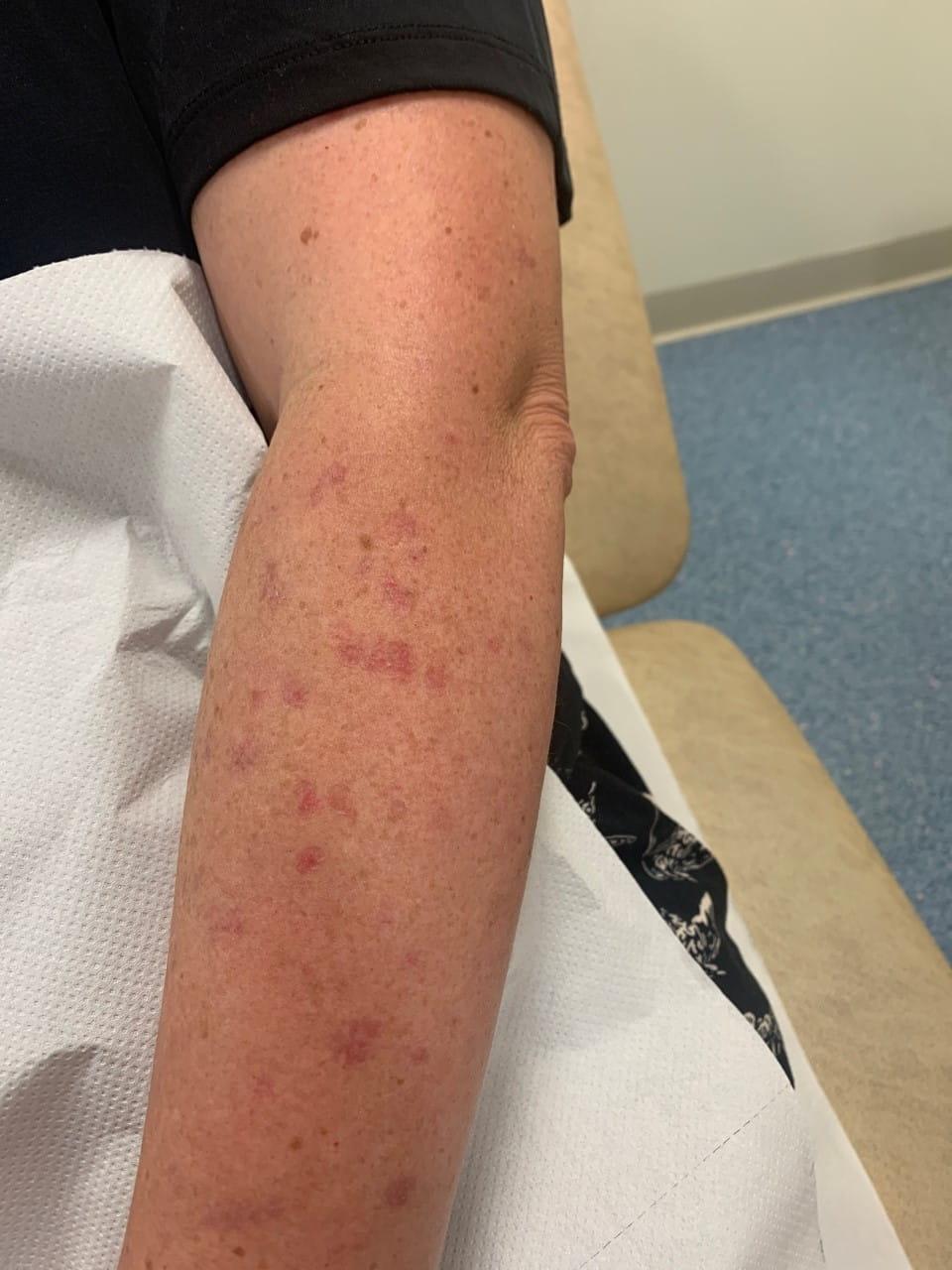
    • This is the most common variety and arises in middle-aged individuals
    • It is an autosomal dominant condition, but a history of chronic UV exposure also appears to be needed
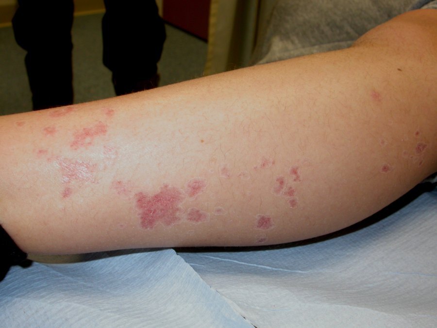
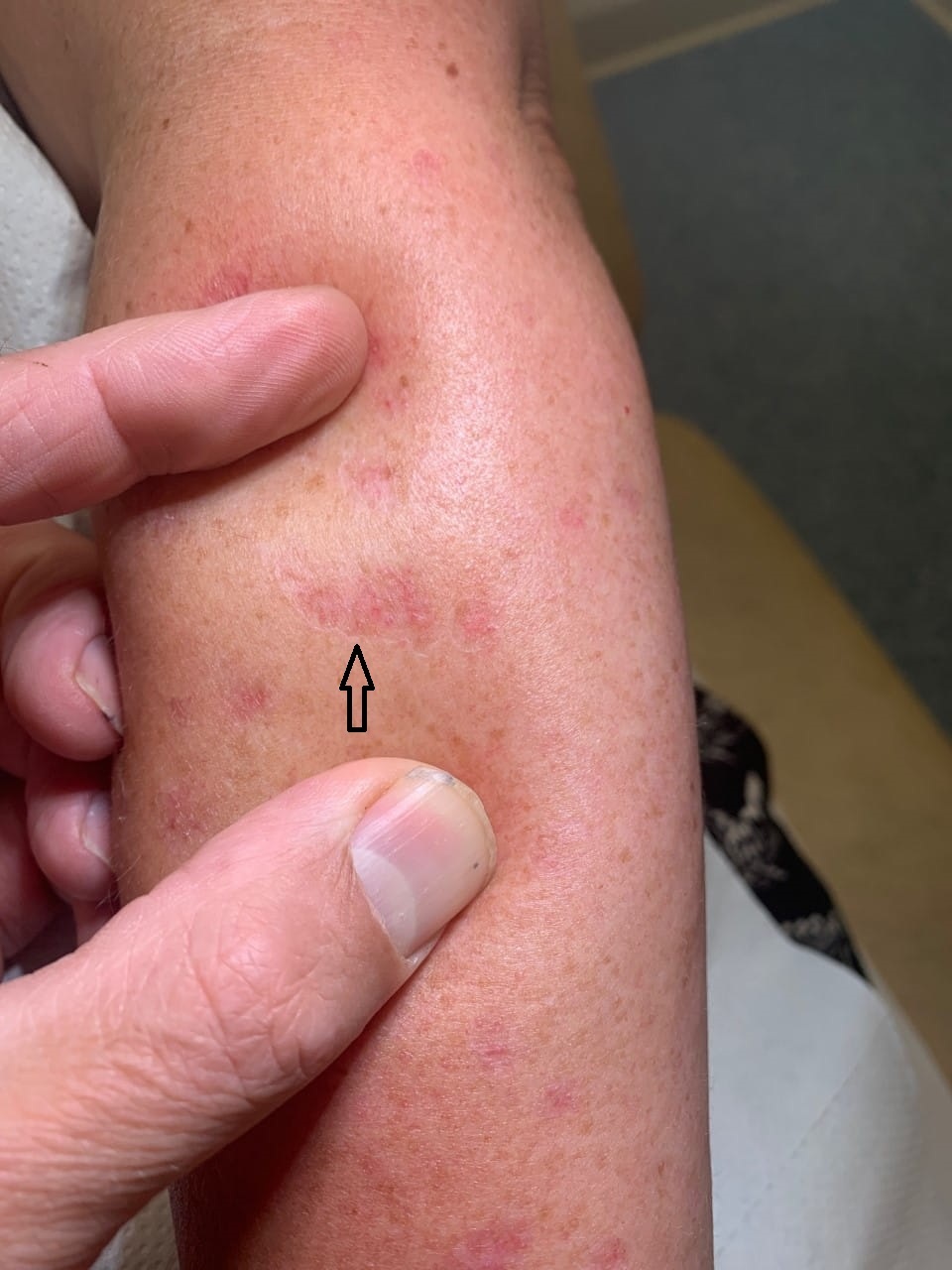
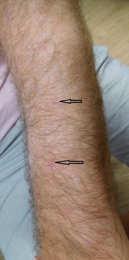
    • Lesions are predominantly found on sun-exposed areas of the legs, and less so the arms

Morphological appearance

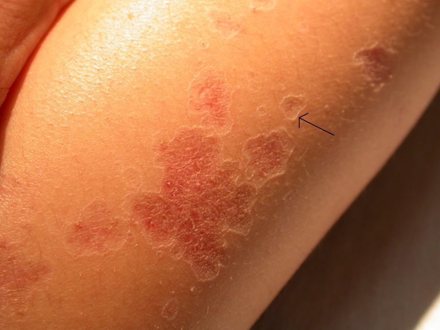
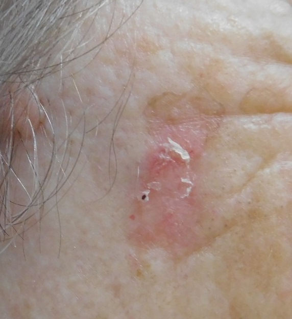
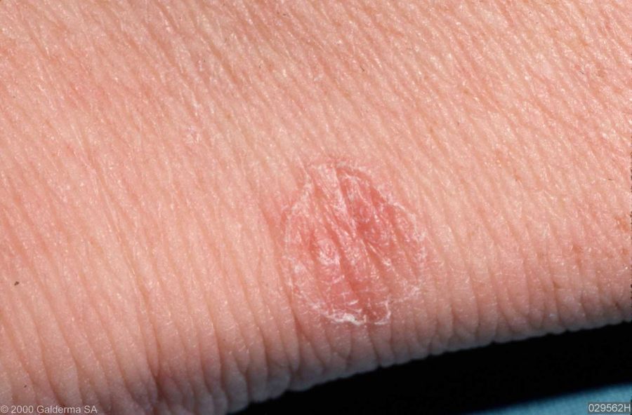
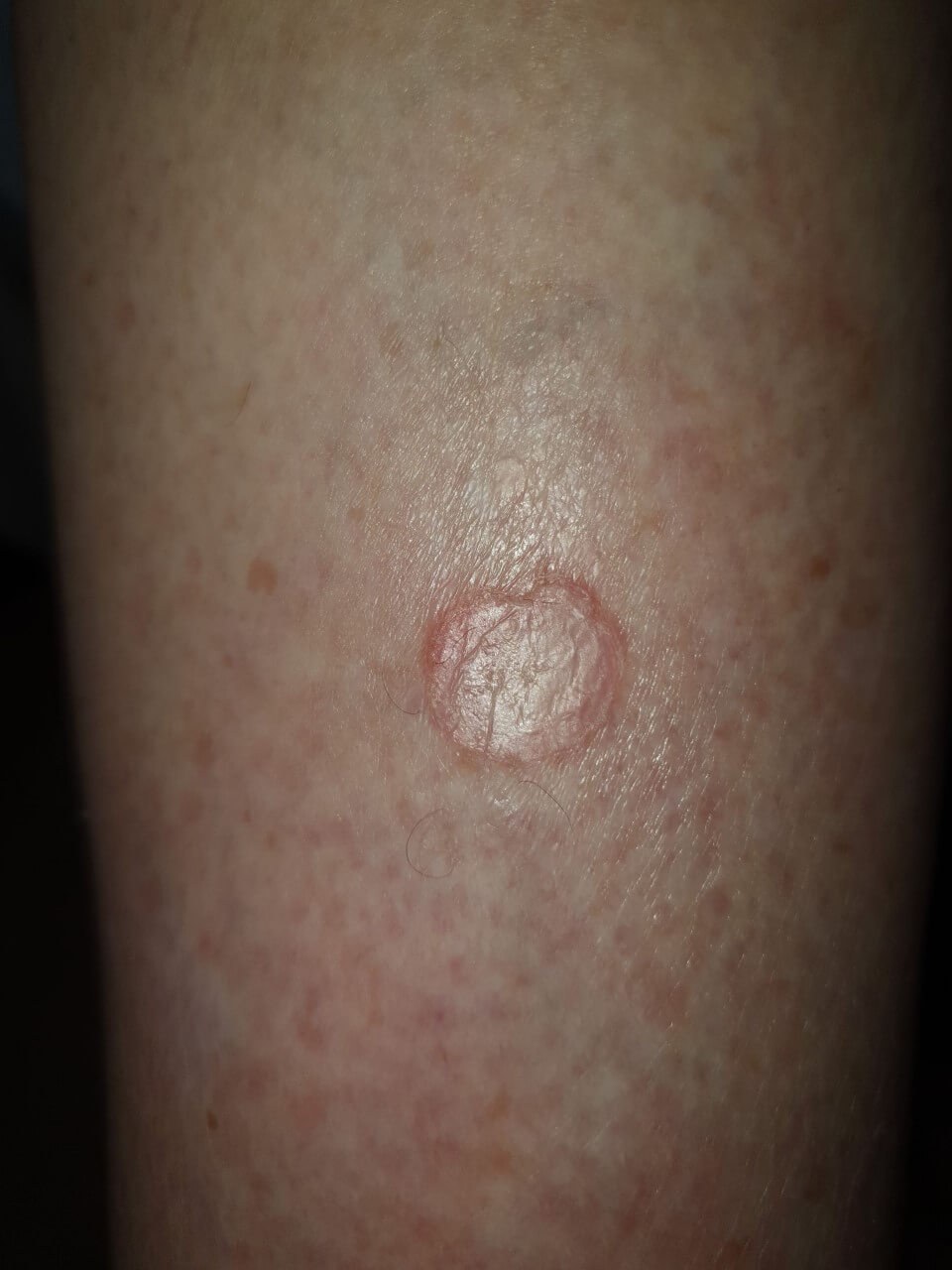
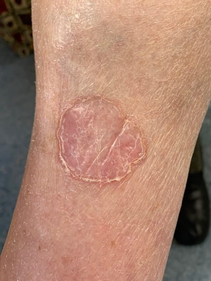
  • Lesions are annular with a well-demarcated, slightly raised keratotic rim known as a cornoid lamella. The centre may be scaly or thin and atrophic
  • They grow gradually to between 3-10 mm. Grouped lesions will appear larger. Rarely, lesions of porokeratosis of Mibelli can be very large and are termed ‘giant’
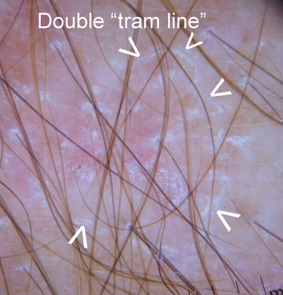
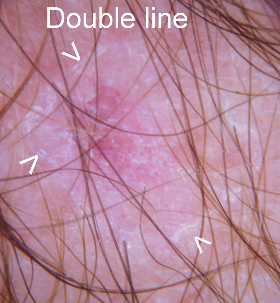
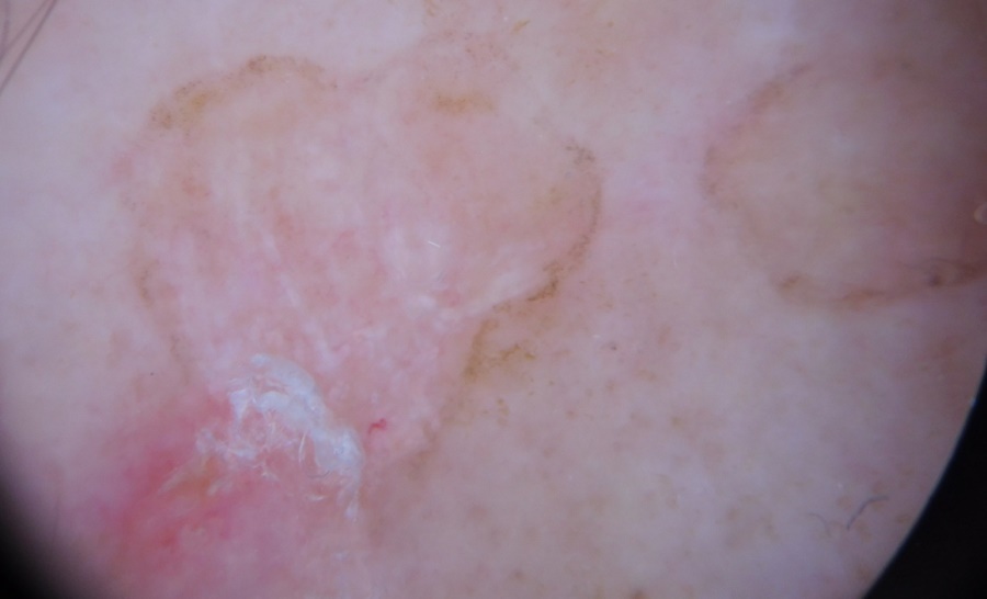
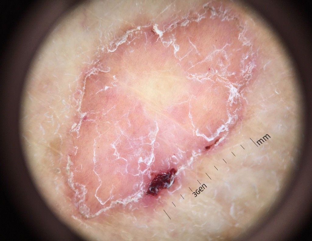
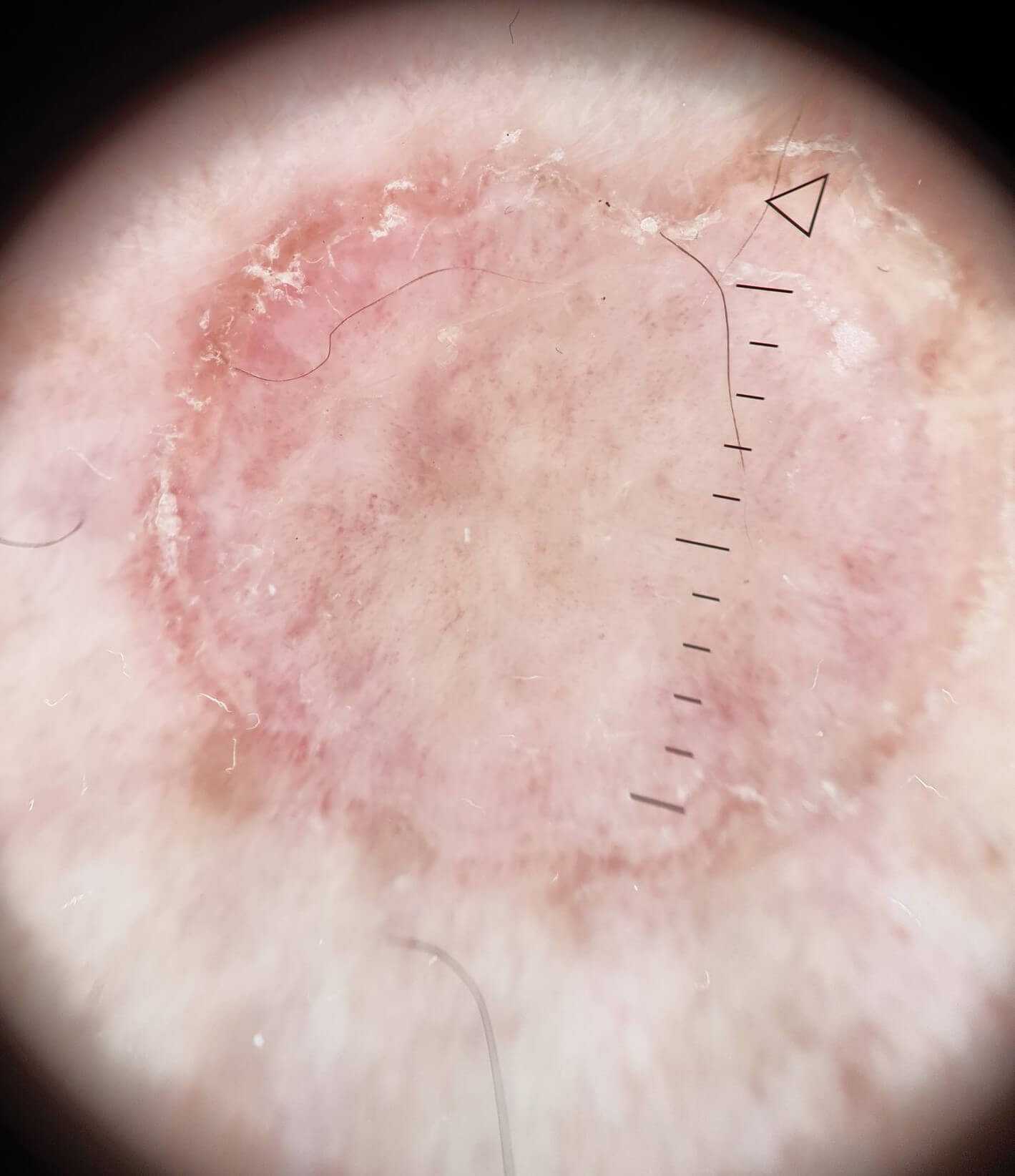
  • Dermoscopically there are parallel lines, sometimes referred to as 'tram lines' at the periphery 

Clinical Images

Please refer to notes on image rights at bottom of the page with regards to individual image ownership.
Investigations

  • The diagnosis is normally clinical, although if a diagnostic biopsy this must be taken from the scaly edge of the lesion. Histology demonstrates the presence of a cornoid lamellae - a thin column of tightly packed parakeratotic keratinocytes within a keratin‐filled invagination of the epidermis through the stratum corneum

Management

General measures 

  • Historically there appears to be no effective treatment, many have been tried. Short-term studies on a small number of patients have shown significant improvement when using a topical mixture of 2% lovastatin (or 2% simvastatin) and 2% cholesterol cream. This is a private therapy, which can be difficult to obtain
  • With regards DSAP:
    • Provide a patient information leaflet
    • UV protection - although half of the children of affected individuals will have a tendency to develop DSAP, only some will go on to develop the condition, the risk is likely to be less for those with better UV protection

Risk of malignant transformation

  • Patients with DSAP appear to have a very low risk of malignant transformation - large lesions are said to have the highest malignant potential, and malignant change has also been reported in linear forms. SCC is the most common tumour
  • Patients should be advised on UV protection and asked to report any significant change 

Disclaimer - the author PCDS cannot accept responsibility for any misleading or incorrect statements, and the management of individual patients remains the direct responsibility of the individual doctor. We do however hope that visitors to this site can contact us regarding comments that are considered misleading or incorrect so that we can continue to improve the site.

Image Rights - The PCDS would like to thank Dermatoweb, DermQuest (Galderma), and others who have contributed images. All named individuals and organisations maintain copyright for the relevant images.

Quick Links

The following pharmaceutical companies have had no involvement in the content of this website or in our conference programmes


Almirall
Galderma
Glenmark
Johnson & Johnson
La Roche-Posay
LEO Pharma
Pierre Fabre
Schuco