Pyoderma gangrenosum
LAST UPDATED: Feb 16, 2026
Introduction
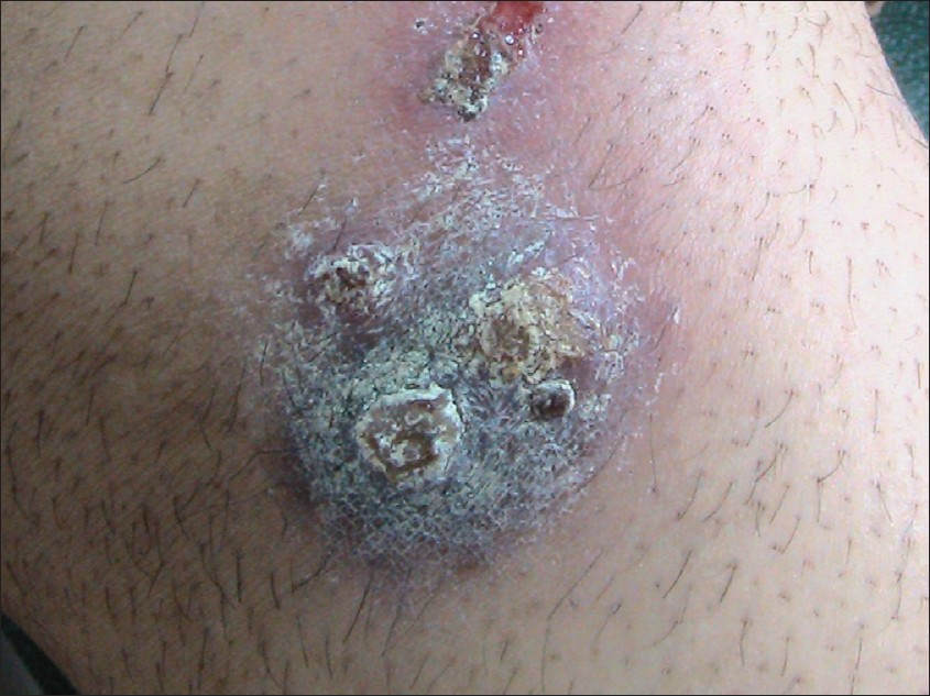
Pyoderma gangrenosum (PG) is a rare, neutrophilic dermatosis, commonly associated with systemic disease. Although there are a number of PG variants, the typical presentation is a painful ulcer (or ulcers) that grows rapidly and develops an irregular, undermined, purple edge. It is important to exclude other causes of ulcers including infection and factitious disease.
This chapter is set out as follows:
Aetiology
50-70% of cases have an underlying cause, approximately equally divided between three groups:
- Inflammatory bowel disease (IBD), especially ulcerative colitis
- Haematological conditions, especially myeloid leukaemia's. Also myelodysplasia and monoclonal gammopathy
- Inflammatory arthritis such as rheumatoid arthritis
Additional causes include chronic active hepatitis and collagen vascular disorders.
History
-
PG affects males and females equally
-
It can arise at any age, but is more common over the age of 50
-
Pain is the predominant complaint and is out of proportion to the clinical findings
-
Arthralgia and malaise are often present
-
A complete history is required to determine any underlying systemic disease
Clinical findings
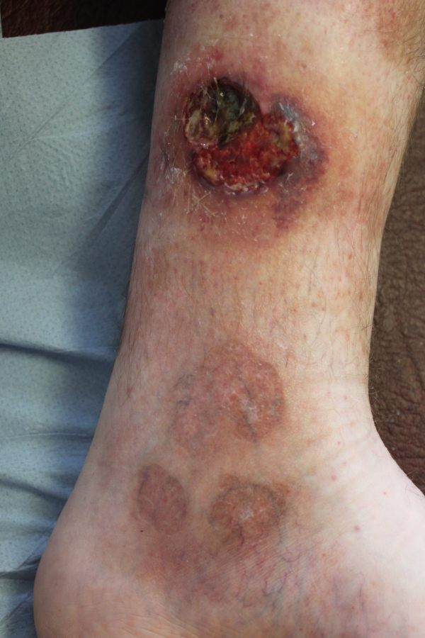
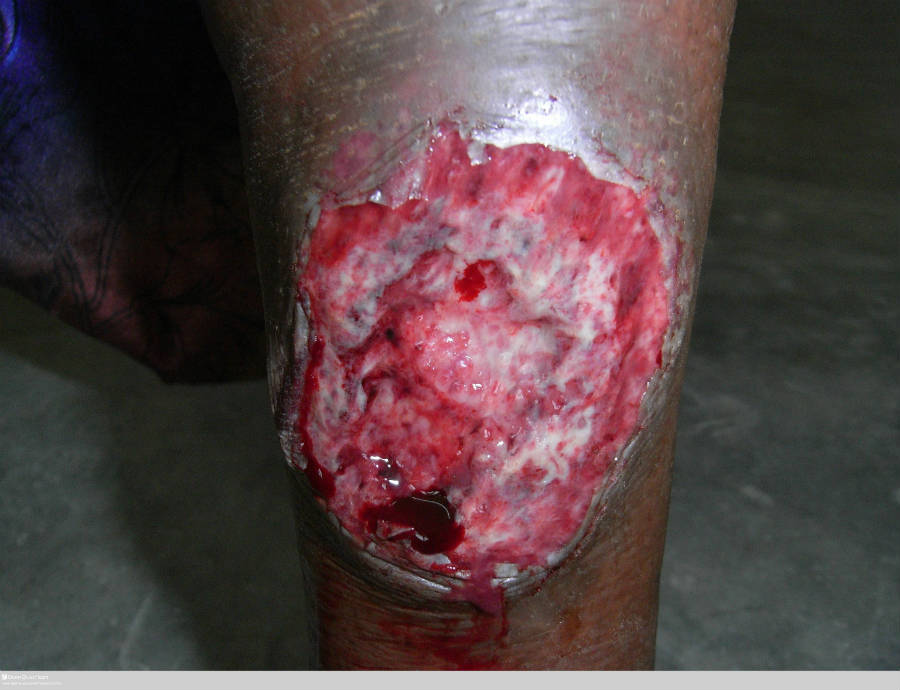
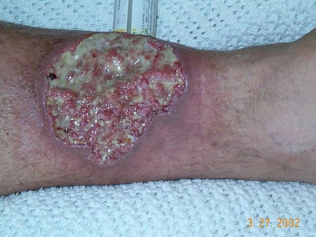
Typical features of PG
- Usually starts quite suddenly, and sometimes at the site of a minor injury
-
Although any body site can be affected, the lower legs are the most common site
-
The initial lesion may be a papule, pustule or bulla, but this soon develops in to an ulcer
-
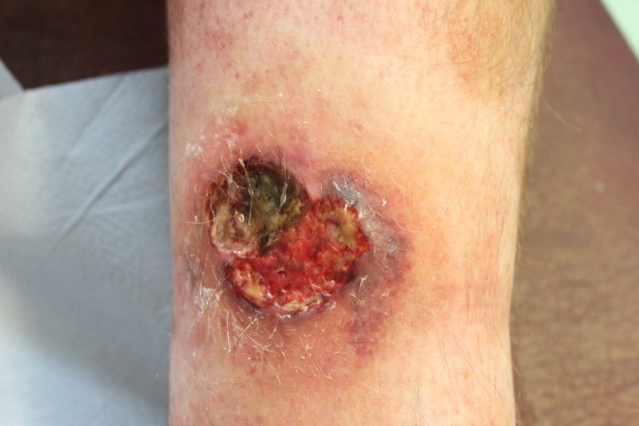
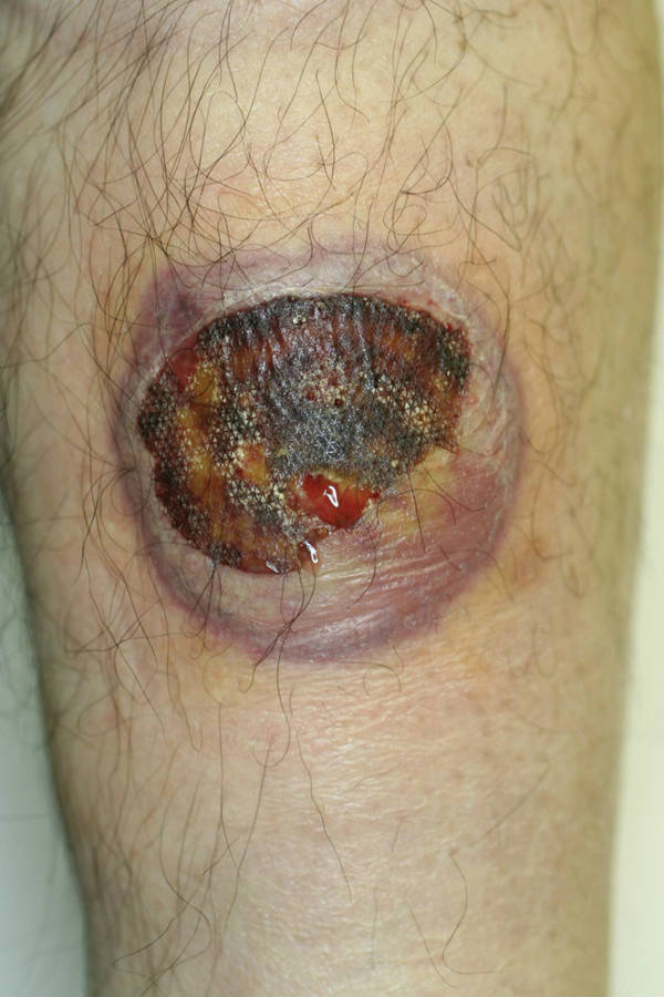
The ulcer (or ulcers) enlarges rapidly (normally > 1 cm per day), with an irregular, undermined, purple-blue edge
-
Untreated, the ulcer may continue to enlarge, persist unchanged or slowly heal. Deep ulcers heal with scarring, sometimes with a cribriform pattern
- Pathergy - patients with PG may demonstrate pathergy, in which an exaggerated skin injury (the development of a papule, pustule or ulcer) within a few days of minor trauma. This can be tested for by using a sterile needle and making pricks in the skin
PG variants
-
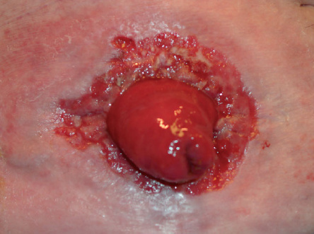
Peristomal PG
-
Comprise about 15% of all PG cases
-
They are situated close to abdominal stomas
-
Most commonly seen in patient with IBD, but can arise secondary to other conditions requiring stomas
-
Pustular PG
-
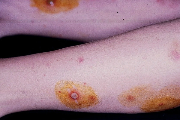
Discrete painful pustular lesions with a surrounding erythematous halo
-
Predominantly on the extensor surfaces of the limbs
-
Lesions tend to scar
-
Tends to flare during exacerbations of IBD, and resolve with control of IBD
-
Bullous PG
-
Concentric, haemorrhagic bullae, which can break down in to more superficial ulcers than in typical PG. Lesions still have the purple-blue undermined edge
-
The upper limbs and face are more common sites
-
Associated predominantly with myeloproliferative conditions
-
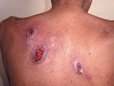
Superficial (or vegetative) PG
-
Often a solitary, slowly progressive ulcer. Less aggressive and painful than other types of PG. May lack the undermined edge
-
The trunk is the most common site
-
Not usually associated with systemic disease
Coexisting conditions
-
PG can occasionally occur along with other neutrophilic dermatoses such as Sweet's syndrome
Clinical Images
Please refer to notes on image rights at bottom of the page with regards to individual image
ownership.
Investigations
Diagnosing pyoderma gangrenosum
-
A diagnosis may be possible based on the clinical appearance, although it is important to exclude other causes of ulcers including those secondary to infection, and factitious disease
-
Skin biopsies, taken from the edge of the lesion, may be required to exclude other conditions. Samples should be sent for both for histopathology and culture, the latter to exclude infections such as unusual fungi and atypical mycobacteria
-
Although PG characteristically results in a neutrophilic infiltrate, this is not always present, and the histopathological findings are often variable and non-specific
Looking for an underlying cause
-
Blood tests include - FBC, inflammatory markers, routine biochemistry, rheumatoid factor, immunoglobulins with plasma protein electrophoresis, and cryoglobulins
- Further investigations may be required
Management
Step 1: general measures
Step 2: topical or intralesional treatments
- Super-potent topical steroids eg Dermovate ® (clobetasol), with or without 0.1% Protopic ointment (tacrolimus)
- Intralesional Adcortyl ® (triamcinolone)
Step 3: moderate to severe cases
- Prednisolone - high doses may be required initially, lower doses for maintenance
- Consider also using high-dose tetracycline's eg lymecycline 408 mg BD
Step 4: other treatments
- Colchicine 500 mg tds
- Ciclosporin appears to have a similar efficacy to prednisolone
- Sulphasalazine (with or without colitis)
- The biologic drug Infliximab ®
Disclaimer - the author PCDS cannot accept responsibility for any misleading or incorrect statements, and the management of individual patients remains the direct responsibility of the individual doctor. We do however hope that visitors to this site can contact us regarding comments that are considered misleading or incorrect so that we can continue to improve the site.
Image Rights - The PCDS would like to thank Dermatoweb, DermQuest (Galderma), and others who have contributed images. All named individuals and organisations maintain copyright for the relevant images.