Radiotherapy of the skin

LAST UPDATED: Jun 08, 2021

Acknowledgements: This chapter was written by Dr Agata Rembielak Clinical Oncology Consultant, The Christie NHS Foundation Trust, Manchester Honorary Senior Lecturer, The University of Manchester

Introduction

Radiotherapy is an important treatment modality in the management of patients with skin malignancies in definitive, adjuvant and palliative settings. The primary role of radiotherapy is to treat certain basal cell carcinomas and squamous cell carcinomas. It can also be applied in other less common cutaneous malignancies such as Merkel call carcinoma, adnexal carcinomas, cutaneous lymphomas, dermal sarcomas and to limited extent in some in-situ or benign conditions such as Bowen’s disease, keloids and cutaneous pseudolymphomas. There is limited use of primary radiotherapy in melanoma with the exception of selected cases of lentigo maligna.

This chapter is set out as follows:


History

Radiotherapy together with surgery and systemic modalities form mainstay treatments used in modern oncology. Radiotherapy utilises high energy X-rays which are a powerful form of ionizing electromagnetic field. Radiation works by damaging the DNA within the cancer cells. Cancer cells have limited mechanisms for repair compared to normal tissues but there are also limitations as to the total dose of radiation that normal tissues can tolerate. In order to allow for normal tissue repair, the total dose of radiation is usually delivered in small portions called fractions, daily from Monday to Friday.

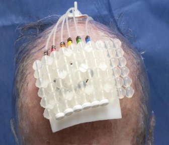
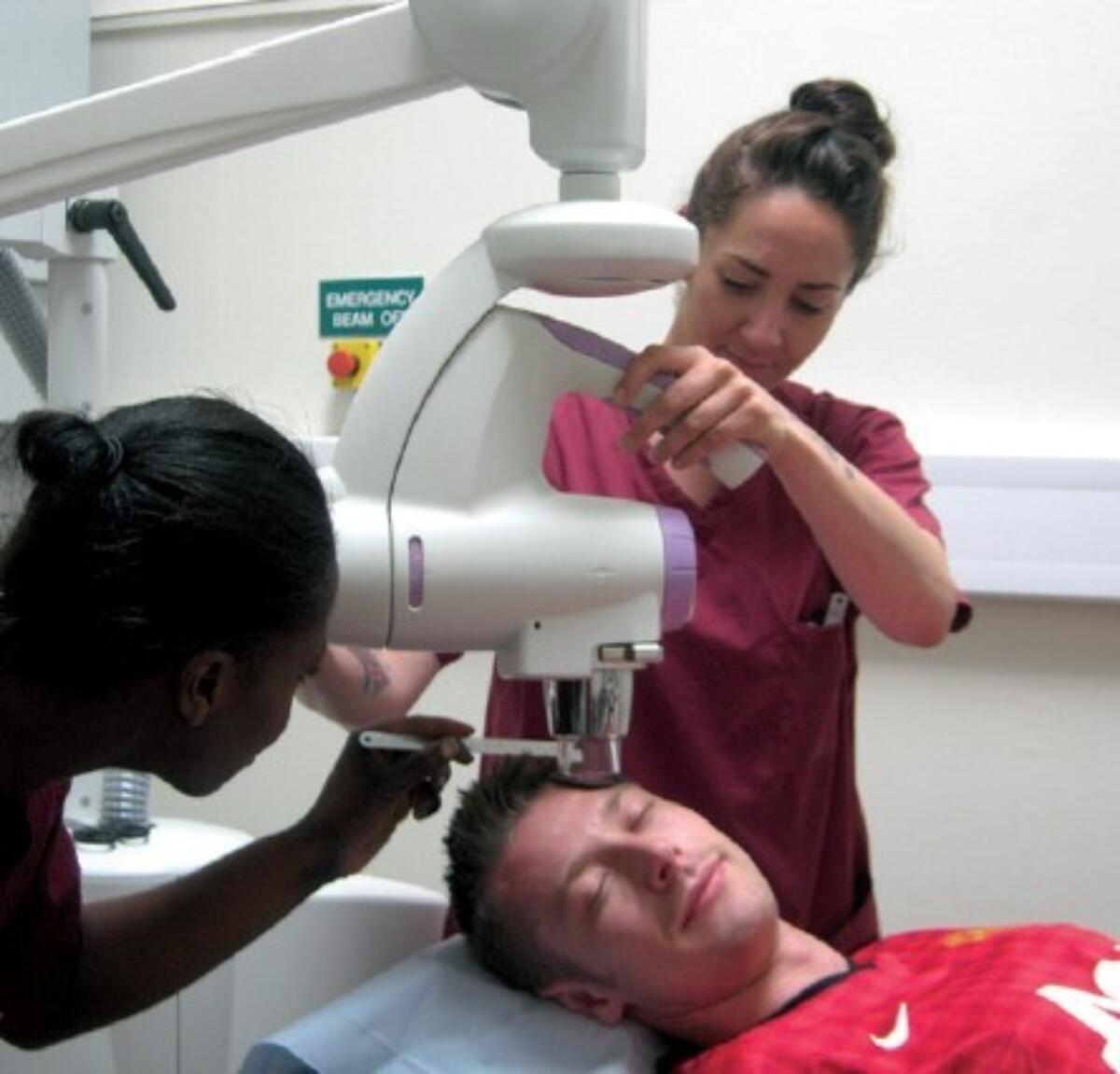
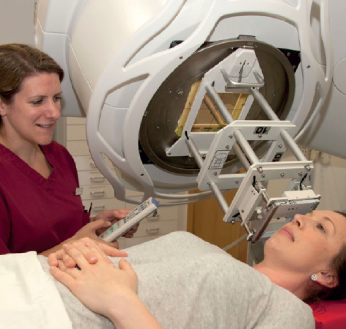
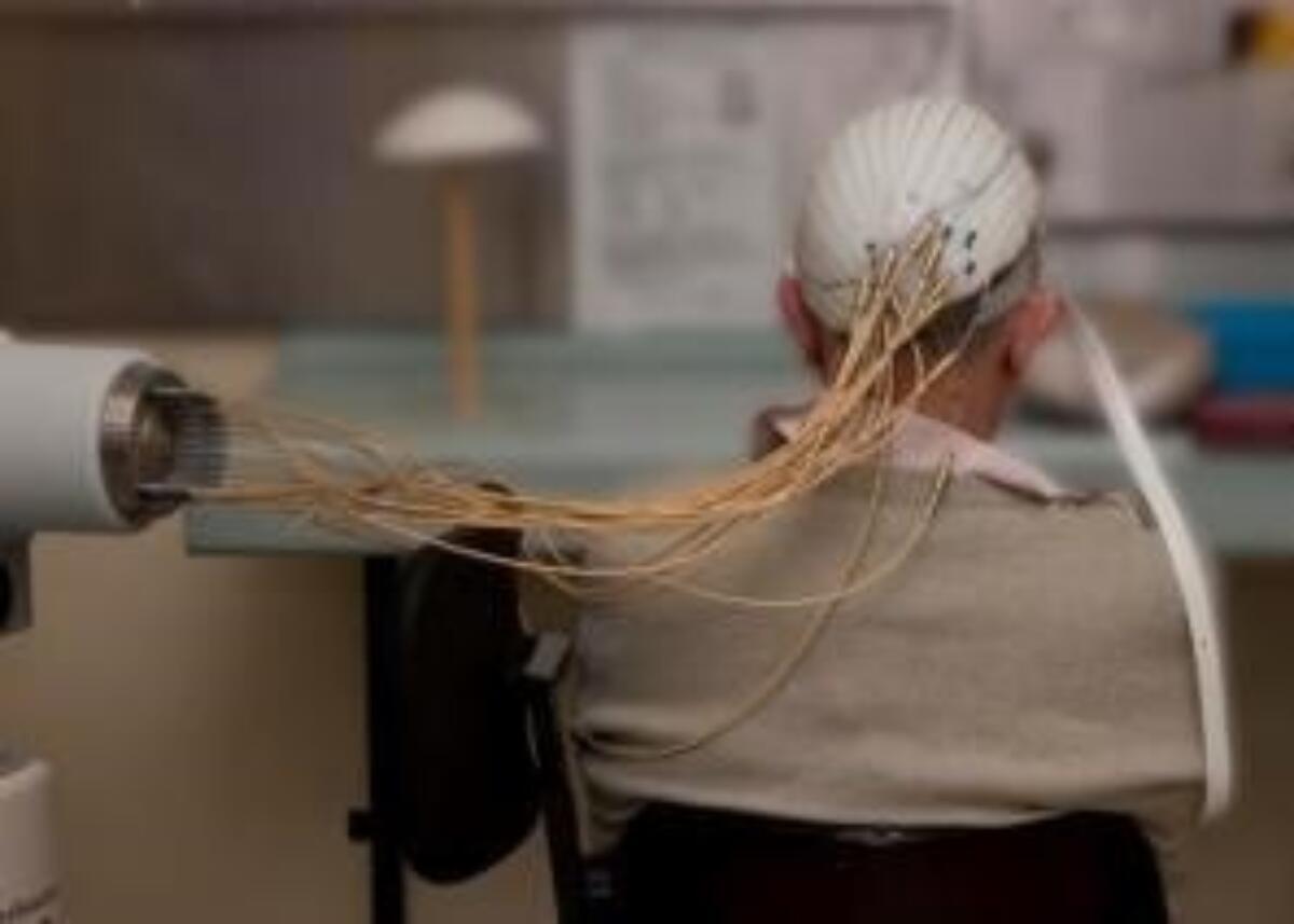
Due to the nature of ionizing radiation, this treatment must be provided in specially shielded treatment rooms, called bunkers. The patient remains in the bunker on their own while the treatment is delivered and needs to remain very still. This requirement has to be carefully considered in less compliant patients e.g.  with severe dementia or Parkinson’s disease. Depending on the type and energy of radiation, the delivery of radiotherapy is done by a superficial / orthovoltage machine or a linear accelerator called linac (Additional images 1 and 2). Both machines produce so called external beam radiotherapy where the source of radiation is placed at a certain distance from the patient. In brachytherapy, the radiation is delivered by direct application of a radioactive source to the malignancy in a specially designed plastic mould placed on the skin (Additional image 3) or through catheters inserted directly into the tumour.


Clinical findings

Skin radiotherapy can be given for the following purposes:

  • Definitive (or primary) treatment - to a skin primary and / or nodal metastases
  • Adjuvant - in postoperative settings in order to reduce risk of local recurrence, including in flaps and grafts, and in significant perineural invasion, particularly in immunocompromised patients. Ideally in SCCs postoperative radiotherapy should be commenced 2-3 months after surgery, providing the surgical site has healed
  • Palliative – to control disease and / or help with symptoms in patients with an advanced or incurable neoplasm
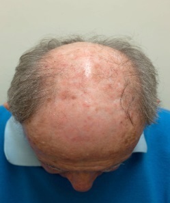
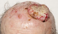
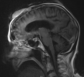
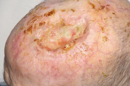
  • Examples of skin cancers treated with external beam radiotherapy and brachytherapy are figures 1-8

While the majority of skin cancers can be successfully treated with surgery or other non-surgical and non-radiation based modalities (e.g. topical treatment, PDT), there are certain situations where definitive radiotherapy may be favored. These include:

  • Elderly and / or frail and infirm patients
  • Large and / or multiple lesions
  • Cosmetically (nose, ear, lip, lower eyelid, medial canthus) or functionally (hand, feet) sensitive areas
  • Avoidance of surgical morbidity, scarring, need for reconstruction
  • Recurrent disease after surgery, incompletely excised lesions (close and involve pathological margins), significant perineural invasion
  • Patients who refuse or are unfit for surgery or are on anticoagulation

Contraindications for skin radiotherapy

There are a few medical conditions where radiotherapy is absolutely contraindicated or should be considered very carefully in the absence of any other treatment options. These include:

  • Xeroderma pigmentosum and Gorlin’s syndrome - due to risk of induction of multiple and recurrent skin cancers
  • Verrucous carcinoma - risk of further anaplastic transformation and metastases
  • Site of previous radiotherapy - increased risk of late damage to normal tissues, including soft tissue and cartilage necrosis due to cumulative effect of radiation 
  • Bone and / or cartilage invasion - such skin tumours should be considered for surgery and adjuvant radiotherapy
  • Multifocal tumours with areas of dysplastic skin
  • Young patients due to risk of late skin complication and carcinogenesis
  • Hair bearing areas
  • Upper (outer) eyelid - due to risk of damage to lacrimal glands with subsequent dry eye with all its consequences
  • Poorly vascularised areas, particularly over bones e.g. anterior shin over tibia (stretched and thin skin) in which radiotherapy is associated with poor and prolonged healing
  • Skin cancers arising in sites of chronic ulceration, trauma or thermal burns

One of the most commonly discussed and feared side effects of skin radiotherapy is radiation-induced in-field malignancy such as another cancer or soft tissue / bone sarcoma. Available data on such risks comes from radiotherapy used in various malignancies and delivered using different techniques and not specifically in skin cancers treated with radiation. The data indicate very small (1 in 1000) risk of carcinogenesis from 10 to 15 or more years after exposure and they likely overestimate the real risk. Nevertheless, younger patients, e.g. below 60, should be appropriately counselled about such risks, if radiotherapy is the preferred treatment option. For the same reason skin lesions overlying important organs such as thyroid, breast, spine or kidney should be treated surgically.


Clinical Images

Please refer to notes on image rights at bottom of the page with regards to individual image ownership.
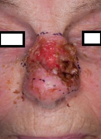
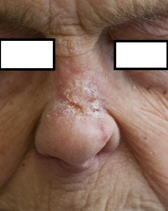
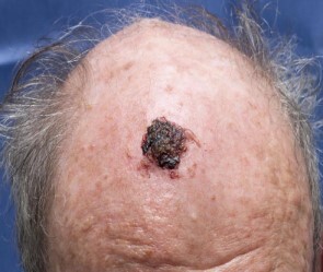
Management

Radiotherapy schedules and shielding

  • “Daily” radiotherapy is delivered from Monday to Friday but treatment can start on any day of the week, not necessary on Monday. The choice of radiotherapy schedule depends on patient and tumour related factors. In general, in definitive skin radiotherapy, for bigger skin lesions (or in postsurgical cases), the more protracted schedules are used (more fractions). For lesions below 2-3 cm in diameter 5-10 daily fractions are usually used. Small skin cancers in elderly and infirm patients may be treated with a single exposure of radiation when cosmesis is of not too much concern. Some elderly and infirm patients with larger lesions may be also treated once a week over 6-7 weeks, depending on their performance status and treatment target. Larger skin lesions are treated with 15 - 20 fractions and very large lesions with 30 daily fractions to optimise local control and achieve acceptable toxicity. Radiotherapy schedules remain the same for different subtypes or grades of BCCs and SCCs
  • Brachytherapy is a particularly useful radiation modality in elderly or infirm patients and / or those with poor compliance (e.g. with severe dementia or Parkinson’s disease) as this treatment delivers larger doses per fraction over a shorter period of time (treatment twice a day over 4 days) or less frequently than standard daily treatments (e.g. weekly, twice or three times a week). The fact that the radiation source is placed directly on the skin or inserted in the tumour significantly improves compliance and any movement during such treatment is of not so much concern as in external beam radiation
  • Patients may be treated using standard applicators or individual Pb cut outs. The latter would be used in irregular skin lesions or close to important structures such as eyes. Skin lesions treated in close proximity to the eye e.g. canthus may require an internal eye shield placed at each fraction
  • Palliative regimes usually include single, five or ten daily doses of radiation or weekly treatments in selected cases. Such treatment is helpful in ulcerated, bleeding or fungating cutaneous primaries and dermal-based metastases or in bone secondaries

Side effects of skin radiotherapy

Side effects of skin radiotherapy are limited to the radiotherapy site only, are usually minor, temporary and well tolerated. Post radiotherapy healing usually takes longer than after surgery and in some cases it may take 6-8 weeks or more for the skin to heal. The common side effects of skin radiotherapy include:

  • In the acute phase skin erythema, dry or moist desquamation, scabbing and bleeding
  • In the long term hypo- or hyperpigmentation or telangiectasia, permanent hair loss in radiotherapy area and very rarely epidermal atrophy or a small ulcer requiring long-term dressing or surgical intervention

In general, the most appropriate sites for skin radiotherapy are those on well vascularised areas, such as the face. Skin radiotherapy on any body parts below neck may not be advisable due to contact trauma (sitting, sleeping, wearing clothes) hence increased risk of non-healing complications. Such patients are much better served by surgery. If a surgical option is not possible, skin lesions on poorly vascularised areas such as limbs (dorsum of hand or foot, shin) should be considered for skin brachytherapy. 

Other considerations during radiotherapy

  • Patients who are pregnant must not receive radiation treatment
  • Patients referred for skin radiotherapy should have pathological confirmation of the type of skin malignancy. In selected clinical cases, such as in elderly and infirm patients, radiotherapy can be delivered on the basis of clinical diagnosis  only, providing that at least two consultants are in agreement as for the malignancy (usually the referring consultant and clinical oncology consultant)
  • Patients with an implanted pacemaker should have their pacemaker checked before and after radiotherapy due to a small risk of pacemaker malfunction as a result of radiotherapy (changes in electromagnetic field).
  • Patients with ICDs need to have their device switched off and on daily for the duration of radiotherapy. Such treatment requires input from a local pacemaker clinic and close monitoring during radiation when the ICD is off.
  • In patients on systemic treatment eg. chemotherapy, novel agents or on methotrexate the timing of their radiotherapy should fall between cycles or at an introduced break, providing such a delay in their systemic treatment is acceptable.  Concurrent radiation and systemic treatment may cause an  increased risk of radiotherapy related skin toxicity

Other resources


Disclaimer - the author PCDS cannot accept responsibility for any misleading or incorrect statements, and the management of individual patients remains the direct responsibility of the individual doctor. We do however hope that visitors to this site can contact us regarding comments that are considered misleading or incorrect so that we can continue to improve the site.

Image Rights - The PCDS would like to thank Dermatoweb, DermQuest (Galderma), and others who have contributed images. All named individuals and organisations maintain copyright for the relevant images.

Quick Links

The following pharmaceutical companies have had no involvement in the content of this website or in our conference programmes


Almirall
Galderma
Glenmark
Johnson & Johnson
La Roche-Posay
LEO Pharma
Pierre Fabre
Schuco