Speckled lentiginous naevus (syn. naevus spilus)
LAST UPDATED: Dec 15, 2025
Introduction
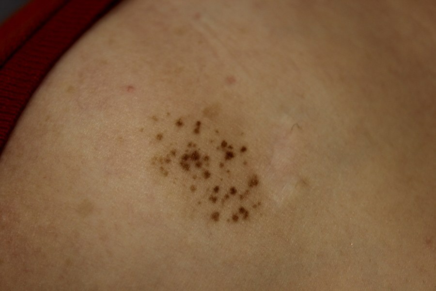
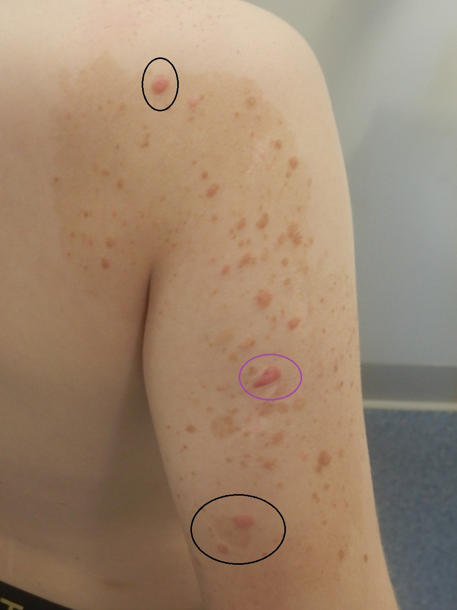
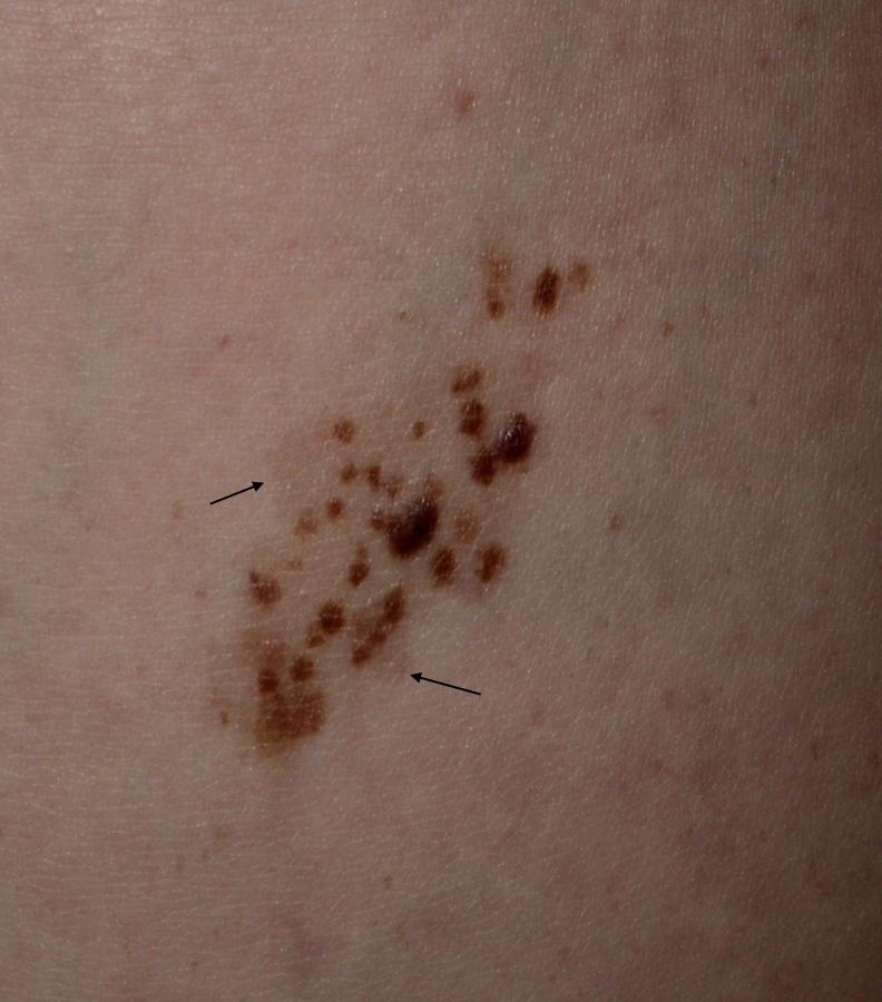
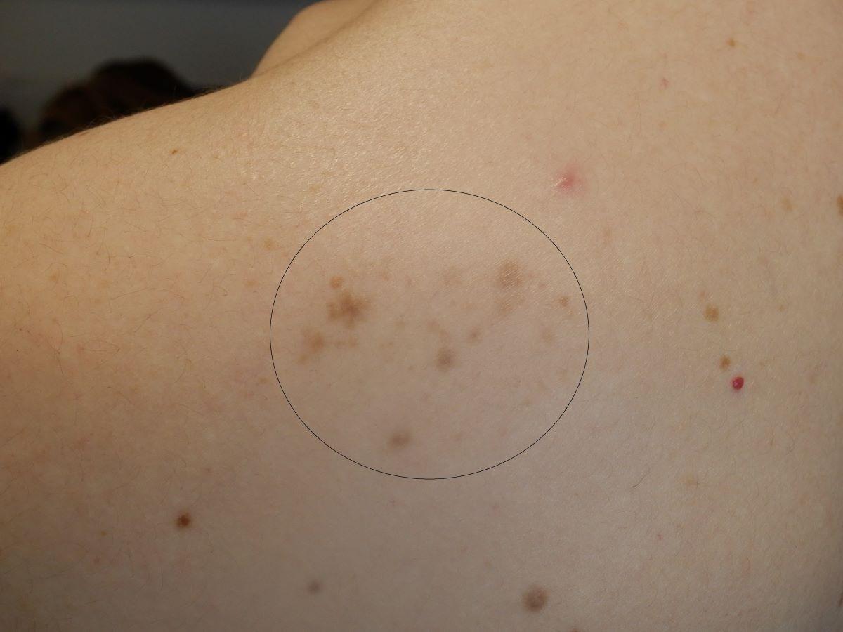
A speckled lentiginous naevus (SLN) is a melanocytic lesion composed of small, dark macules or papules within a larger but more lightly pigmented patch. Some consider SLN to be subtypes of congenital melanocytic naevi. The macules or papules within the patch are naevi, typically common naevi and less often blue or Spitz naevi.
This chapter is set out as follows:
History
- There is no gender preference; they are more common in individuals with lighter skin tones
- Lesions generally arise in the first year of life
- Initially the lesion is a pigmented patch (lentiginous) resembling a cafè-au-lait patch
- The lesion develops darker palpable components usually around puberty, although sometimes in adults, in a 'speckled' distribution
- As with other benign melanocytic naevi, SLN tend to fade as individuals get older
- Associations - while a SLN usually exists as solitary lesion, rarely, it can appear with other skin conditions including complex syndromes. In the Naevus Spilus syndrome the skin lesions are linked to ipsilateral dysesthesia, muscular weakness and hyperhidrosis; in phakomatosis pigmentokeratotica there are associations with neurological, skeletal or ocular alterations and to epidermal naevi, often of sebaceous differentiation
Clinical findings
Distribution
Clinical features
- Morphologically the lesion has two components:
- A patch of skin that is only subtly darker than the surrounding skin and can reach several cm in diameter
- Within the patch develop numerous darker macules and papules, 1-3 mm in diameter
Dermoscopic features
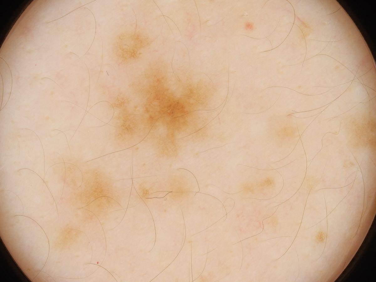
- Typical lesions have a light brown homogenous background, sometimes with a subtle pigment network, and superimposed darker macules or papules that can have reticular, globular, or mixed patterns
Clinical Images
Please refer to notes on image rights at bottom of the page with regards to individual image
ownership.
Management
- The risk of malignant transformation into a melanoma is, for the majority, very low and as such prophylactic excision is not recommended. Patients should be taught on self-examination, asked to take photographic records, and report any changes
- Patients developing any suspicious change should be referred on an urgent skin cancer pathway
- Individuals with naevi in cosmetically sensitive areas may wish to use cosmetics for camouflage or be referred to a camouflage service
Disclaimer - the author PCDS cannot accept responsibility for any misleading or incorrect statements, and the management of individual patients remains the direct responsibility of the individual doctor. We do however hope that visitors to this site can contact us regarding comments that are considered misleading or incorrect so that we can continue to improve the site.
Image Rights - The PCDS would like to thank Dermatoweb, DermQuest (Galderma), and others who have contributed images. All named individuals and organisations maintain copyright for the relevant images.