Tuberous sclerosis
LAST UPDATED: Nov 19, 2021
Introduction
Tuberous sclerosis is an autosomal dominant condition characterised by skin anomalies, epilepsy and developmental difficulties. Other commonly affected organs include the eyes, kidney and heart. This chapter discusses the dermatological manifestations of tuberous sclerosis, and is set out as follows:
History
-
Onset before the age of five years with cutaneous changes or epilepsy is usual, but the disease may remain latent (or unrecognised) until adolescence or adult life
-
Cutaneous features are found in approximately 70% of all cases
Clinical findings
-
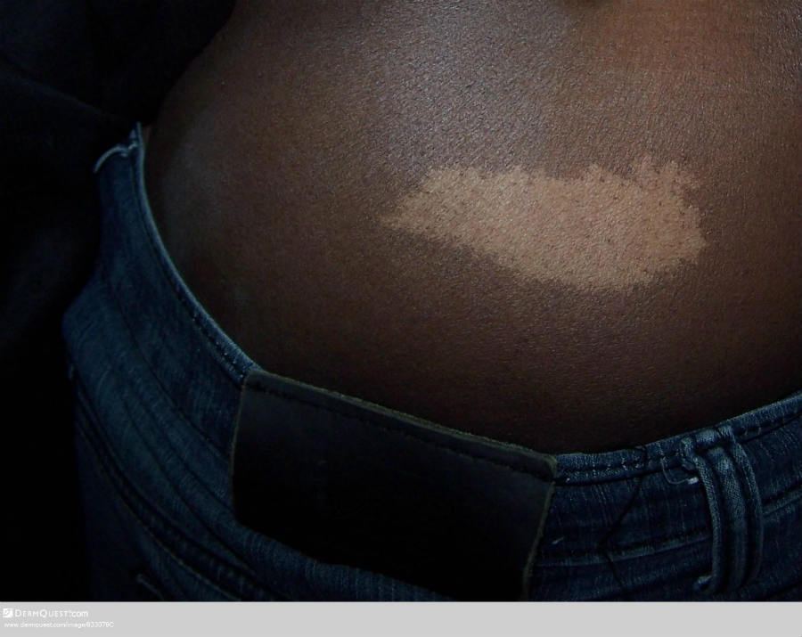
Ash-leaf spots
-
Age - usually the first cutaneous sign of tuberous sclerosis, presenting in the first year of life
-
Distribution - trunk and limbs
-
Morphology - ash-leaf or ovoid shaped, hypopigmented macules / patches, 1-3 cm in diameter. The lesions are more easily identified with a Wood's lamp
-
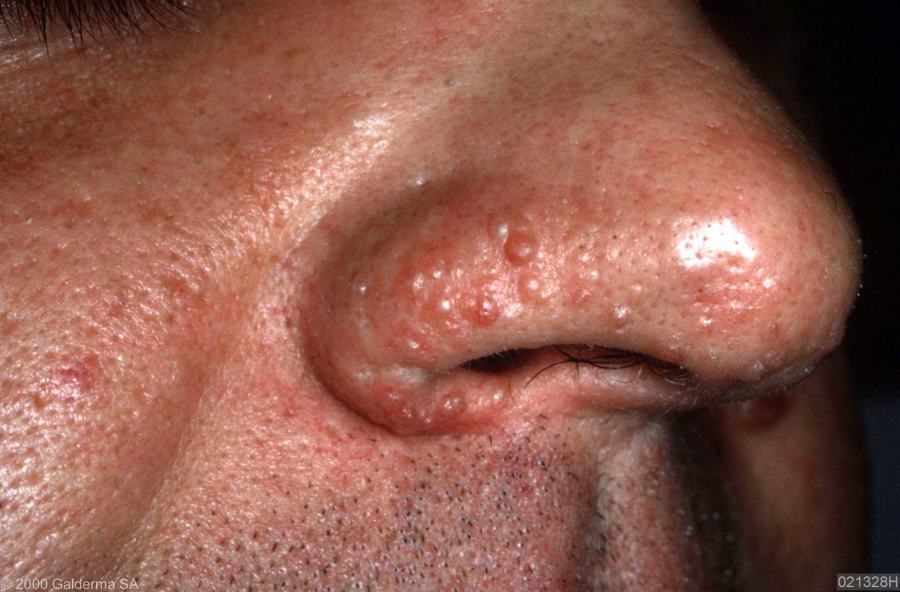
Angiofibromas
-
Age - normally arise in childhood, often between the ages of 3-10 years. Lesions tend to become more extensive at puberty and then remain unchanged
-
Distribution - predominantly the central face (nose, cheeks and chin). They can occasionally be found at other sites such as the scalp
-
Morphology - multiple, small, red / brown, telangiectatic papules
-
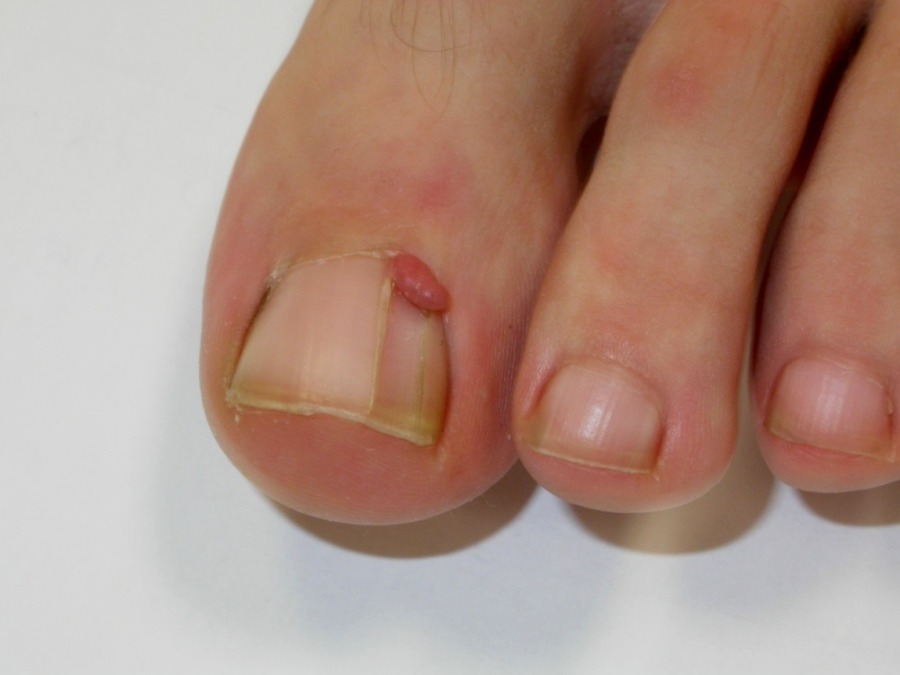
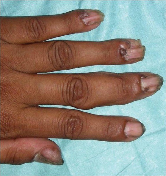
Periungual fibromas
-
Age - appear at or after puberty
-
Morphology - smooth, firm, fleshy lesions arising from the nail folds. Usually 5-10 mm in size but sometimes larger
-
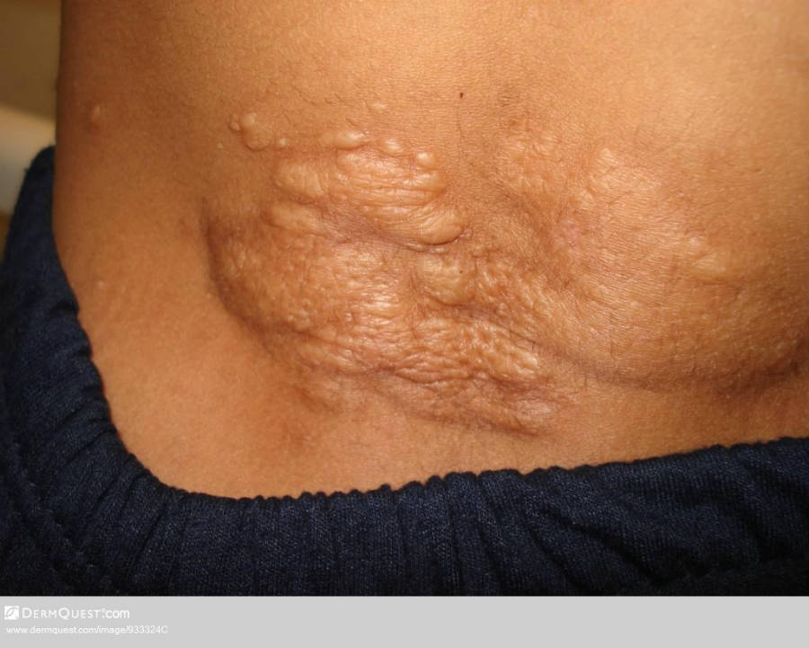
Shagreen patch
-
Distribution - lower back
-
Morphology - an irregularly elevated soft, skin-coloured plaque with an 'orange-peel' appearance
Clinical Images
Please refer to notes on image rights at bottom of the page with regards to individual image
ownership.
Management
Step 1: general measures
-
A multi-disciplinary team approach is needed
Step 2: monitoring of renal function in adults
-
The main risk in adults is the development of renal hamartomas - patients should have their renal function checked every two years
Step 3: angiofibromas
- Rapamycin gel (Secondary Care only) is an effective treatment and should be used in preference to CO2 laser
- Repeat treatments are usually required as lesions gradually recur
Other resources
Disclaimer - the author PCDS cannot accept responsibility for any misleading or incorrect statements, and the management of individual patients remains the direct responsibility of the individual doctor. We do however hope that visitors to this site can contact us regarding comments that are considered misleading or incorrect so that we can continue to improve the site.
Image Rights - The PCDS would like to thank Dermatoweb, DermQuest (Galderma), and others who have contributed images. All named individuals and organisations maintain copyright for the relevant images.