Clear cell acanthoma of Degos

LAST UPDATED: Mar 01, 2026
Introduction

A clear cell acanthoma of Degos is an uncommon benign epidermal lesion containing clear cells with a glycogen-rich cytoplasm. It most commonly arises as a solitary lesion on the lower legs.

The chapter is set out as follows:


History

  • Age - usually affects adults with a peak incidence between 50 and 60 years of age
  • Can be asymptomatic although surface oozing is not uncommon and occasionally lesions can bleed

Clinical findings

Distribution

  • Normally solitary but occasionally multiple lesions
  • Lower legs most commonly affected site

Morphology

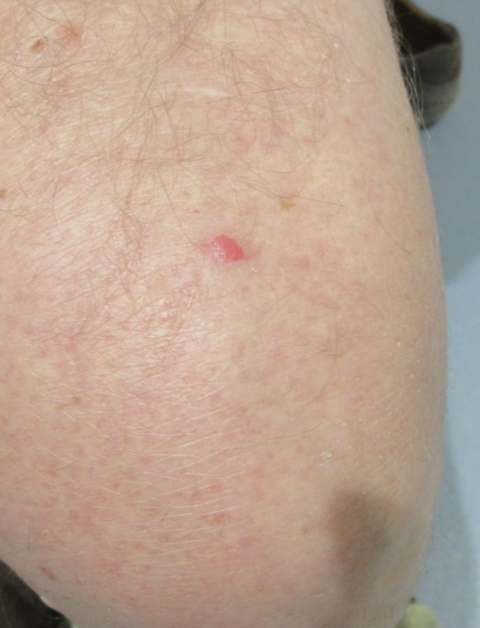
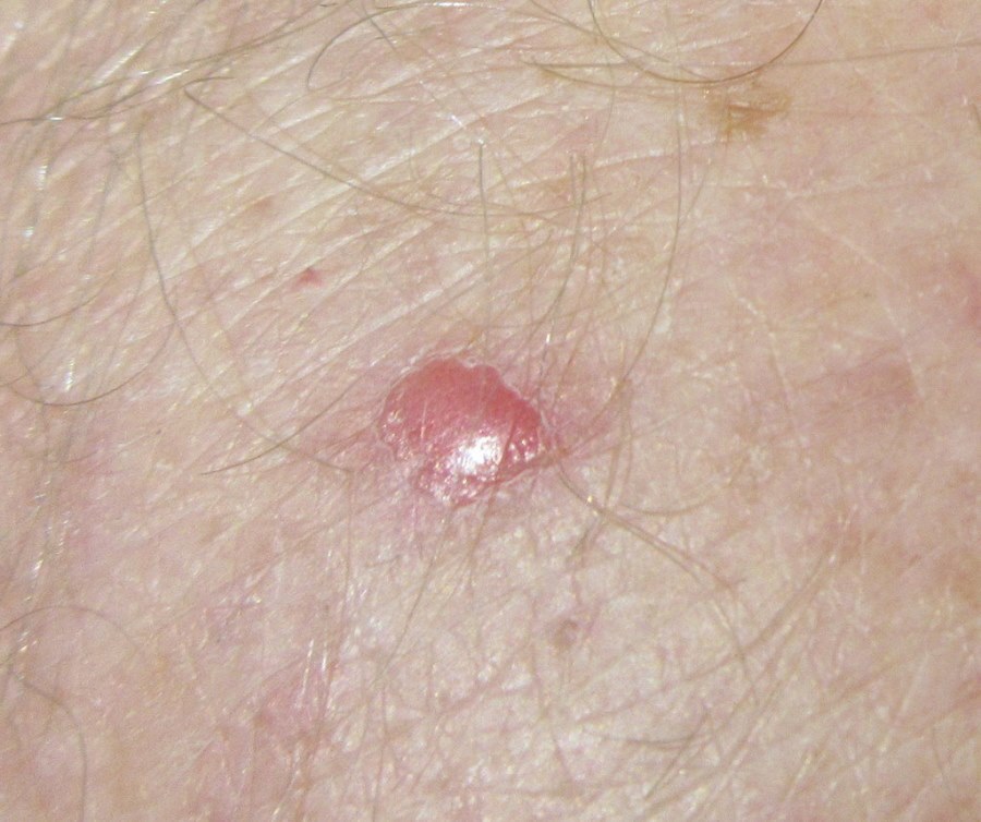
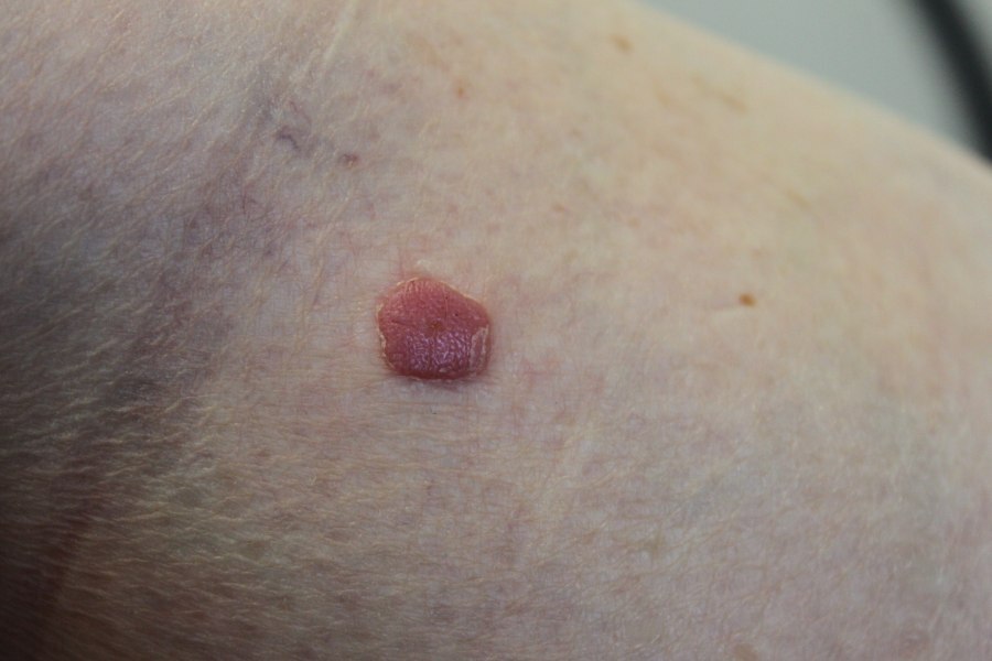
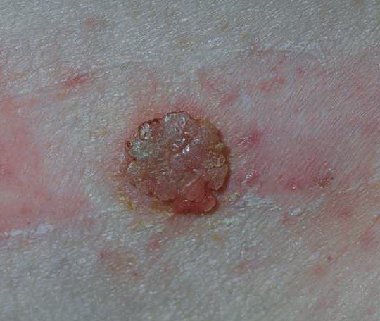
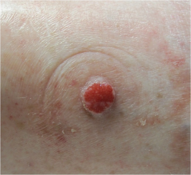
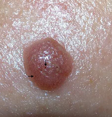
  • Size varies from 3-20 mm in diameter
  • Pink-red, occasionally brown
  • Plaque-like or nodular
  • Abrupt margin
  • A thin surface scale that is adherent peripherally and leaves a moist or bleeding surface when removed
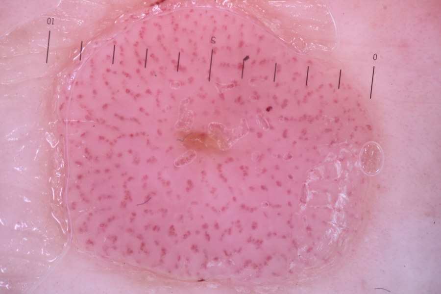
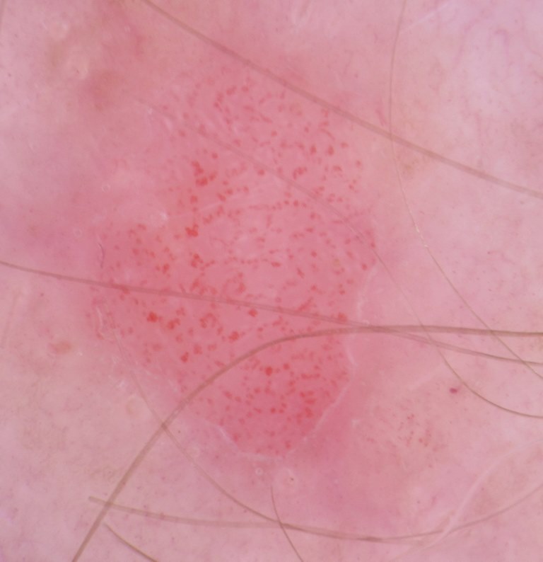
  • Vascular punctae may be visible on the surface

Dermoscopic appearance (hover over terminologies for description)

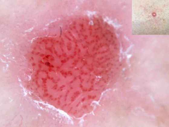
  • The most characteristic feature of CCA is the ''string of pearls'' vascular pattern in which dot or coiled (glomerular) vessels are arranged in serpiginous pattern (for more information on vessels refer to the chapter Vessel patterns in dermoscopy)
  • The background colour is a homogenous pale pink or light red
  • Shiny white lines and orange crusts may be present

Clinical Images

Please refer to notes on image rights at bottom of the page with regards to individual image ownership.
Investigations

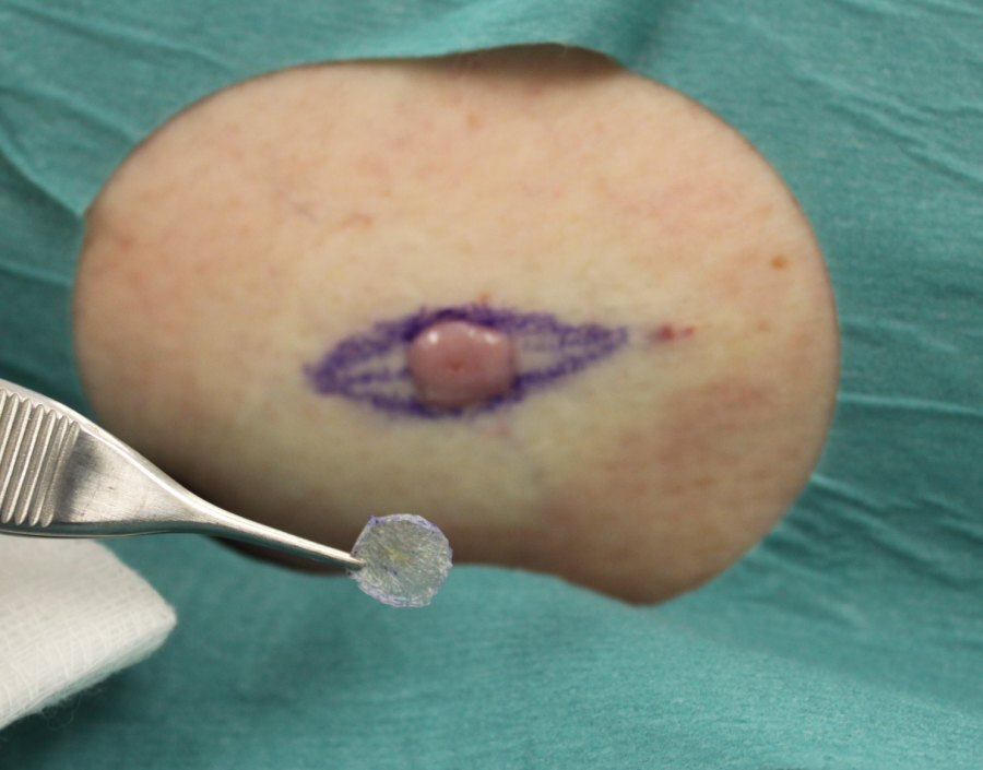
  • If there is diagnostic uncertainty a biopsy will be required - the histopathological features are characteristic. These include psoriasiform acanthosis with pale periodic acid–Schiff (PAS) positive glycogen‐rich keratinocytes (clear cells). The cytoplasm of the clear cells contains an abundance of glycogen, which on electron microscopy is seen to displace tonofibrils. There is intercellular oedema and an infiltrate often containing many polymorphonuclear leukocytes forming parakeratotic microabscesses. The papillary dermis is oedematous and the superficial capillaries and veins are increased in number (reference Rook's Textbook of Dermatology) 

Management

  • Lesions are benign and so once diagnosed no further treatment is required 

Disclaimer - the author PCDS cannot accept responsibility for any misleading or incorrect statements, and the management of individual patients remains the direct responsibility of the individual doctor. We do however hope that visitors to this site can contact us regarding comments that are considered misleading or incorrect so that we can continue to improve the site.

Image Rights - The PCDS would like to thank Dermatoweb, DermQuest (Galderma), and others who have contributed images. All named individuals and organisations maintain copyright for the relevant images.

Quick Links

The following pharmaceutical companies have had no involvement in the content of this website or in our conference programmes


Almirall
Galderma
Glenmark
Johnson & Johnson
La Roche-Posay
LEO Pharma
Pierre Fabre
Schuco