Fibrous tumours

LAST UPDATED: Nov 22, 2021
Introduction

There are several types of fibrous tumours that can affect the nails, fingers, toes and much less commonly others sites.

This chapter is set out as below:


Clinical findings

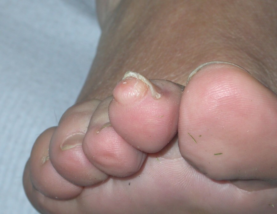
Acquired (digital) fibrokeratoma
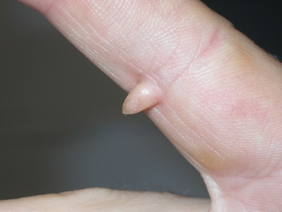
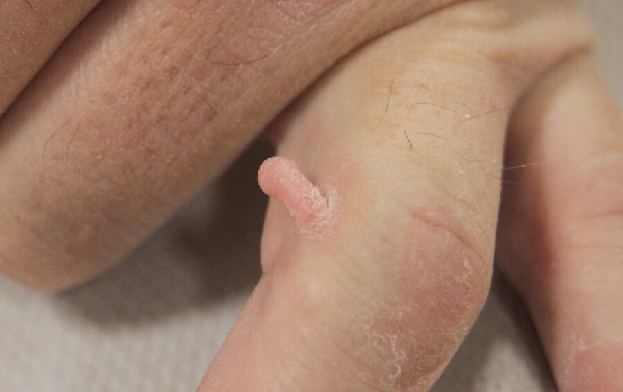
  • Lesions manifest as solitary, skin-coloured, dome-shaped papules or tall fingerlike protrusions with a hyperkeratotic surface. Most are less than 1.5 cm in height or diameter
  • A distinguishing feature is that of a collarette of slightly raised skin that encircles the base of the lesion 
Periungual fibromas of tuberous sclerosis
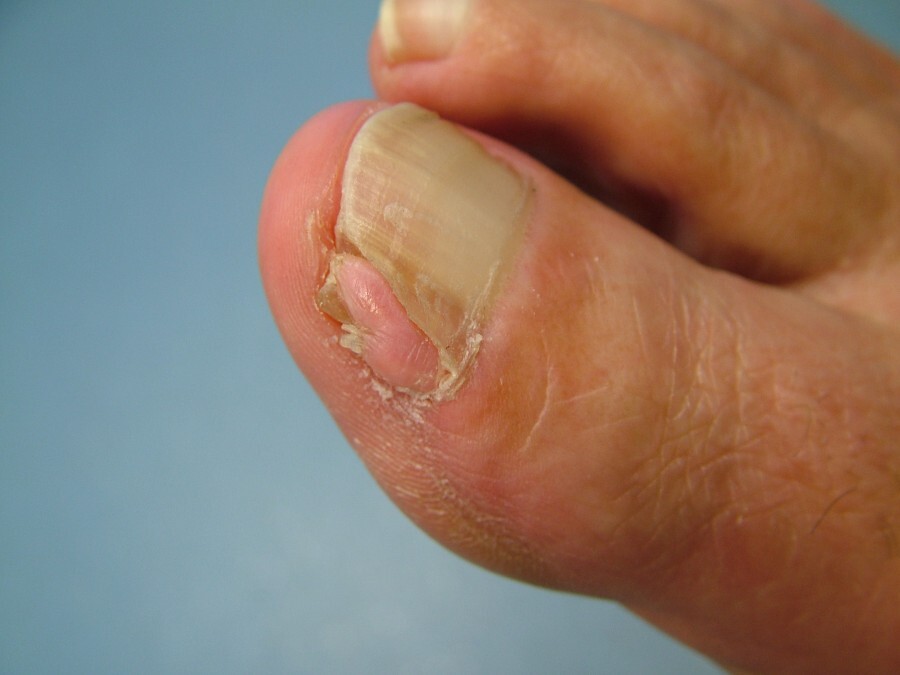
  • Similar growths to the above are found in the subungual or periungual region of patients with tuberous sclerosis, and are referred to as Koenen tumours or garlic clove fibromas
  • Lesions develop around puberty and in young adults
  • They tend to be multilobulated, involve several digits and, histologically, may have atypical stellate myofibroblasts
  • Nail involvement is common
  • Refer to the related chapter on tuberous sclerosis for more information 
Acquired fibrous nodule
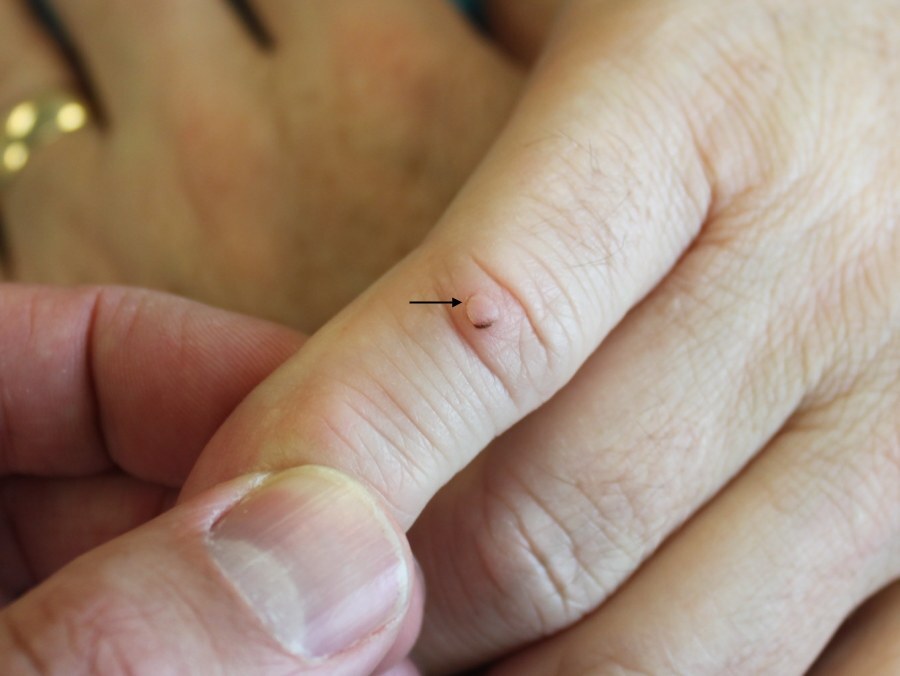
  • Is a true fibroma
  • It usually presents as a smooth-edged lesion, commonly in the periungual tissue as opposed to within the nail unit
  • Lesions are generally devoid of a hyperkeratotic surface 
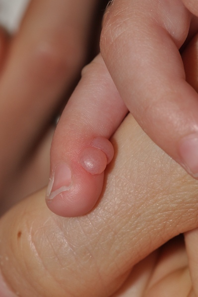
Infantile digital fibromatosis
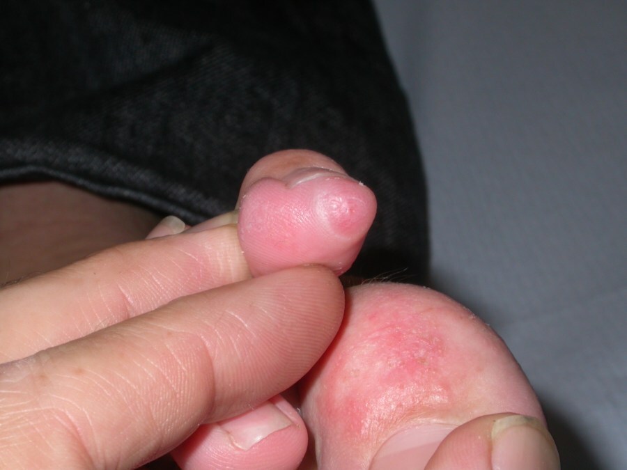
  • This is a rare condition, manifested by the benign, asymptomatic, nodular proliferation of fibrous tissue occurring almost exclusively on the dorsal and lateral aspects of the fingers or toes
  • Most nodules appear in the first few months of life; one third are congenital, and 75-80% are noted during the first year of life
  • Clinical features
    • Single or multiple, firm, erythematous, dermal nodules with a smooth dome-shaped surface on the dorsolateral aspect of the distal phalanges of the digits
    • There is sparing of the thumbs and the great toes
    • Lesions can grow up to 2 cm in diameter, yet they rarely cause functional impairment or deformity
  • Because of the benign nature of infantile digital fibromatosis, frequent recurrence after surgery, and a tendency towards spontaneous resolution over two to three years, observation is suggested as the best form of management

Clinical Images

Please refer to notes on image rights at bottom of the page with regards to individual image ownership.
Management

  • Asymptomatic lesions are best left alone
  • Symptomatic lesions can be removed by shave excision or curettage and sent for histology
  • Infantile digital fibromatosis - refer to the section above on clinical findings

Disclaimer - the author PCDS cannot accept responsibility for any misleading or incorrect statements, and the management of individual patients remains the direct responsibility of the individual doctor. We do however hope that visitors to this site can contact us regarding comments that are considered misleading or incorrect so that we can continue to improve the site.

Image Rights - The PCDS would like to thank Dermatoweb, DermQuest (Galderma), and others who have contributed images. All named individuals and organisations maintain copyright for the relevant images.

Quick Links

The following pharmaceutical companies have had no involvement in the content of this website or in our conference programmes


Almirall
Galderma
Glenmark
Johnson & Johnson
La Roche-Posay
LEO Pharma
Pierre Fabre
Schuco