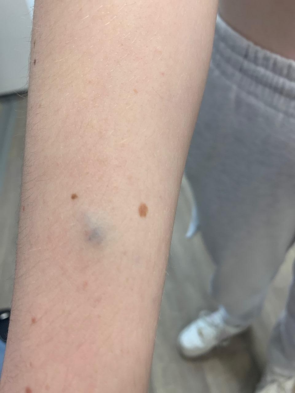
Glomulovenous malformation (syn. glomangioma)
LAST UPDATED: Nov 07, 2022
Introduction
A glomulovenous malformation (GVM) is an inherited, autosomal dominant, venous anomaly characterised by multifocal bluish purple papules or nodules.
This chapter is set out as follows:
Aetiology
- GVMs are a type of vascular malformation made of abnormal accumulations of blood vessels and glomus body cells. Glomus body cells are a normal part of the skin and are associated with temperature regulation - in cold weather they regulate blood flow away from the surface to keep the body warm, in warm weather they improve blood supply to the skin surface for cooling
- GVMs are rare, accounting for about 5% of venous anomalies
- Single GVMs are more common than multiple ones
- The cause of GVMs is unknown
- More than 80% of GVMs are inherited as an autosomal dominant trait
- There is no sex preponderance
History
- Most present at birth or later in infancy or childhood, although they can arise at any age
- Lesions tend to be painless unless knocked or when there is an extreme change in temperature, which differs to the Blue Rubber Bleb Naevus syndrome in which lesions are characteristically painful (especially at night)
Clinical findings
- GVMs can be cutaneous, subcutaneous, or involve mucous membranes inside the mouth and eyelids. They occasionally affect the muscles but no other organs, unlike some cases of the Blue Rubber Bleb Naevus syndrome
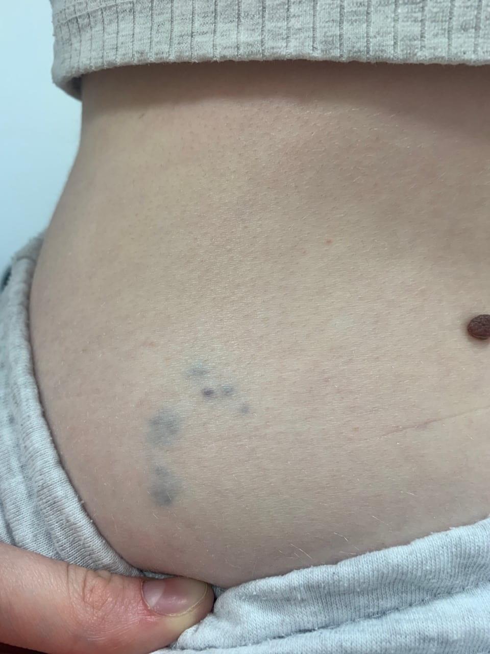
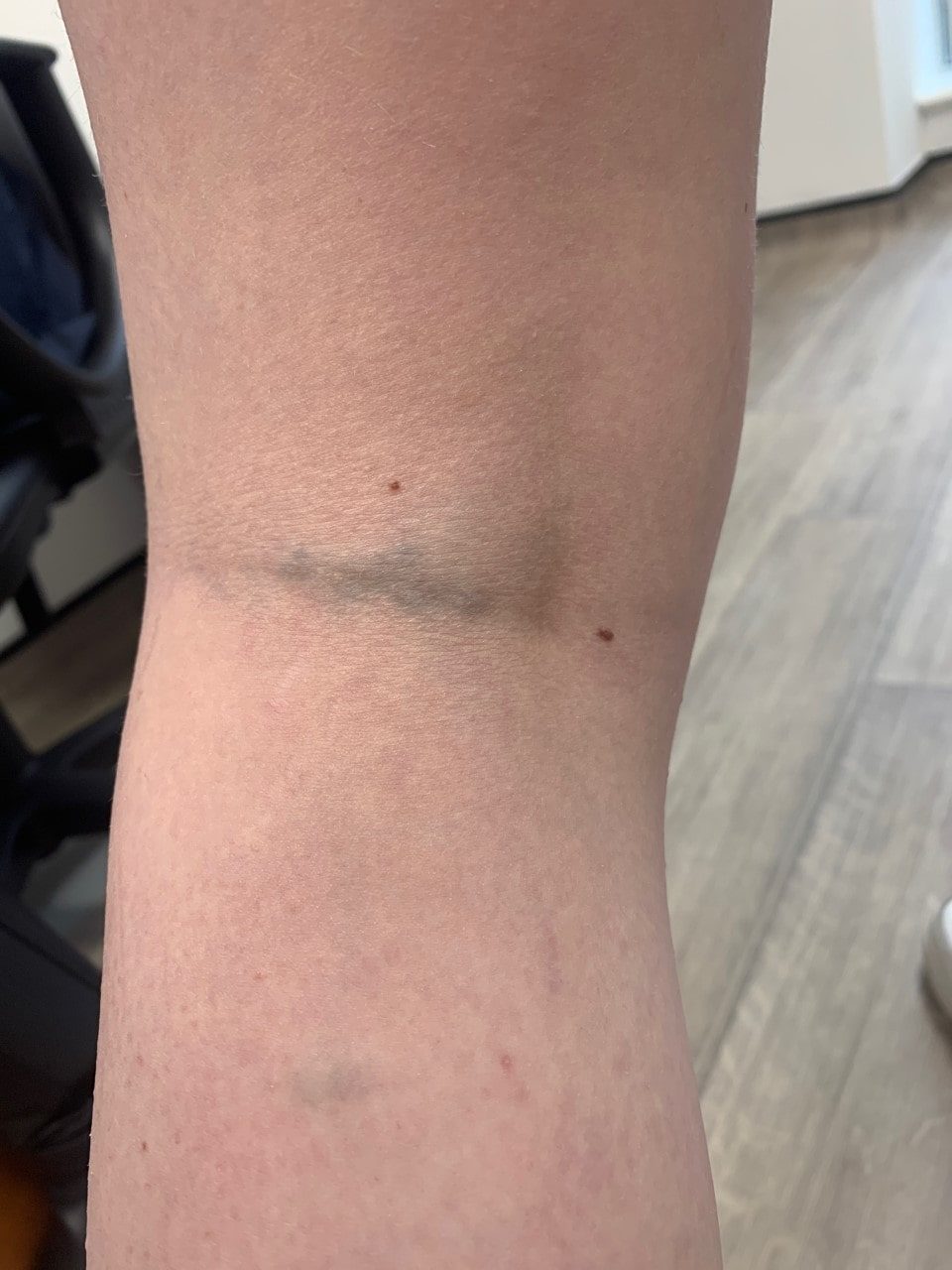
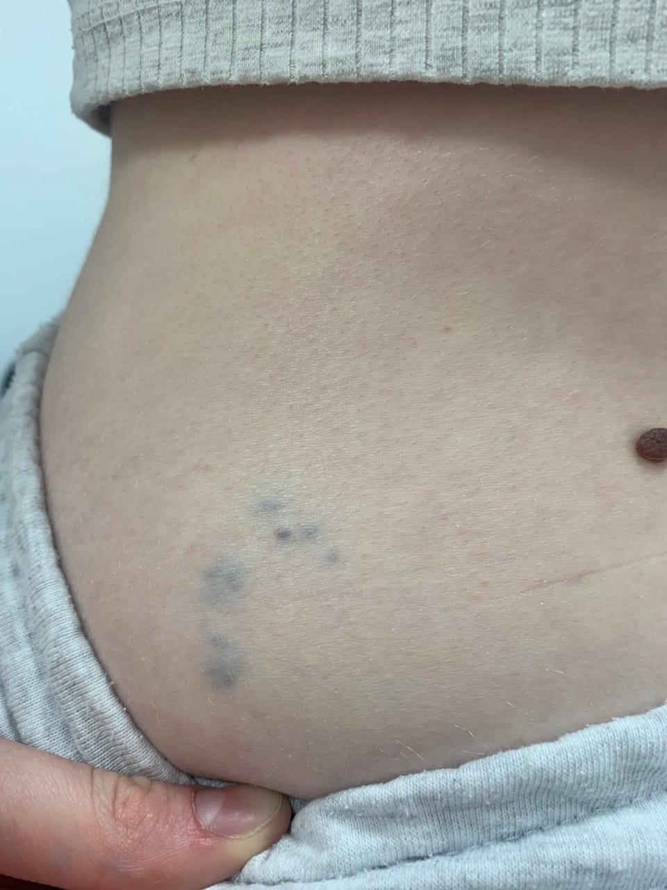
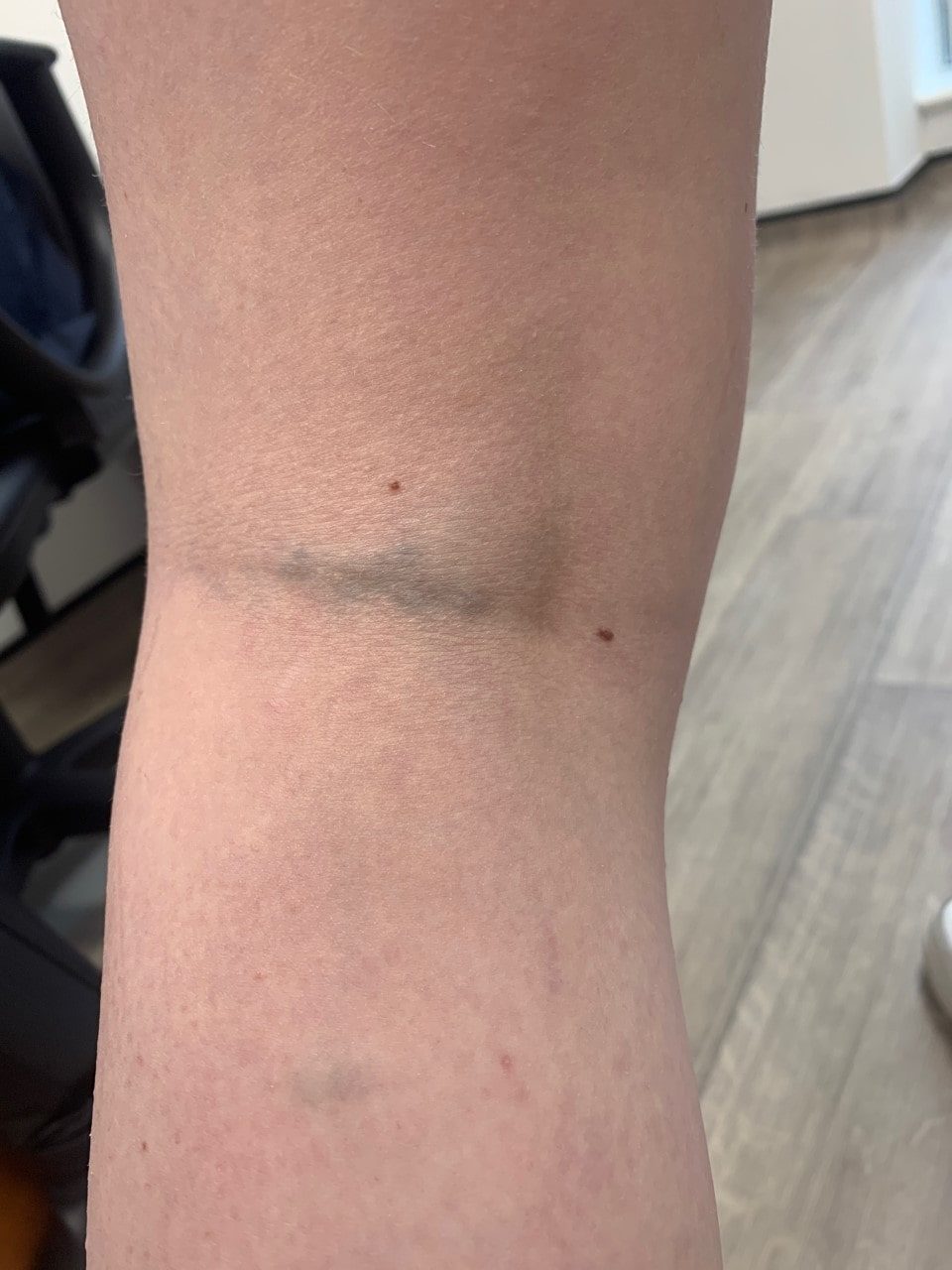
- There is great variability in the number, size and location of lesions between affected individuals. Some have a few inconspicuous papules (often grouped), whereas others have extensive lesions. They can be localised or segmental
- GVMs are usually small, pink-red or blue, papules / nodules. With age, lesions tend to become more widespread, turn darker blue and more uneven in texture
- Lesions are less compressible than other venous malformations
Clinical Images
Please refer to notes on image rights at bottom of the page with regards to individual image
ownership.
Investigations
- Investigations are not always needed
- GVMs are not associated with coagulation abnormalities
- If a skin biopsy is taken it will demonstrate the presence of glomus body cells
- If there is concern about the size / extent of a GVM, imaging will be required
Management
- Lesions never regress spontaneously
- Treatment should only be considered if the lesions are problematic
- Tailored compression garments are contraindicated as they increase pain
- Surgical excision of GVMs is difficult. Other treatment options include laser (for smaller lesions) and sclerotherapy
- Genetic counselling is indicated
Disclaimer - the author PCDS cannot accept responsibility for any misleading or incorrect statements, and the management of individual patients remains the direct responsibility of the individual doctor. We do however hope that visitors to this site can contact us regarding comments that are considered misleading or incorrect so that we can continue to improve the site.
Image Rights - The PCDS would like to thank Dermatoweb, DermQuest (Galderma), and others who have contributed images. All named individuals and organisations maintain copyright for the relevant images.