Venous malformations - cutaneous

LAST UPDATED: Nov 07, 2022
Introduction

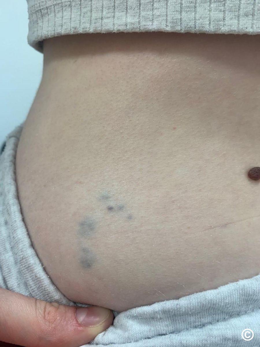
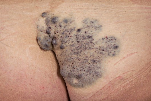
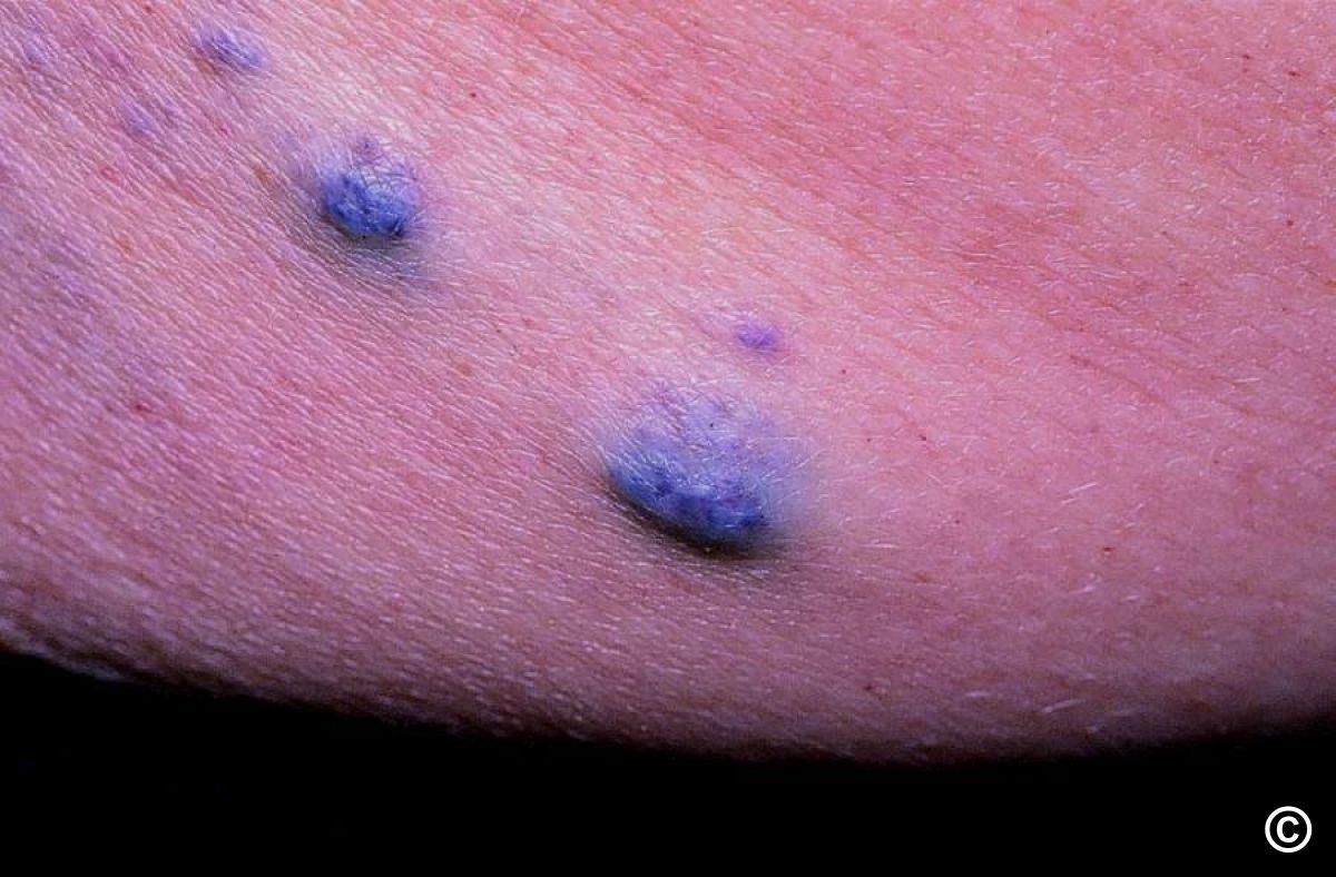
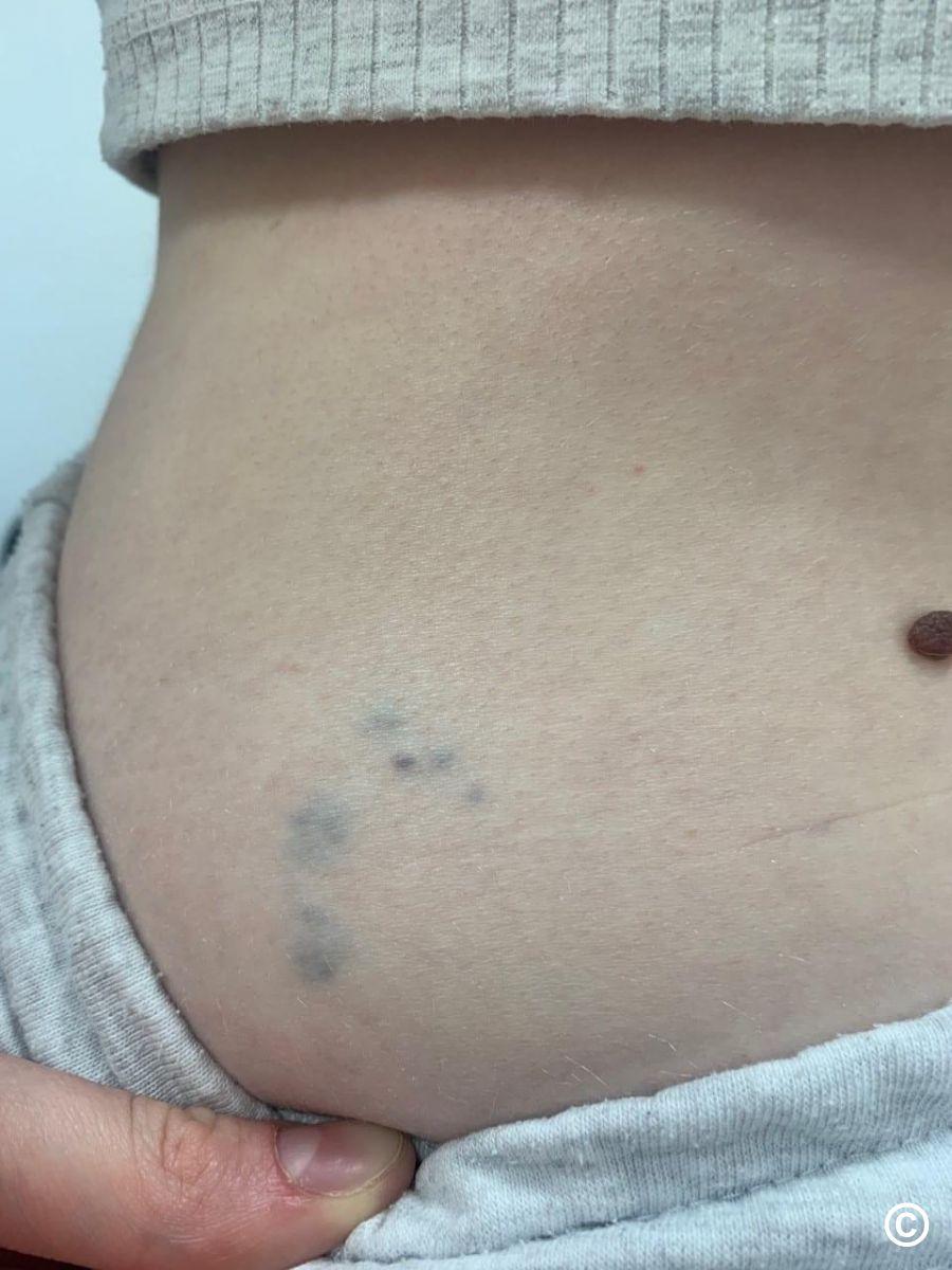
Venous malformations (VMs) are sporadic, caused by malformed dilated veins, and can affect any tissue or organ. They appear as skin-coloured, blue or purple swellings, often with prominent veins near the skin surface. VMs are always present at birth, although may not become clinically apparent until later. They may vary in size from small to occasionally involving a whole limb. Unlike infantile haemangiomas, they grow in proportion to the child's general growth.

This chapter is set out as follows:


Aetiology

  • Venous malformations are rare with have an incidence estimated around 1:10 000
  • Males and females are equally affected

History

  • Grow proportionately with the individual
  • They never regress spontaneously
  • Although often asymptomatic, they can be painful, cause bleeding and/or functional impairment - on the extremities can cause muscle weakness
  • Pulmonary thromboembolism has been reported in some patients with extensive VMs of the extremities

Clinical findings

  • Mainly solitary, <1% are multifocal
  • Variable size, from small blebs to large, bluish lesions
  • There is no thrill or bruit, and the malformation is not warmer than non‐lesional areas
  • Associations - VMs can be associated with various conditions such as Maffucci syndrome, characterised by enchondromas, bone deformities, and venous malformations
  • The Blue Rubber Bleb Naevus syndrome is a rare syndrome comprising of multiple venous malformations of the skin, gastrointestinal tract and other organs (refer to the related chapter for more information)
  • Glomulovenous malformation (GVMs) can have a similar appearance to the above, but differ in that they are inherited, lesions tend to be painless unless knocked or during an extreme change in temperature, and they do not involve internal organs (refer to the related chapter for more information)

Clinical Images

Please refer to notes on image rights at bottom of the page with regards to individual image ownership.
Investigations

  • Doppler ultrasound (sometimes with MRI) is the best examination to confirm the vascular nature of the lesion (slow flow) and to identify the anatomy of the draining vessels 
  • Larger and more extensive lesions can be associated with coagulation abnormalities 

Management

Prognosis

  • Lesions never regress spontaneously
  • Aesthetic and functional impairment worsen with time because of expansion or bleeding
  • Depending on location and size, they can occasionally be life‐threatening

Management

  • Patients require referring to Secondary Care, where a multidisciplinary approach is required
  • Treatment strategies depend on the type of malformation and may include any of: tailored compression garments, aspirin, low‐molecular‐weight heparin if d‐dimer levels elevated, sclerotherapy, surgical resection, and laser therapy

Disclaimer - the author PCDS cannot accept responsibility for any misleading or incorrect statements, and the management of individual patients remains the direct responsibility of the individual doctor. We do however hope that visitors to this site can contact us regarding comments that are considered misleading or incorrect so that we can continue to improve the site.

Image Rights - The PCDS would like to thank Dermatoweb, DermQuest (Galderma), and others who have contributed images. All named individuals and organisations maintain copyright for the relevant images.

Quick Links

The following pharmaceutical companies have had no involvement in the content of this website or in our conference programmes


Almirall
Galderma
Glenmark
Johnson & Johnson
La Roche-Posay
LEO Pharma
Pierre Fabre
Schuco