Granuloma annulare
LAST UPDATED: Feb 22, 2024
Introduction
Granuloma annulare (GA) is a relatively common condition that occurs in all age groups, but is rare in infancy. Although there are several clinical patterns, the typical lesion of GA is a smooth, annular plaque with a bumpy (papular) edge. The precise cause of GA is unknown. Histological examination reveals foci of degenerative collagen associated with palisaded granulomatous inflammation.
This chapter is set out as follows:
Aetiology
-
Although the cause of GA is unknown it does appear to be a delayed hypersensitivity reaction to a number of triggering factors in the dermis
-
Generalised GA is sometimes associated with diabetes mellitus
History
-
GA affects all age groups, but is rare in infancy. It is most common in young adults
-
It is twice as common in women as in men
-
Lesions evolve slowly and tend to be asymptomatic
Clinical findings
Typical GA
-
Distribution
-
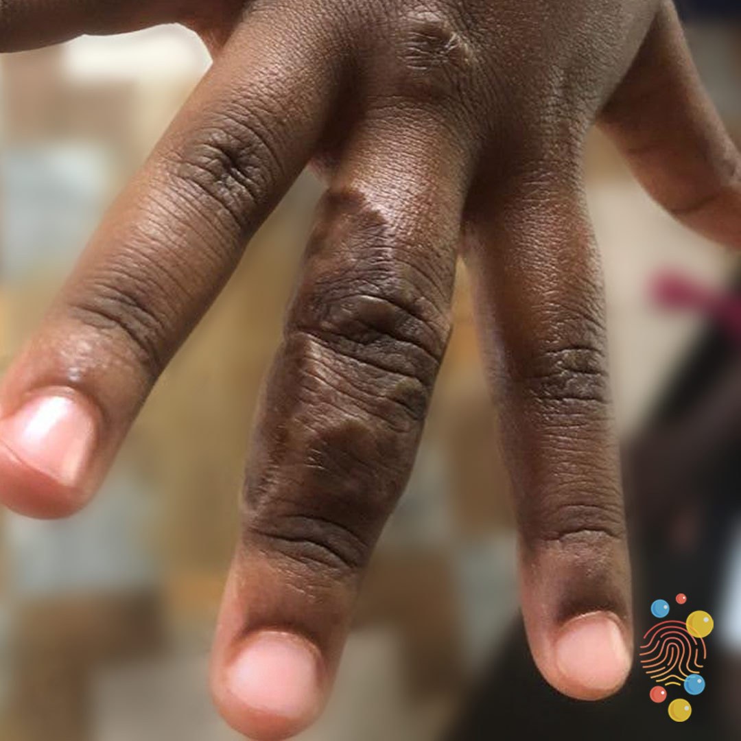
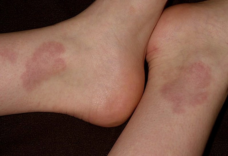
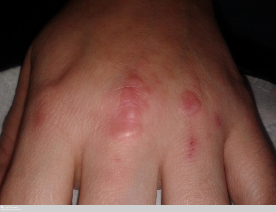
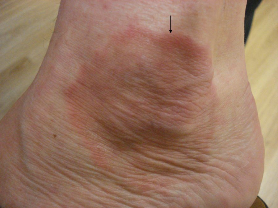
GA can affect any body site, although the dorsa of the hands and feet, as well as other extensor surfaces, are most commonly affected
-
There tends to be one to several lesions, although occasionally they can be numerous and widespread
-
Morphology
-
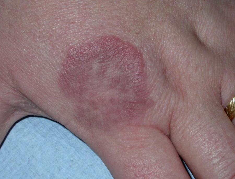
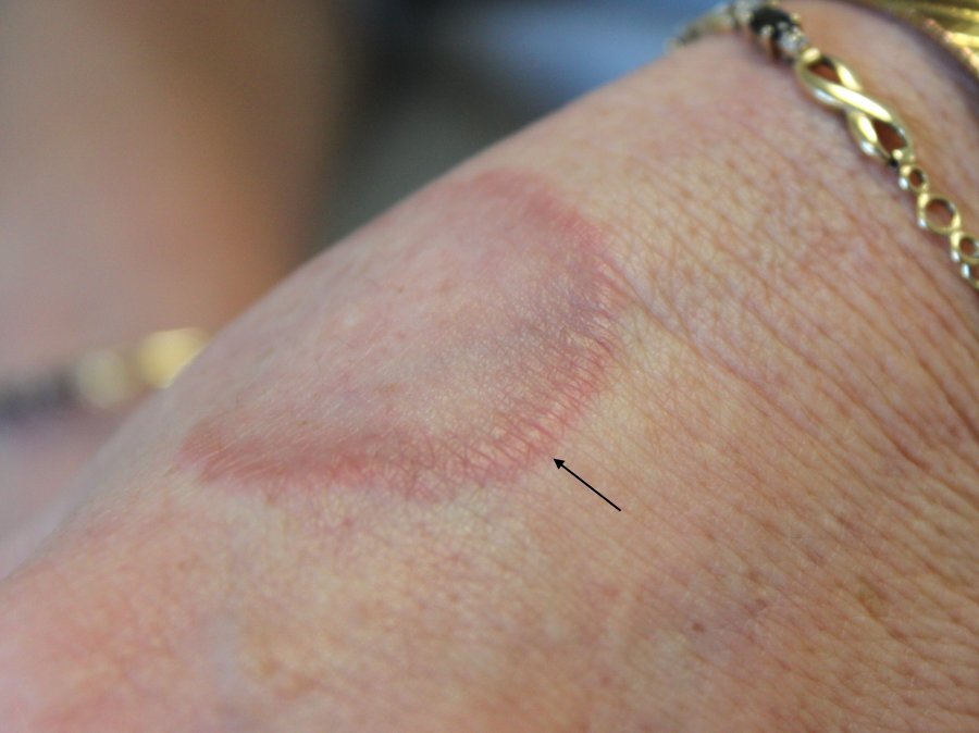
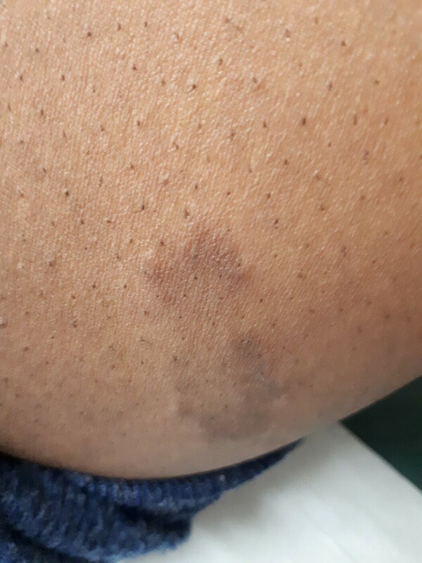
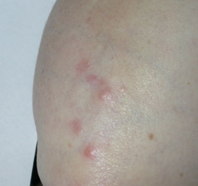
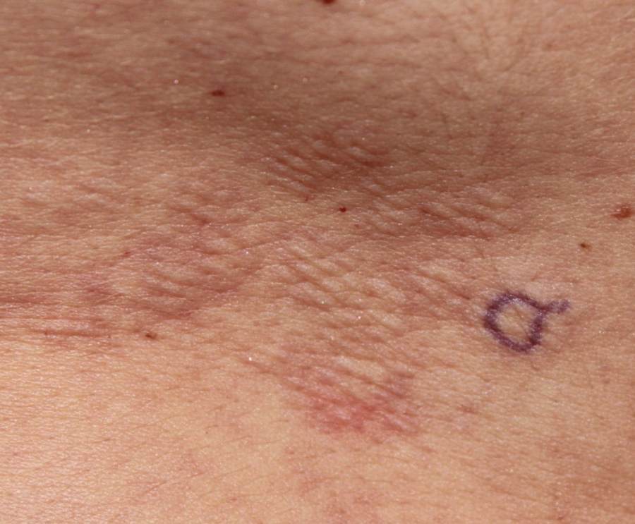
Lesions are made up of smooth, skin-coloured, or pink-red papules with no scale or other surface change
-
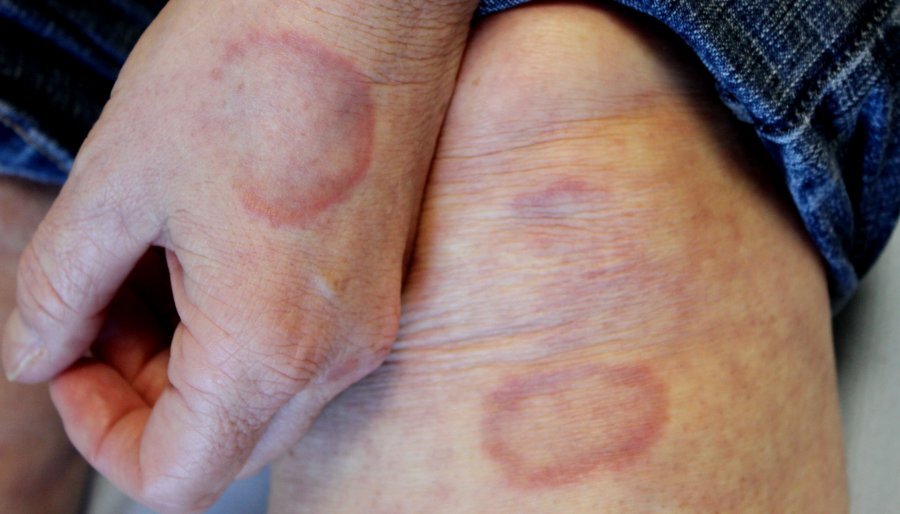
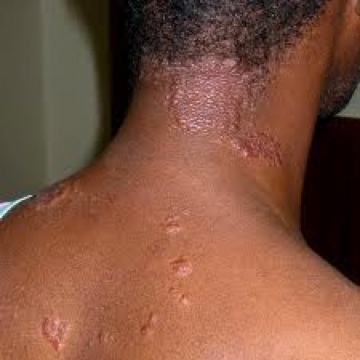
The characteristic lesion is a smooth, annular plaque that enlarges centrifugally with 'bumpy' papules around the periphery. Lesions may reach several cm in diameter
-
Differential diagnosis
-
GA is sometimes mistaken for tinea or an annular erythema, although these are more inflammatory and have epidermal changes such as scaling
-
Other annular lesions, such as annular lichen planus, and occasionally sarcoid, can take on a similar appearance
Other forms of GA
Other forms of GA may occur alone or in association with the characteristic annular lesions, and include:
-
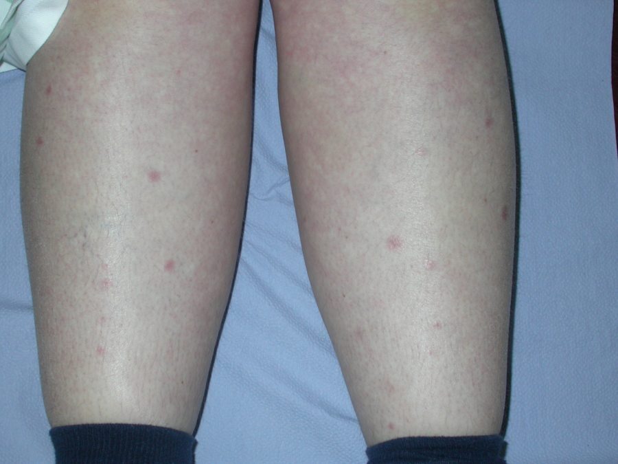
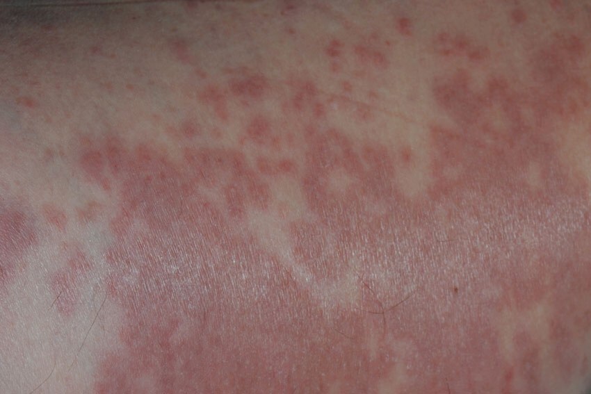
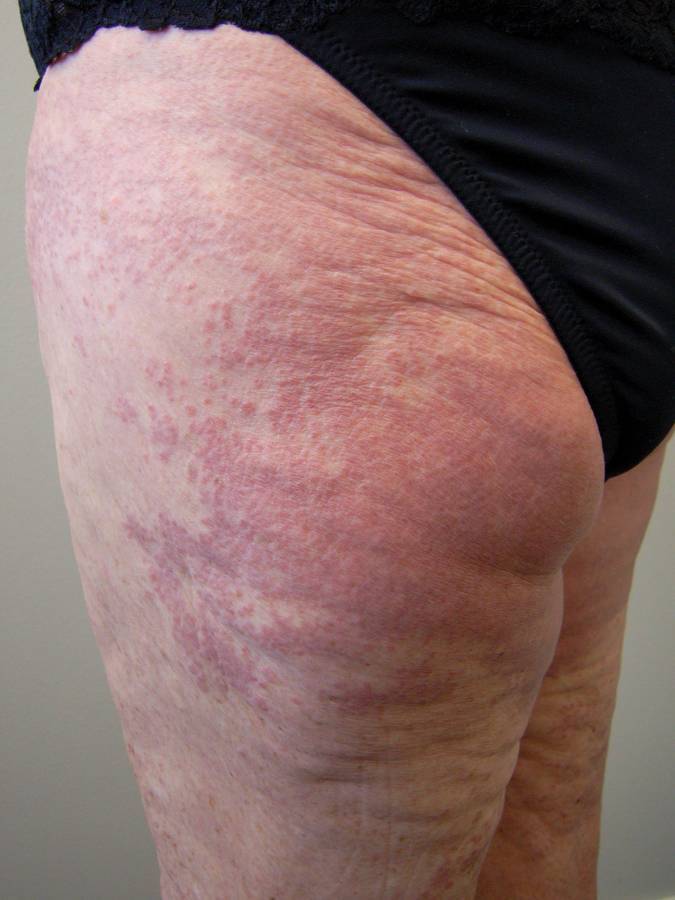
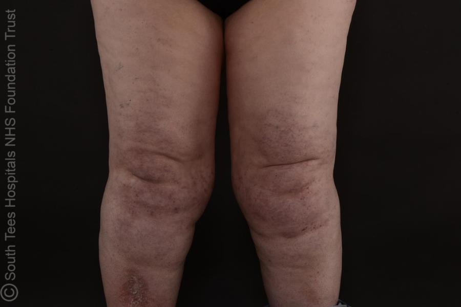
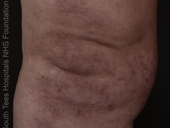
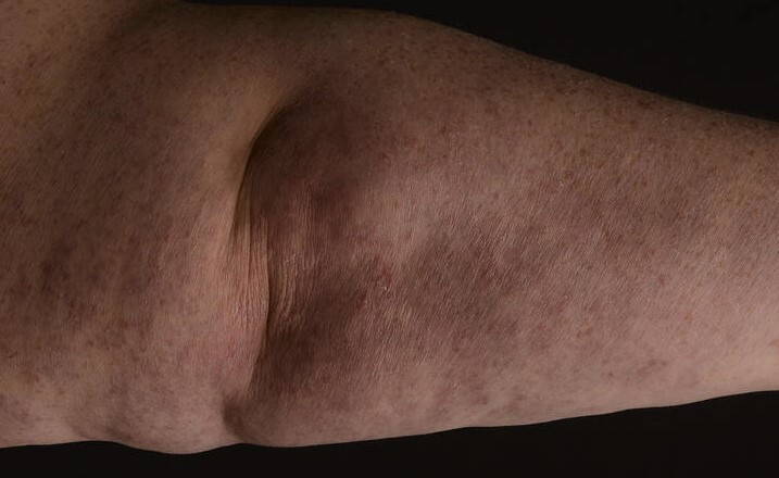
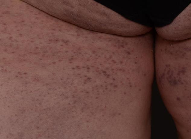
Generalised (disseminated) GA
-
Occurs more commonly in women, with a mean age of approximately 50 years
-
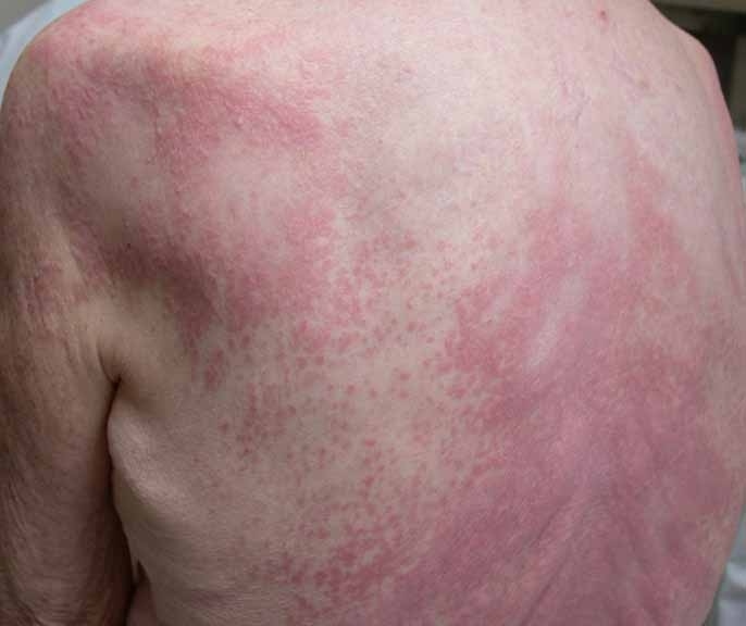
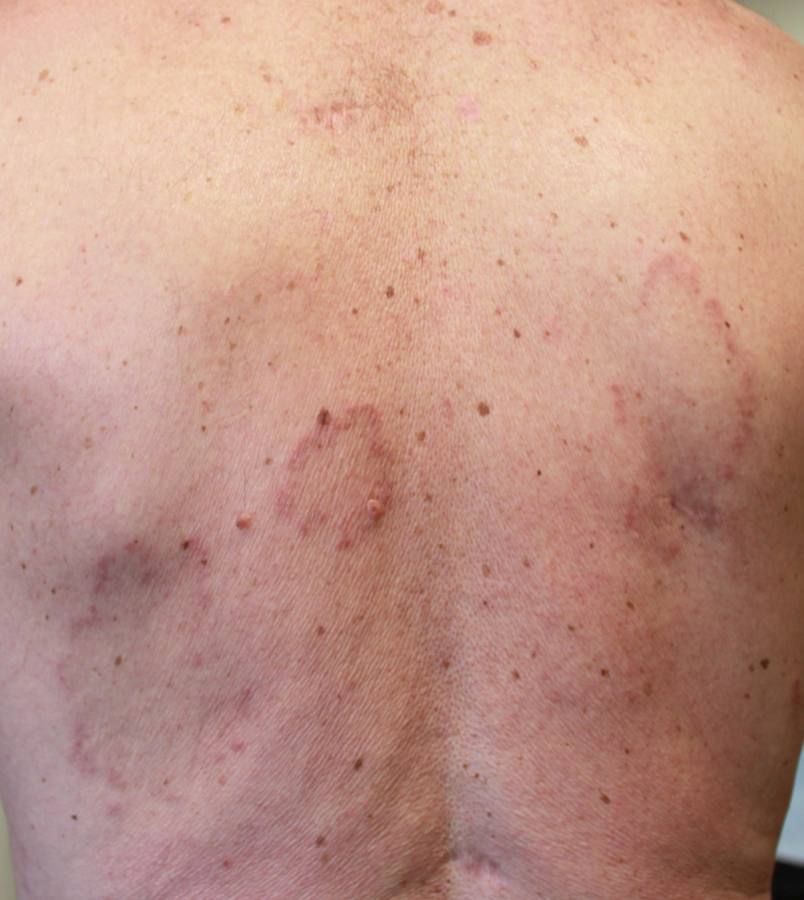
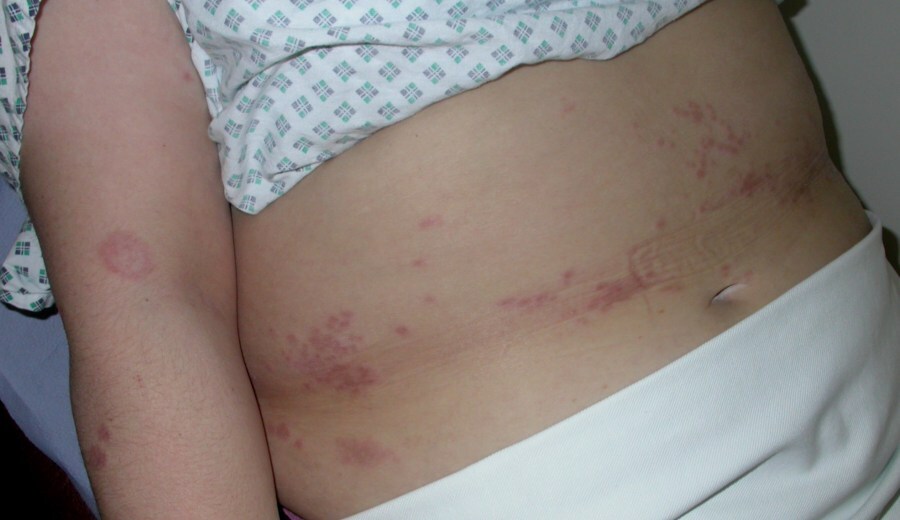
The trunk is usually involved, and other sites may be affected
-
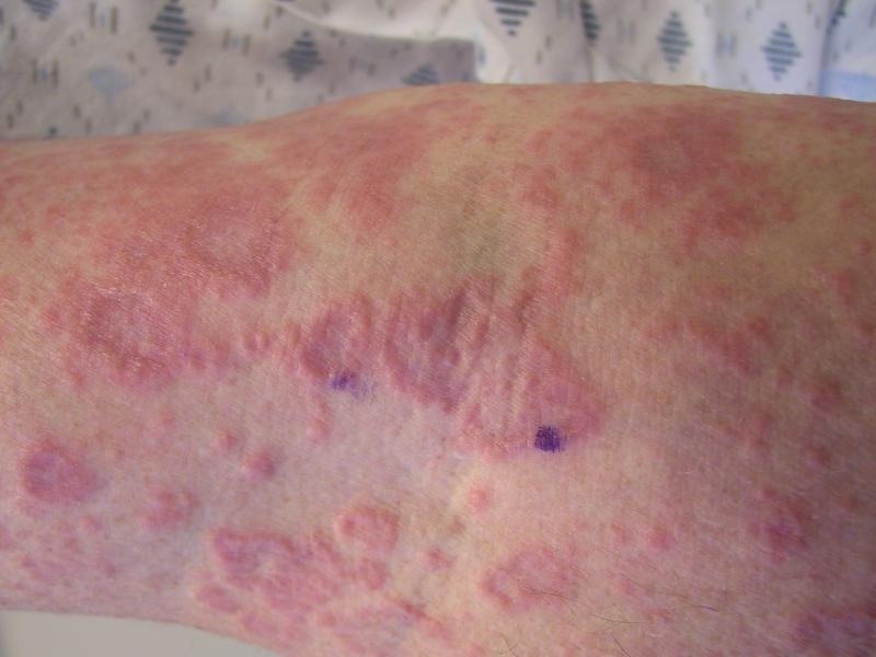
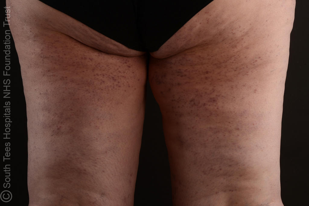
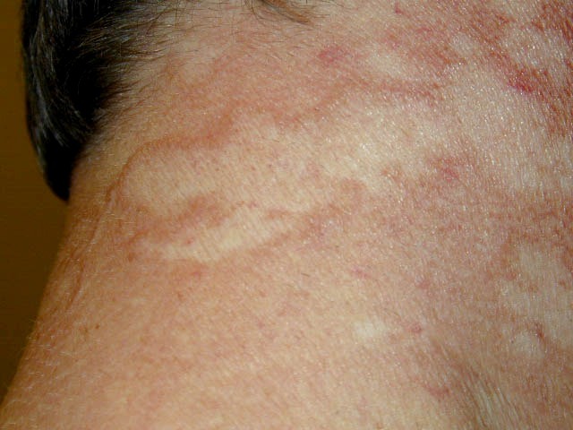
Morphologically there are many skin-coloured or pink-red, smooth papules which may be grouped, develop annular configurations or coalesce in to larger patches with a variety of colouration from yellow to violaceous
-
Cases of anetoderma, and mid-dermal elastolysis, have been reported in association with generalised GA
-
Subcutaneous GA
-
Is uncommon, and arises most commonly in children
-
Lesions are firm, ranging in size from 5 mm to several cm, and are found most commonly on the scalp, lower extremities and hands, although any site may be affected
-
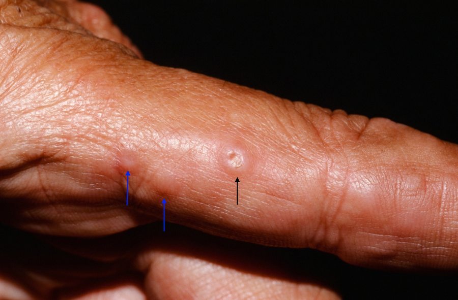
Perforating GA
-
Is very uncommon, and is usually localised to the fingers and dorsal hands although it can affect any body site and may be generalised
-
Lesions are umbilicated papules and may discharge small amounts of clear viscous fluid
-
Depending on the site, lesions can be tender
-
Actinic granuloma
- Lesions develop on UV-exposed sites
-
There is debate around whether it is a variant of granuloma annulare or a distinct entity - refer to the related chapter for more information
Many other GA variants have been described, but these are very rare and as such fall outside the remit of this chapter.
Clinical Images
Please refer to notes on image rights at bottom of the page with regards to individual image
ownership.
1
2
3
4
5
6
7
8
9
10
11
12
13
14
15
16
17
18
19
20
21
22
23
24
25
Investigations
-
GA is usually diagnosed clinically, however, where there is diagnostic uncertainty a skin biopsy may be required
- The most characteristic histological lesion in GA is the necrobiotic granuloma, but there are three histological patterns that may occur: (i) necrobiotic palisading granulomas; (ii) an interstitial form; and (iii) granulomas of sarcoidal or tuberculoid type. There is some variation in the literature in relation to the prevalence of each of these types in the different clinical patterns of disease (reference: Rook's Textbook of Dermatology)
-
An association between generalised GA and diabetes mellitus remains controversial
Management
-
Provide a patient information leaflet
-
For the vast majority of cases no treatment is required, based on the following:
-
The natural tendency for lesions is to resolve spontaneously - in 50% of patients lesions resolve within two years, although they can recur
-
There is limited evidence that treatment is beneficial
-
The potential adverse effects of treatments such as potent topical steroids
-
Potent (or super-potent) topical steroids and/or intralesional steroids
-
Have been used for very visible and persistent lesions, however, often with little benefit
-
With prolonged use may be associated with skin atrophy
- Referral to dermatology - more generalised GA
- For generalized disease, PUVA (a type of phototherapy) appears to give the best results with one retrospective study of 33 patients showing 50% clearance and a further 31% good to moderated improvement. UVA1 has also been reported to be effective in two case series
- Of the systemic therapies, dapsone, retinoids, antimalarials, fumaric acid esters, and methotrexate have been most extensively reported. None of these have been shown to be reliably beneficial
Disclaimer - the author PCDS cannot accept responsibility for any misleading or incorrect statements, and the management of individual patients remains the direct responsibility of the individual doctor. We do however hope that visitors to this site can contact us regarding comments that are considered misleading or incorrect so that we can continue to improve the site.
Image Rights - The PCDS would like to thank Dermatoweb, DermQuest (Galderma), and others who have contributed images. All named individuals and organisations maintain copyright for the relevant images.