HIV and AIDS - cutaneous features
LAST UPDATED: Aug 08, 2021
Introduction
Cutaneous manifestations of human immunodeficiency virus (HIV) disease may result directly from HIV infection, from opportunistic disorders secondary to immunosuppression, or as drug reactions resulting from treatment of HIV.
This chapter, which is set out as below, provides a brief introduction into the cutaneous features of HIV.
Clinical findings
Acute primary HIV infection and seroconversion
-
Once HIV infection has occured the immune system starts to develop antibodies, a process called seroconversion
-
Seroconversion occurs in most people within 45 days of infection, but can take upto three months
-
During this stage approximately 90% of infected individuals will be symptomatic
-
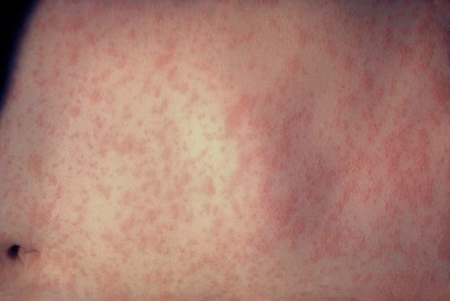
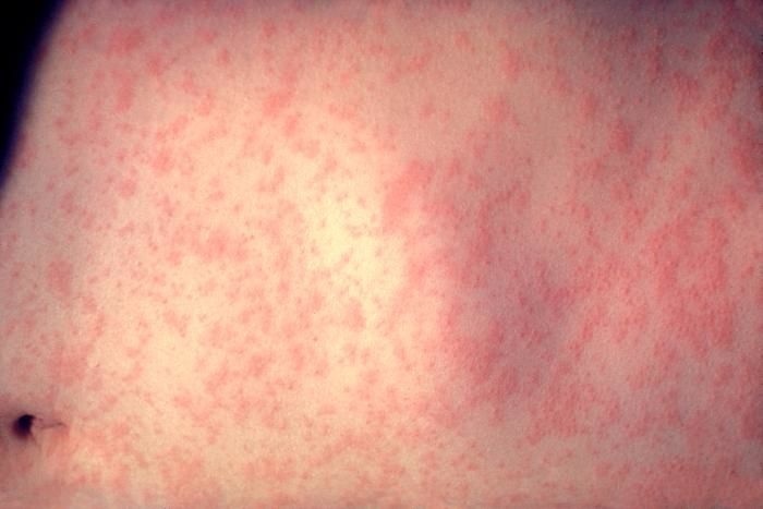
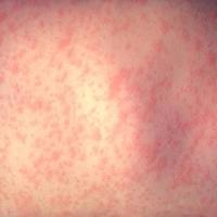
Symptoms are often non-specific with a transient, generalised, maculopapular eruption, associated with systemic symptoms such as fever, lethargy, arthralgia, gastrointestinal upset, headache, and lymphadenopathy
-
Other cutaneous features occasionally seen at this stage include urticaria, toxic urticated erythema, erythema multiforme and oral/genital ulceration
HIV and indicator conditions
NICE guidance and British HIV Association guidance recommends a HIV test is offered to patients presenting with indicator conditions, this includes specifically:
- Herpes zoster
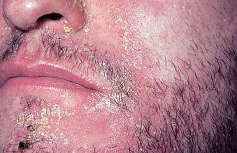
- Seborrhoeic eczema/dermatitis
- Severe or atypical psoriasis
- Exanthema
As well as these AIDS defining conditions:
- Herpes simplex ulcer(s) >1 month
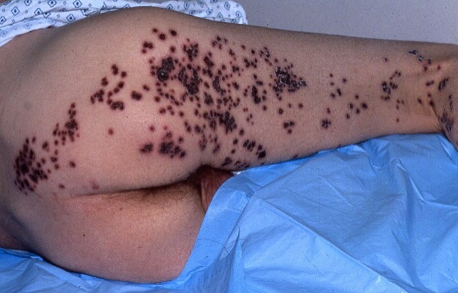
- Kaposi’s sarcoma
Dermatological conditions where HIV infection should form part of the differential diagnosis
-
Clinical features of seroconversion
-
Syphilis, genital herpes simplex and other sexually transmitted disease. Scabies, which can be sexually transmitted, is also more common and troublesome in patients with HIV
-
Mucosal changes
-
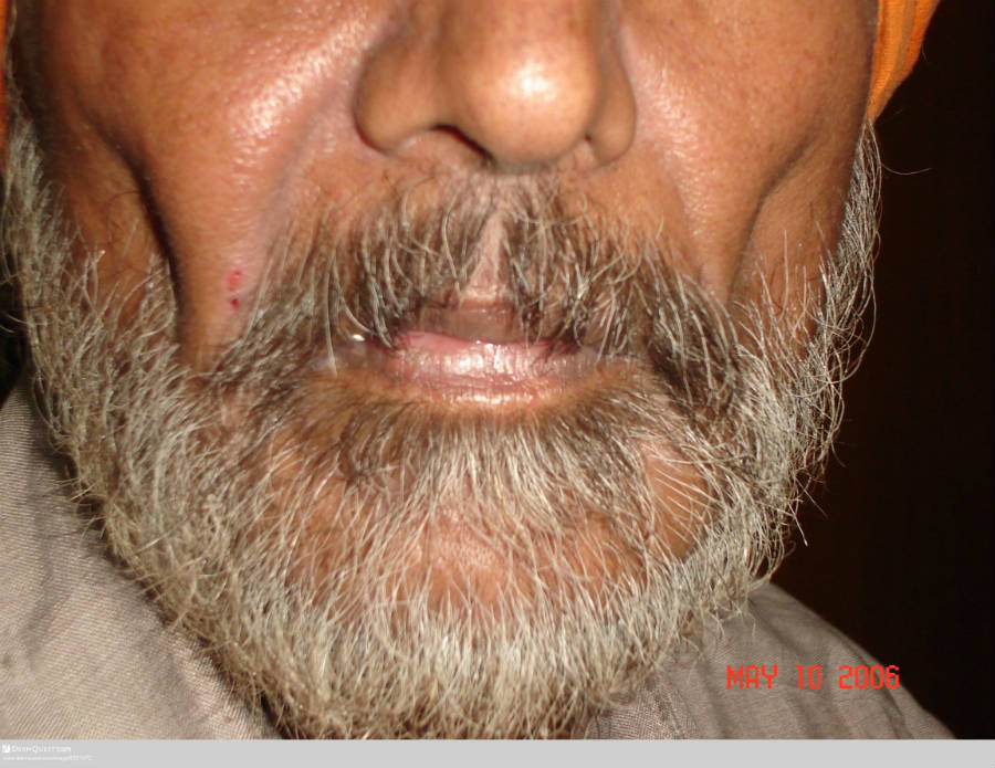
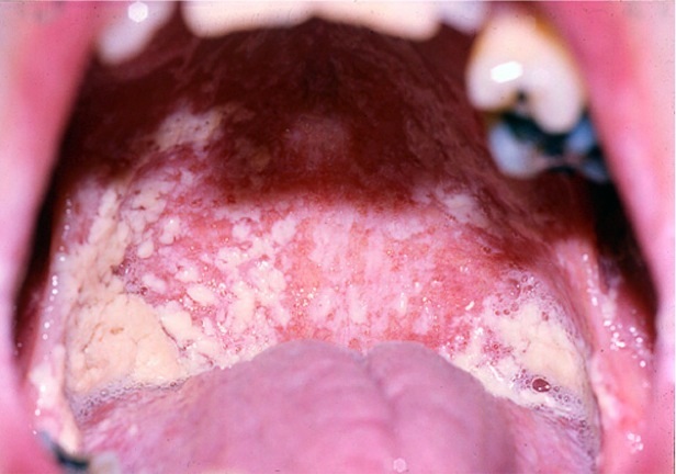
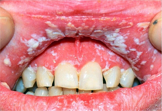
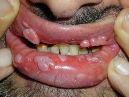
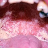
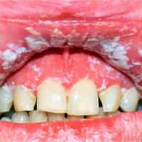
Persistent oral, pharyngeal, and sometimes genital, candida
-
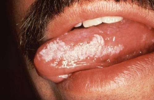
Oral hairy leukoplakia
-
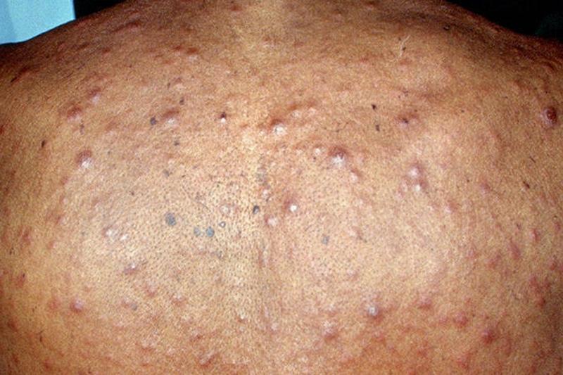
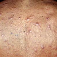
Pruritic, papular or papulo-pustular, follicular eruptions - the most closely associated of this group of conditions is eosinophilic folliculitis, which causes itchy, perifollicular papules/pustules on the face and trunk. Swabs are negative, and histology may be non-specific, showing a perifollicular polymorphic infiltrate, although sometimes it is rich in eosinophils. Persistent staphylococcal and pityrosporum folliculitis have also been associated with HIV infection
-
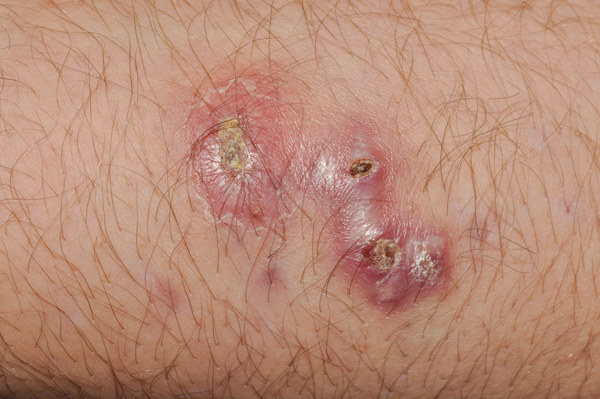
Opportunistic bacteria - tuberculosis, especially disseminated atypical mycobacteria (mycobacterium avium-intracellulare is the type most closely associated with HIV), ecthyma gangrenousm, and bacillary angiomatosis (a similar appearance to Kaposi's sarcoma with purple papules, nodules or plaques). Pneumocystis carinii can also occasionally present in the skin
-
Opportunistic mycoses (usually disseminated) - including cryptococcus (causing necrotic papules and nodules), histoplasmosis (a wide range of morphological features), coccidiodomycoses and penicilliosis
-
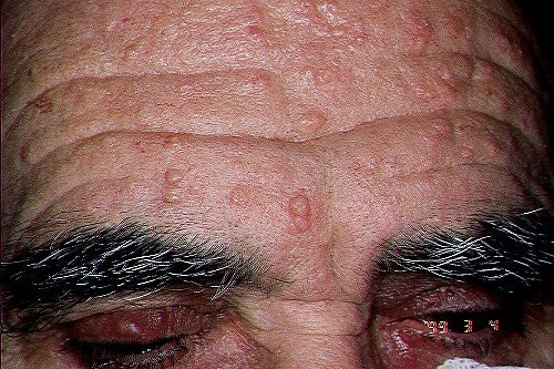
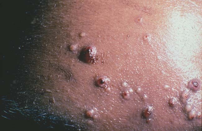
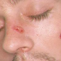
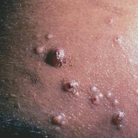
Certain viral infections - disseminated herpes zoster, chronic herpes simplex, widespread and recalcitrant warts, atypical presentations of molluscum (many larger lesions, often most numerous on the head and neck). Cytomegalovirus is rarely found in the skin
-
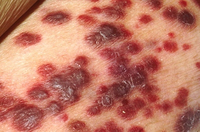
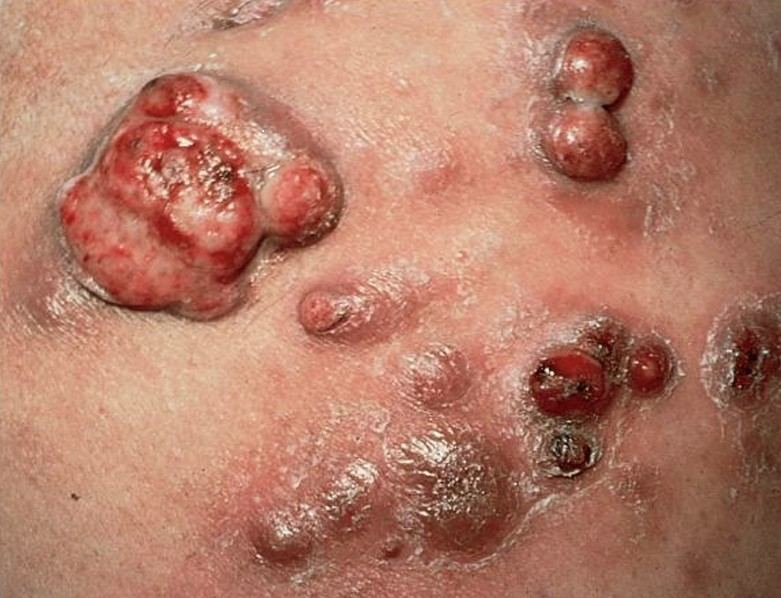
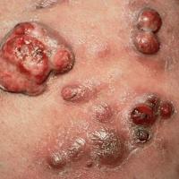
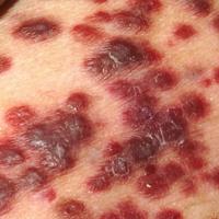
Kaposi's sarcoma - the incidence of which has decreased significantly. Lesions arise as dark blue / purple plaques and nodules
-
HIV-associated liposdystrophy - this is characterised by disappearance of adipose tissue, leading to sunken areas of skin, particularly of the face
-
Pruritus / dry skin - HIV has been associated with a number of such conditions. Xerosis and acquired ichthyosis affect 20-30% of HIV patients
-
Many others dermatological conditions, including moderate-severe cases of skin infections, psoriasis, seborrhoeic eczema, vasculitis and Sweet's syndrome have all been associated, indeed, HIV should be considered in any patient with a troublesome, and sometimes recalcitrant, skin condition, especially if associated with systemic features such as:
-
Weight loss
-
Chronic diarrhoea
-
Persistent generalised lymphadenopathy
-
Haematological abnormalities such as neutropenia and / or unexplained anaemia (Hb < 8)
-
Skin cancer - the risk of SCC and BCC is significantly raised, and both melanoma and cutaneous lesions of lymphoma are likely to have an increased incidence
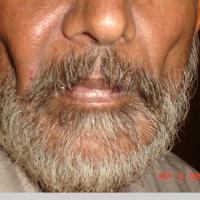
Clinical Images
Please refer to notes on image rights at bottom of the page with regards to individual image
ownership.
1
2
3

4
5
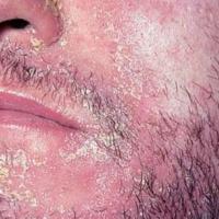
6
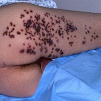
7
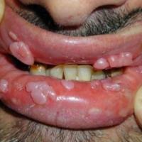
8

9
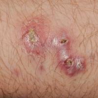
10
11
12
13
14
15
Investigations
-
There are two different types of tests which can detect HIV
-
Antibody test - a blood sample
-
Antigen test - sometimes called an antigen test, or a p24 test. The test uses a small sample of blood or saliva
-
Using both of those tests, the overwhelming majority of people who have HIV can be diagnosed within one month of being infected
-
For a person whose last possible exposure to HIV was at least one month ago, a negative result should be very reassuring. Nonetheless, for a very small number of people, it can take up to three months for the test to give an accurate result
Management
-
The life expectancy for patients with HIV has increased dramatically
-
The best prognosis is in those diagnosed and treated early
-
More patients, with relevant dermatological presentations, should be tested for HIV
-
Patients found to have the infection need to be referred urgently to an appropriate specialist
Disclaimer - the author PCDS cannot accept responsibility for any misleading or incorrect statements, and the management of individual patients remains the direct responsibility of the individual doctor. We do however hope that visitors to this site can contact us regarding comments that are considered misleading or incorrect so that we can continue to improve the site.
Image Rights - The PCDS would like to thank Dermatoweb, DermQuest (Galderma), and others who have contributed images. All named individuals and organisations maintain copyright for the relevant images.