Opportunistic skin infections
LAST UPDATED: Jan 20, 2021
Introduction
Opportunistic infections are those that arise as a result of a weakened immune system. They can be relatively localised or disseminated ie they spread to, and involve, other parts of the body.
This chapter, which is set out as below, provides a brief overview of some of the less well-known opportunistic infections.
Aetiology
The infections described in this chapter may occur as a result of:
-
HIV infection
-
Haematological malignancy
-
Immunosuppressive therapy eg for organ transplants
-
Other causes of immunosuppression
-
Occasionally arise in healthy individuals
-
May be endemic in some parts of the world
Clinical findings
The list below is not comprehensive in terms of the number of conditions and detail provided.
Mycoses (fungal infections)
-
Cryptococcus
-
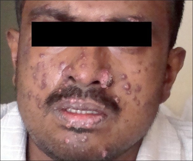
Infection with the encapsulated yeast Cryptococcus neoformans can result in harmless colonisation of the airways, but it can also lead to meningitis or disseminated disease, especially in immunosuppressed patients
-
The respiratory tract is the usual portal of entry, but primary cutaneous lesions can occur
-
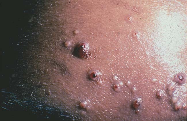
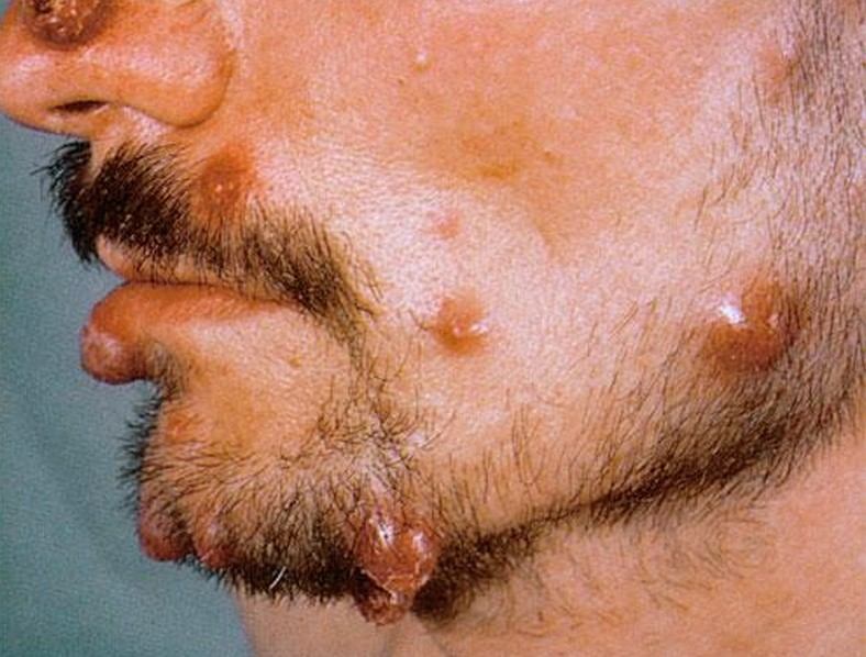
Cutaneous manifestations occur in 10%-15% of cases and usually take the form of slow-growing, firm/cystic, papules and nodules, some of which will ulcerate and become necrotic. Acneiform papules and pustules are characteristic of widespread systemic infection
-
Aspergillus
-
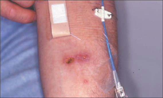
Infections can be primary and occur by direct inoculation (eg sites of trauma, burns, intravenous infusion sites, surgical wounds, and other wounds where there are contaminated dressings or splints), or result from dissemination (eg aspergillus from the lung)
-
Disseminated disease is more likely to be associated with immunosuppression
-
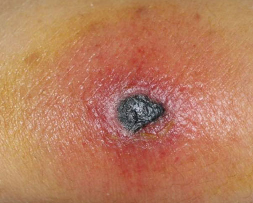
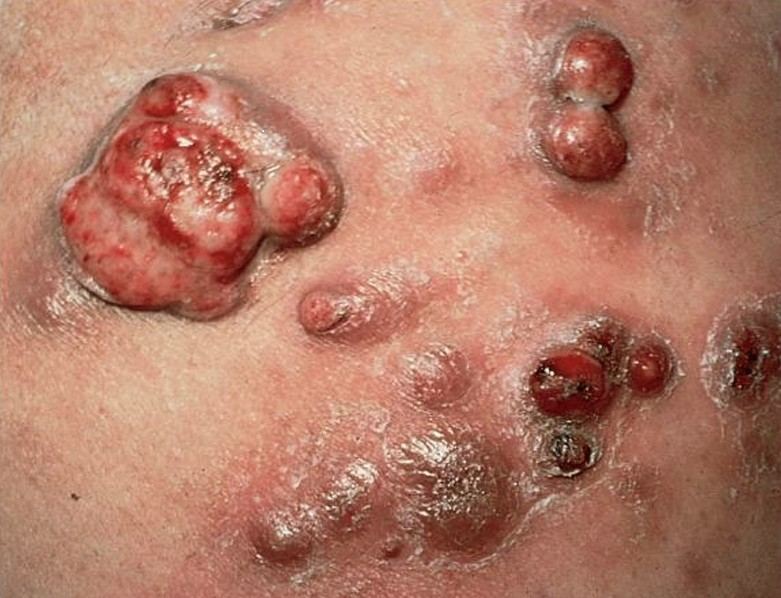
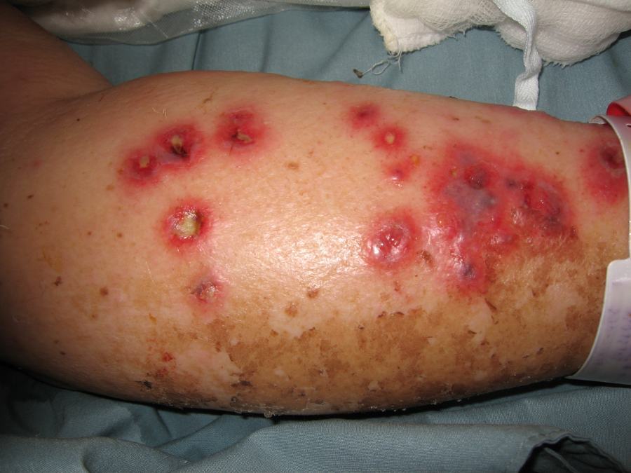
Clinical features - often multiple erythematous papules or plaques, which become pustular or haemorrhagic, and then quickly develop into a necrotic ulcer or eschar
-
Zygomycosis (syn. Mucormycosis)
-
Zygomycosis refers to several different diseases caused by infection with fungi in the order of Mucoral
-
Most zygomycotic infections are life-threatening, and due to underlying immunosuppression, especially neutropenia
-
Cutaneous disease manifests as cellulitis, which progresses to dermal necrosis and black eschar formation, similar in appearance to necrotising fasciitis
-
Histoplasmosis
-
A highly infectious mycosis caused by Histoplasma capsulatum, which primarily affects the lungs
-
Most individuals with histoplasmosis are asymptomatic. Those who develop clinical manifestations are usually immunosuppressed
-
Cutaneous features generally results from dissemination, primary skin inoculation is rare
-
Cutaneous involvement takes on a number of different appearances including papules, nodules, ulcers, granulomas and pigmentary change. Both erythema muliforme and erythema nodosum have been associated with acute pulmonary histoplasmosis
-
There are several other mycoses that can arise in immunosuppressed patients, and the possibility of a disseminated mycosis should always be considered as part of the differential diagnosis in patients developing multiple cutaneous lesions
Bacillary angiomatosis
-
A vascular, proliferative form of Bartonella infection that occurs primarily in immunosuppressed patients
-
It often has a similar clinical appearance to Kaposi's sarcoma with purple papules, nodules or plaques
-
Other cutaneous presentations include hyperpigmented, hyperkeratotic, indurated plaques, typically on the extensor surfaces of the extremities
Mycobacterial infections
-
These may be cutaneous, pulmonary or disseminated
-
The most common mycobacterial infection associated with HIV is mycobacterium avium-intracellulare
- Refer to the chapter Mycobacterial infections for detailed information
Ecthyma gangrenosum
-
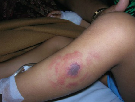
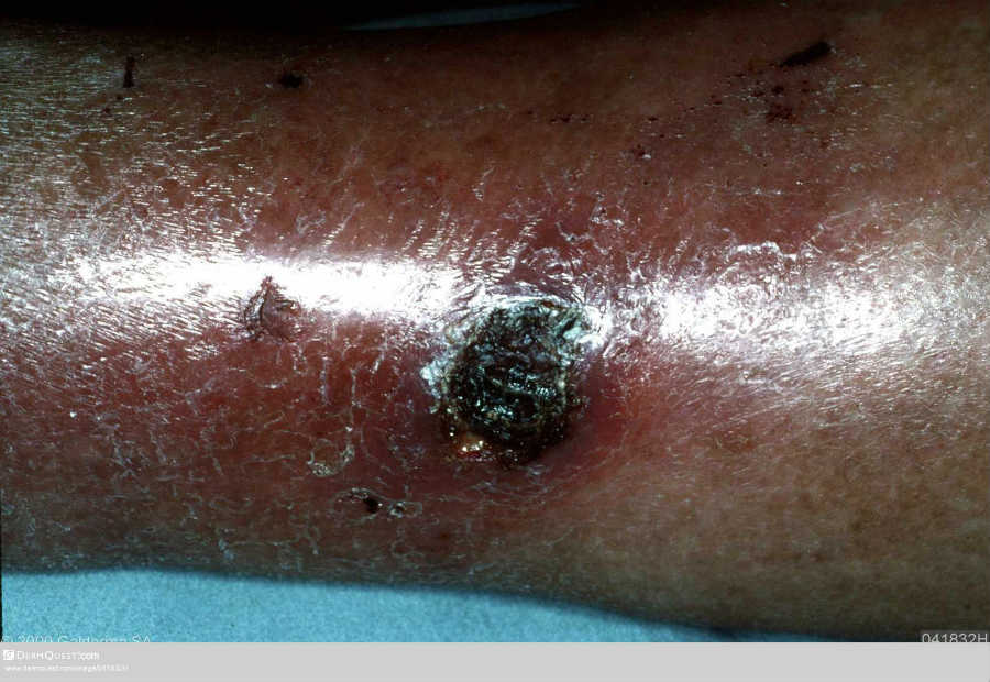
Ecthyma gangrenosum is the cutaneous manifestation of Pseudomonas aeruginosa septicaemia, typically affecting immunosuppressed patients, particularly those with neutropenia
-
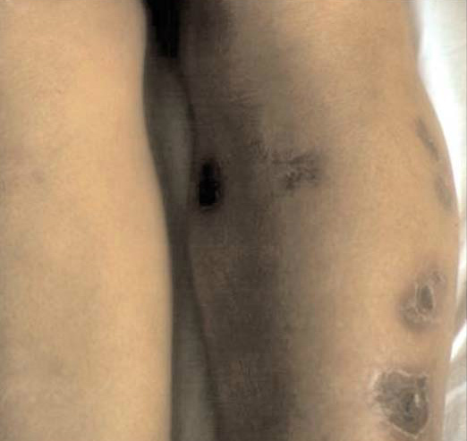
Lesions first presents as a pustule or nodule, which quickly develops a central haemorrhagic area that subsequently breaks down to form a large necrotic ulcer with a tender erythematous border
-
For more information refer to the related chapter Ecthyma (including ecthyma gangrenosum)
Cytomegalovirus (CMV)
-
CMV disease is a severe complication among immunosuppressed patients, it tends to be associated with very few cutaneous features
Cryptosporidiosis
-
Is caused by infection with apicomplexan protozoans of the genus Cryptosporidium
-
In some parts of the world it is a common self-limiting diarrhoeal illness of healthy individuals, mainly children
-
It is recognised as causing prolonged diarrhoea in patients with HIV, and can occasionally cause cutaneous symptoms
Clinical Images
Please refer to notes on image rights at bottom of the page with regards to individual image
ownership.
Investigations
-
Patients require extensive investigations including blood tests, blood cultures, sputum samples, urinalysis and a CXR
-
Cutaneous lesions may need to be biopsied - one biopsy should be sent for standard histological examination (light microscopy), and a second biopsy for tissue culture (a plain tube with a small amount of saline in the base)
Management
-
Management depends on the type of infection and any underlying cause
Disclaimer - the author PCDS cannot accept responsibility for any misleading or incorrect statements, and the management of individual patients remains the direct responsibility of the individual doctor. We do however hope that visitors to this site can contact us regarding comments that are considered misleading or incorrect so that we can continue to improve the site.
Image Rights - The PCDS would like to thank Dermatoweb, DermQuest (Galderma), and others who have contributed images. All named individuals and organisations maintain copyright for the relevant images.