Lichen simplex (syn. circumscribed neurodermatitis)

LAST UPDATED: Dec 18, 2023

Patient Information Leaflet
Link: Lichen simplex (lichen simplex chronicus)

https://www.pcds.org.uk/patient-info-leaflets/lichen-simplex-lichen-simplex-chronicus
Introduction

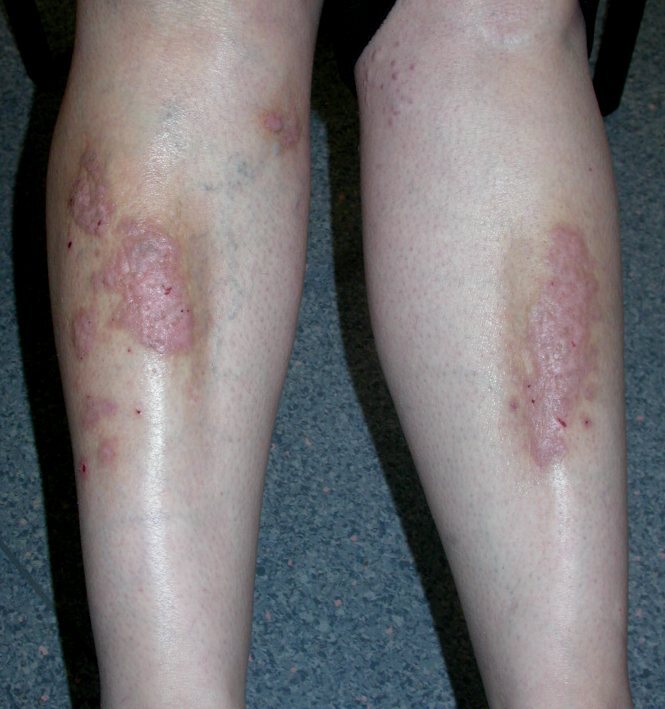
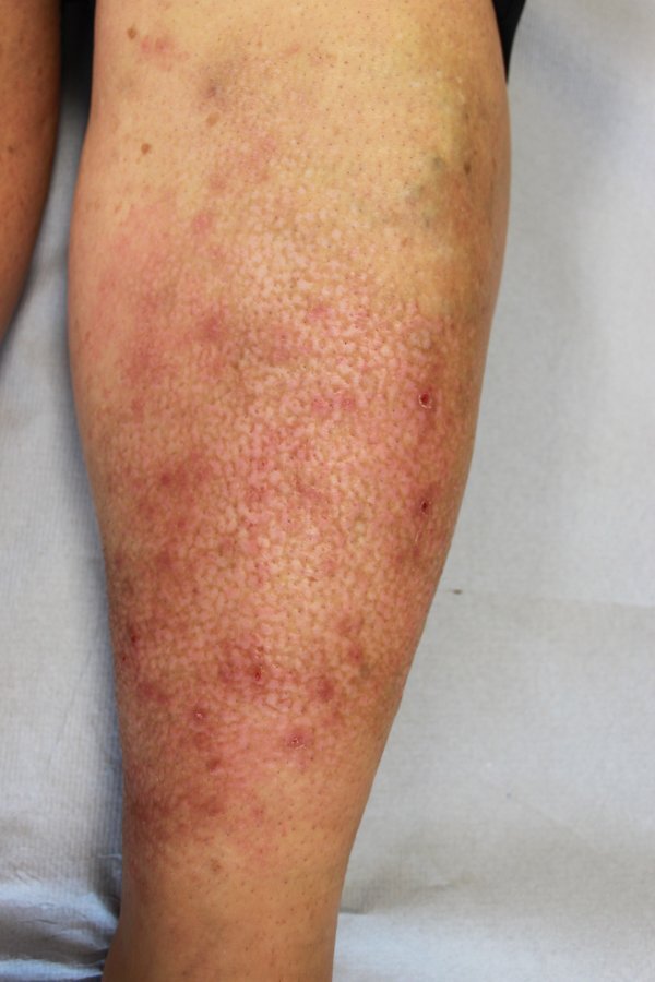
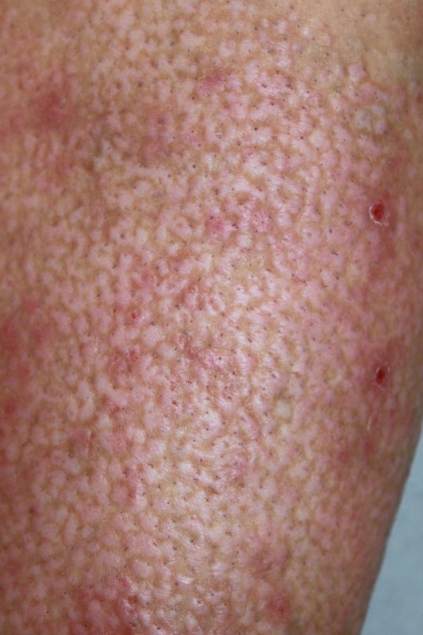
Lichen simplex is an eczematous dermatosis characterised by a small number of pruritic, heavily lichenified plaques or, very often, a single lesion.

This chapter is set out as follows:


Aetiology

  • Not all individuals are capable of developing lichenification, but for those that are, the following are contributing factors to the development of lichen simplex:
    • Patients are more readily conditioned to scratch when itchy
    • Stress
    • An atopic background

History

  • Lichen simplex normally affects adults with a peak incidence of 30-50 years of age
  • Woman are affected more than men
  • Itch is often intense and can be out of proportion to the clinical findings

Clinical findings

Distribution

Lesions can arise on any location that the patient can reach, however, the following are the most common sites:

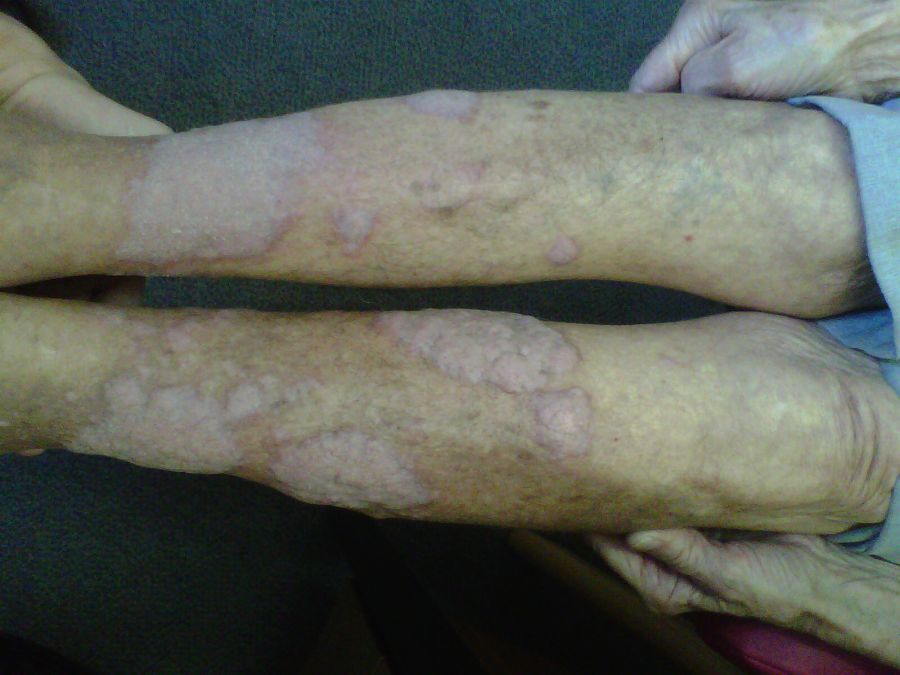
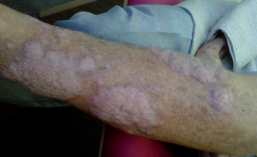
  • Extensor forearms / elbows
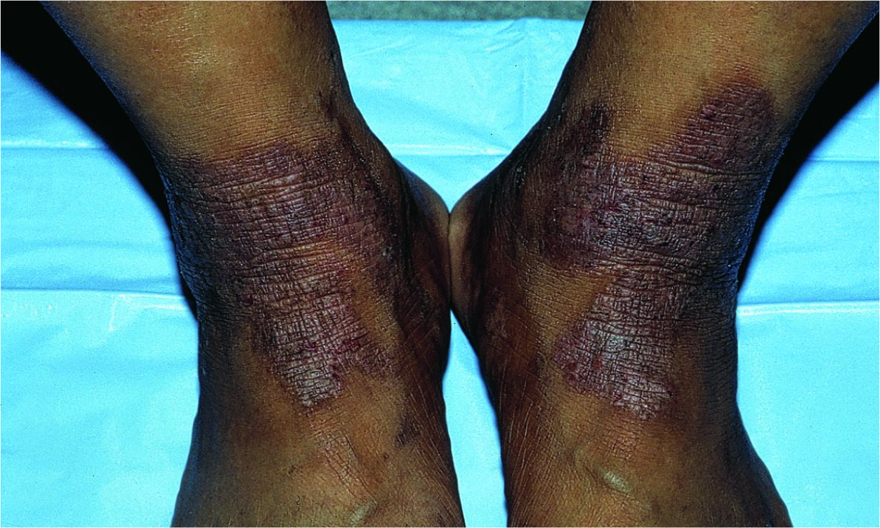
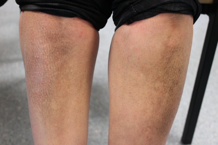
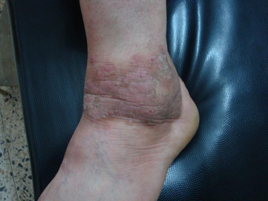
  • Lower legs / ankles
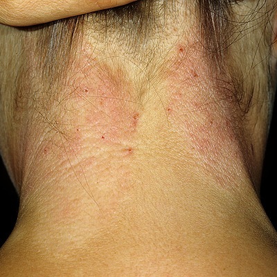
  • The sides and nape of the neck, the latter is found predominantly in women. The scalp may also be involved
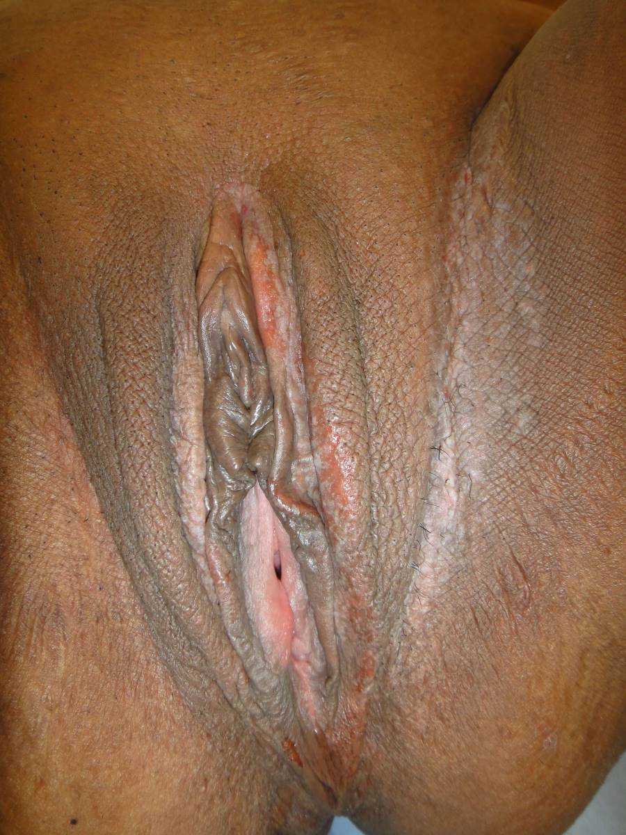
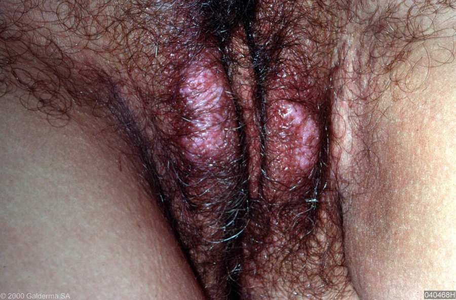
  • Vulva - which often presents with a symmetrical thickening, however the findings can be more pronounced on one side such that in right handed people the changes are often worse on the left and visa versa
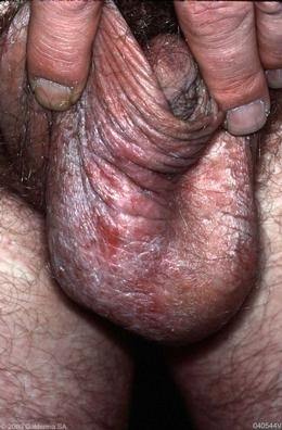
  • Scrotum

Morphology

  • Erythema is present in the early stages
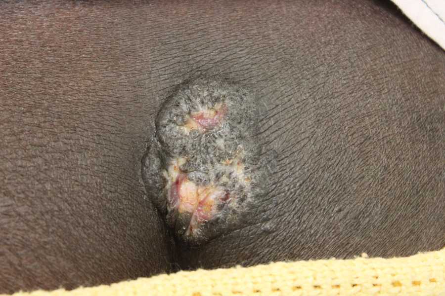
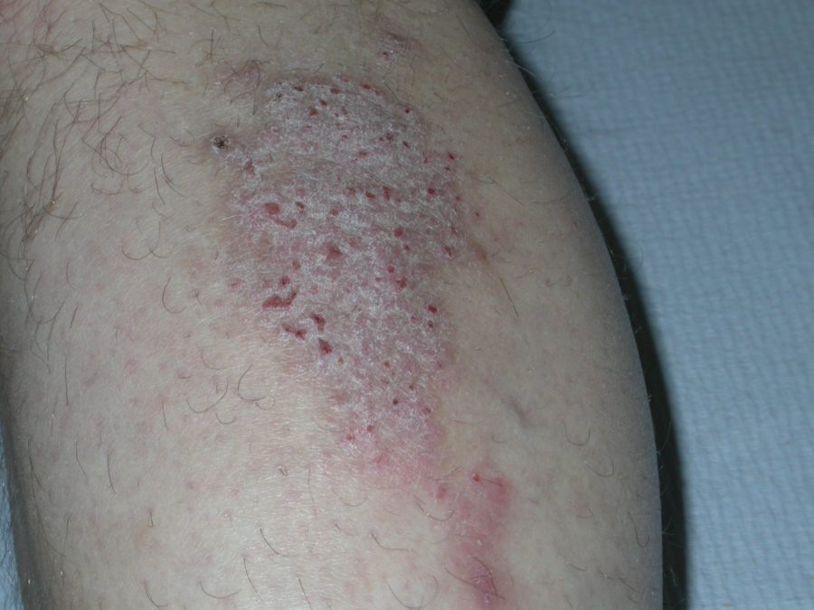
  • Over time the skin becomes thickened (lichenified) centrally to form a scaly plaque, which may become hyperpigmented. Thickening of the skin can be substantial, sometimes developing warty, cribriform surfaces
  • Surrounding the central plaque are a group of firm, lichenoid papules
  • Erosions and ulceration can develop from scratching

Clinical Images

Please refer to notes on image rights at bottom of the page with regards to individual image ownership.
Investigations

Investigations are not usually needed, however, the following can be considered:

  • Mycology - skin scrappings can be sent if there is any suggestion of tinea. Indeed, lichenification can sometimes occur in chronic, untreated tinea
  • Patch testing - if there is a possibility of a contact allergic dermatitis
  • Skin biopsy - to rule out more serious skin pathology (eg a cutaneous lymphoma) if the history / examination are not typical. Histologically there is epidermal hyperplasia and hyperkeratosis. The rete ridges are lengthened. Spongiosis is sometimes present, and small areas of parakeratosis are occasionally seen. The dermis contains a chronic inflammatory cell infiltrate. In very chronic lesions there may be some fibrosis (reference: Rook's Textbook of Dermatolo

Management

Step 1: general 

  • Provide a patient information leaflet
  • Address any exacerbating factors such as stress
  • Encourage patients not to rub / scratch the skin
  • Advice on the regular use of soothing emollients eg menthol in aqueous cream
  • Consider the use of sedating anti-histamines where practical eg Atarax ® (hydroxyzine) tablets

Step 2: potent or super-potent topical steroids 

  • 0.1% Betnovate ® cream or Dermovate ® cream / ointment applied thinly once daily until the plaques flatten and the itch settles. If the site allows, it can be very useful to occlude the area once the treatment has been applied - one of the most effective occlusive agents is Clingfilm, tubular bandages are an alternative
  • Steroid impregnated treatments - there are two types, both of which can be left on for up to 24 hours before reapplying:
    • Haelan ® tape (fludroxycortide tape) - attached is a video demonstrating how to use the tape
    • Betesil ® medicated plaster (betamethasone valerate 2.25mg) - attached is a video demonstrating how to use the plaster
  • For stubborn patches or those lesions that are moderate-severe at onset, the most useful treatment is the use of a potent or super-potent topical steroid under medicated bandages eg ZIPZOC ® or Viscopaste ®. The bandages should be replaced once or twice a week and the treatment continued until the plaques flatten and the itch settles. Please click on the following link for demonstrations on how to apply these bandages/dressings

Step 3: intralesional steroid therapy

  • Steroid therapy, eg triamcinolone acetonide, can be given directly into lesions. A high pressure is often needed for effective infiltration of the lesion, which can be achieved by using a 1 ml insulin syringe. Several treatments may be required, each given approximately 3-6 weeks apart  

Step 4: patient expectation

  • Once the skin has settled, patients should continue on with the regular use of emollients
  • Lichen simplex can frequently recur, especially if the patient is unable to address exacerbating factors and the habit of scratching

Disclaimer - the author PCDS cannot accept responsibility for any misleading or incorrect statements, and the management of individual patients remains the direct responsibility of the individual doctor. We do however hope that visitors to this site can contact us regarding comments that are considered misleading or incorrect so that we can continue to improve the site.

Image Rights - The PCDS would like to thank Dermatoweb, DermQuest (Galderma), and others who have contributed images. All named individuals and organisations maintain copyright for the relevant images.

Quick Links

The following pharmaceutical companies have had no involvement in the content of this website or in our conference programmes


Almirall
Galderma
Glenmark
Johnson & Johnson
La Roche-Posay
LEO Pharma
Pierre Fabre
Schuco